

Author(s) :
Raquel Sánchez1, Raquel Ciérvide1, Beatriz Alvarez1, Mercedes Lopez1, Bruno Esteban1, Daniel Zuca2, Carmen Rubio1
1Radiation Oncology Department, University Hospital HM Sanchinarro, Madrid, Spain
2Medical Physics Department, University Hospital HM Sanchinarro, Madrid, Spain
Corresponding author: Raquel Sanchez, Email: raquelsc53@gmail.com
Publication History: Received - 20 November 2025, Revised - 3 August 2026, Accepted - 30 August 2026, Published Online - 30 September 2026.
Copyright: © 2026 The author(s). Published by Casa Cărții de Știință.
User License: Creative Commons Attribution – NonCommercial (CC BY-NC)
 Share
Highlights
- Primary cardiac sarcomas are rare, aggressive tumors with frequent local recurrence and limited options once surgery is no longer feasible.
- Stereotactic radiotherapy delivered with VMAT under deep inspiration breath-hold enables safe, high-precision treatment near the heart.
- High-dose irradiation adjacent to a mechanical mitral valve is feasible without acute cardiac or pulmonary toxicity or valve dysfunction.
- VMAT-based SABR with respiratory control offers a practical alternative for centers without MR-guided radiotherapy, though longer follow-up is needed.
Abstract
Primary cardiac sarcomas are extremely rare and aggressive cancers with a generally poor prognosis and limited treatment options. Surgery remains the mainstay of management, but local recurrence is frequent even after apparently complete resection.
We describe the case of a 56-year-old woman with a recurrent dedifferentiated liposarcoma of the left atrium following mitral valve replacement. The recurrence was treated with stereotactic body radiotherapy (SABR) delivered by volumetric modulated arc therapy (VMAT) under deep inspiration breath-hold (DIBH). A total dose of 32.5 Gy in five fractions was administered. The treatment was well tolerated, with no acute cardiac or pulmonary toxicity and preserved valve function. Two months after treatment, lesion remains stable and no altered cardiac function. This case supports the feasibility and safety of SABR-VMAT with DIBH for recurrent cardiac sarcoma, offering an effective local option when surgery or MR-guided RT are not available.
1. Introduction
Primary cardiac sarcomas (PCS) are exceedingly rare tumors, accounting for less than 0.1% of all cardiac malignancies. They arise most commonly within the atrial chambers—particularly the left atrium—and tend to follow an aggressive clinical course characterized by early local invasion and a high risk of recurrence after resection. Angiosarcoma is the most frequently reported histological subtype, followed by intimal sarcoma and undifferentiated pleomorphic sarcoma; cardiac liposarcoma, by contrast, is rarely encountered. (1-5)
Surgical resection remains the cornerstone of management and the only potentially curative approach. Complete resection, however, is often precluded by anatomical constraints and the proximity of the tumor to critical cardiac structures, and local recurrence is common even after apparently complete excision. Systemic therapy with anthracycline- and ifosfamide-based regimens may offer benefit in selected patients, yet overall outcomes remain poor.
Radiotherapy has historically played a limited role in the management of cardiac sarcomas, owing to concerns about cardiac tolerance, cardiorespiratory motion and the difficulty of accurately targeting tumors arising in or adjacent to the heart. In recent years, however, advances in image guidance, motion management and dose delivery have expanded its therapeutic potential. Techniques such as stereotactic ablative radiotherapy (SABR), volumetric modulated arc therapy (VMAT), CyberKnife-based radiosurgery and magnetic resonance–guided adaptive radiotherapy (MRgRT) now enable highly conformal dose distributions while limiting exposure to adjacent organs at risk. (2,5) More recently, MR-guided and adaptive approaches have further demonstrated the feasibility of high-precision radiotherapy in challenging cardiac locations, including cases involving prosthetic valves. (1,4,6-8)
Here, we describe a case of recurrent dedifferentiated liposarcoma of the left atrium treated with SABR-VMAT under deep inspiration breath-hold (DIBH) and review the current evidence supporting this strategy.
2. Case Report
A 56-year-old woman was initially evaluated in 2019 for fever and joint pain. Transthoracic echocardiography revealed a lobulated mass arising from the anterior mitral leaflet, producing severe mitral regurgitation. Staging CT and coronary angiography were unremarkable. She underwent surgical resection of the mass with mechanical mitral valve replacement in January 2020. Histology showed a total resection (R0) of a dedifferentiated liposarcoma with MDM2 gene amplification, overexpression that leads to tumorigenesis and cancer progression by allowing cells to evade normal growth control and apoptotic mechanisms. The patient received six courses of ifosfamide–doxorubicin adjuvant chemotherapy and started follow-up with good general conditions.
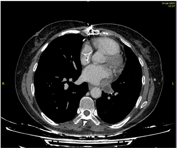
In late 2023, MRI detected a recurrent mass within the left atrium, extending into the left pulmonary veins which were partially occluded by thrombosis (Fig.1). After retreatment with ifosfamide–doxorubicin, a second surgery was performed with positive surgical margins. Consequently, the patient received a chemotherapy-based treatment, starting with trabectedin as second-line therapy for relapse, followed by eribulin as a third-line treatment. However, no response was observed. What is more, by mid-2025, imaging revealed a slow enlargement beside the pulmonary veins, with no evidence of distant metastases (Fig. 2). After careful evaluation, the multidisciplinary tumor board recommended stereotactic ablative radiotherapy (SABR) for definitive local control.
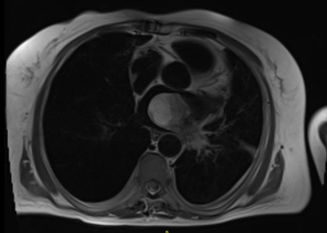
Figure 1. Cardiac MRI performed in December 2023 showing evidence of disease recurrence.
A. Chest CT demonstrating interval enlargement of the lesion (14/07/2025)
B. Findings compatible with post-radiotherapy alterations (14/10/2025)
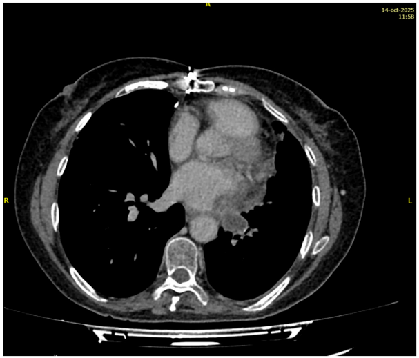
Figure 2. Thoracic CT scan in axial view (A. and B.)
Radiotherapy planning and delivery:
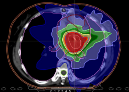
Simulation CT was performed under deep inspiration breath-hold (DIBH) with the patient in supine position and arms raised above her head. Target structures and organs at risk were contoured on a contrast-enhanced planning CT scan with 2-mm slice thickness. The GTV (gross tumor volume) corresponding to the recurrence of the cardiac liposarcoma was delineated, and a 5-mm margin was added to generate the PTV (planning target volume). Planning was carried out with the Collapsed Cone algorithm (Monaco v5.6), incorporating the iBEvo couch model. Two coplanar dynamic arcs were used. A total dose of 32.5 Gy in five daily fractions (6.5 Gy/fx) was prescribed to 85% of the planning target volume (PTV). The mean PTV dose was 33.7 Gy (D95 = 32.6 Gy). Dose constraints were achieved for all organs at risk: heart Dmax < 55 Gy, lung V15 < 15%, esophageal Dmax < 55 Gy and spinal cord Dmax < 28 Gy.
Treatment was delivered using a Versa HD linear accelerator (Elekta AB, Sweden) with 6 MV flattening filter-free (FFF) photons and VMAT (Volumetric Modulated Arc Therapy).
SABR was delivered in August 2025 over five consecutive treatment days. Each fraction was delivered under DIBH system and daily cone-beam CT and surface guided radiation therapy (Catalyst, C-Rad) verification. Average session time was around 15 minutes, requiring three to four breath-hold cycles per fraction. The procedure was well tolerated, without acute side effects. Post-treatment echocardiography confirmed normal prosthetic valve function
At the most recent follow-up in October 2025, approximately two months after SABR, the patient was in good clinical condition, without any symptomatology attributable to disease or treatment.
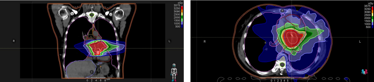
Figure 3. Axial and coronal views of the SABR–VMAT plan delivered under DIBH
Follow-up:
Cardiac MRI one month after treatment showed a stable residual thickening at the left pulmonary vein confluence, with preserved biventricular function and no pericardial effusion. At the most recent follow-up (October 2025) (Fig.2) the patient was in a good clinical condition, without any symptomatology attributable to disease or treatment. Additionally, a CT evaluation was performed, revealing no radiological evidence of either local or distant recurrence.
3. Discussion
Primary cardiac sarcomas are rare, aggressive tumors with poor outcomes. Surgery offers the best chance of prolonged survival, but complete resection is often not feasible and recurrences are common. Chemotherapy provides limited benefit, and radiotherapy has traditionally been avoided due to concerns about cardiac motion and radiosensitivity. However, recent advances in image-guided and highly conformal radiotherapy techniques (e.g., SABR, VMAT, MRgRT) now allow precise high-dose treatment while sparing normal tissue, opening new therapeutic possibilities. Although clinical experience is still limited, early results are encouraging.
Table 1 summarizes clinical results with radiation therapy for sarcomas arising at the heart. Corradini et al.1 reported four inoperable primary cardiac sarcomas treated with MR-guided SABR, achieving local control without relevant toxicity. Pomp et al.8 and Noyan et al.7 described similar experiences using MR-adaptive radiotherapy with daily replanning. Later, Hinojosa-González et al.6 and Batumalai et al.1 confirmed that ablative MR-guided treatment can be safely delivered even in patients with prosthetic valves, maintaining function and disease stability.
Encouraging results have also come from non-MR platforms. Blanc et al.2 published the first case of SABR delivered with VMAT (35 Gy in five fractions) for a primary cardiac angiosarcoma, with complete local response and no toxicity. Shah et al.9 reported successful salvage radiosurgery using CyberKnife (25 Gy in one fraction) after prior RT, achieving durable control. Stergioula et al.10 combined image-guided IMRT (60 Gy in 30 fractions) with weekly paclitaxel in an unresectable angiosarcoma, resulting in significant tumor regression that allowed resection.
| Author / Year | Tumor type / Location | Technique | Dose / Fractionation | Outcome / Follow-up |
|---|---|---|---|---|
| Blanc J et al., 2020 | Primary angiosarcoma (left atrium) | SABR-VMAT | 35 Gy / 5 fractions | Complete local control ; follow-up: not specified; no toxicity reported. |
| Present case (2025) | Recurrent dedifferentiated liposarcoma (left atrium) | SABR-VMAT + DIBH | 32.5 Gy / 5 fractions | Stable disease ; follow-up: 2 months; no acute cardiac or pulmonary toxicity; preserved valve function |
| Stergioula A et al., 2023 | Unresectable angiosarcoma (right atrium) | IG-IMRT + Paclitaxel | 60 Gy / 30 fractions + weekly paclitaxel | >50% tumor reduction ; follow-up: not specified; no treatment-related toxicity reported. |
| Corradini S et al., 2021 | 4 primary cardiac sarcomas (various) | MR-guided SABR | 30–41 Gy / 5 fractions | Local control ; follow-up: ≥6 months; no ≥ grade 2 toxicity. |
| Pomp J et al., 2021 | Recurrent intimal sarcoma (left atrium) | SMART (MR-adaptive RT) | 60 Gy / 12 fractions | Stable 6 months, no toxicity |
| Noyan A et al., 2022 | Angiosarcoma (right atrium, pediatric) | MR-adaptive SABR (SIB) | 25–30 Gy / 5 fractions | Stable disease; follow-up: 6 months; no toxicity reported. |
| Hinojosa-González DE et al., 2023 | Recurrent cardiac sarcoma near mitral bioprosthesis | MR-guided SABR | 36 Gy / 6 fractions | Local control ; follow-up: >12 months; no valve dysfunction reported. |
| Batumalai V et al., 2023 | Recurrent cardiac sarcoma adjacent to mechanical mitral valve | MR-Linac SABR | ~35 Gy / 5–6 fractions | Stable disease; follow-up: 12 months; no valve dysfunction. |
| Bonomo P et al., 2015 | Primary cardiac malignancies | CyberKnife SABR | 24–30 Gy / 3–5 fractions | Local control ; follow-up: 6 months; toxicity minimal or not reported. |
| Gabani P et al., 2019 | Cardiac angiosarcoma / metastases | CyberKnife SABR | 24–30 Gy / 3–5 fractions | Feasible treatment; follow-up: not specified; minimal toxicity reported. |
| Shah SN et al., 2023 | Recurrent pleomorphic sarcoma (left atrium) | CyberKnife SRS (salvage) | 25 Gy / 1 fraction (after 72 Gy / 15 fx) | Local control; follow-up: >12 months; no toxicity reported. |
Table 1. Published cases of radiotherapy for primary or recurrent cardiac sarcomas
In our patient, relapse occurred near the left pulmonary veins and mechanical mitral valve. Re-operation was excluded, and prior systemic therapies had failed. SABR planned with VMAT and delivered under DIBH was selected to achieve high-precision treatment in a mobile and anatomically complex target. DIBH reduced thoracic motion, improving treatment reproducibility and target accuracy. At the most recent follow-up, two months after SABR, the patient remained clinically well, with stable disease, no acute cardiac or pulmonary toxicity, and preserved mechanical mitral valve function.
The fractionation schedule of 32.5 Gy in five fractions was selected to balance the need for a dose-intense hypofractionated local treatment with the need to minimize the risk of toxicity to adjacent critical structures. Given the proximity of the lesion to the mechanical mitral valve, left atrium and pulmonary veins, further dose escalation was not considered appropriate. The selected regimen allowed adequate target coverage while respecting all predefined organ-at-risk constraints. It was also consistent with previously published SABR experiences in cardiac sarcomas, in which hypofractionated schedules of approximately 30–40 Gy delivered in five to six fractions have been reported with acceptable short-term toxicity.
Given the singularity of this tumor entity and its location, we consider that one of the strengths of this case lies in the feasibility of safely delivering high radiation doses near the heart, provided that the technical conditions under which treatment is administered allow for high precision. However, to determine whether this approach provides the required degree of radicality, longer follow-up is necessary.
Taken together, these experiences, modern, image-guided radiotherapy approaches can provide meaningful benefit for selected patients with cardiac sarcomas. Hypofractionated SABR regimens around 30–40 Gy in five to six fractions appear effective and safe. VMAT-based SABR combined with respiratory control offers a practical alternative to MR-guided treatments. Further studies with larger patient cohorts and longer follow-up are still needed to establish long-term cardiac tolerance and optimal planning parameters.
This report has several limitations. First, it describes the outcome of a single patient, which precludes any definitive conclusions regarding efficacy or generalizability. Second, follow-up remains relatively short, limiting assessment of long-term local control and late cardiac toxicity. Finally, the use of SABR for primary or recurrent cardiac sarcomas requires highly specialized planning, image guidance and motion-management techniques, which may limit its applicability across centers. Nevertheless, given the extreme rarity of these tumors, carefully documented case reports may help to refine patient selection, treatment planning and safety considerations in this challenging clinical setting.
4. Conclusion
This case demonstrates the feasibility and safety of delivering stereotactic body radiotherapy with VMAT under deep inspiration breath-hold for recurrent cardiac sarcoma adjacent to a mechanical mitral valve. The approach achieved excellent target coverage with minimal exposure to surrounding tissues and no treatment-related adverse effects.
In patients who are not candidates for surgery, SABR represents a valuable local treatment option. VMAT-based SABR with respiratory control offers precision comparable to MR-guided techniques and can be applied in centers without MR-Linac systems. Continued clinical experience and longer follow-up will be essential to refine dose constraints and further define the role of SABR in cardiac sarcomas.
Abbreviations
SRS – stereotactic radiosurgery
SIB – simultaneous integrated boost
MR – magnetic resonance
Gy – Gray
Mo – month
IG – image guided
VMAT – volumetric arc therapy
DIBH – deep inspiration breath hold
Statements
Conflict of interest: The authors declare no conflict of interest regarding any aspect of this manuscript
Funding sources: No funding was received for this work.
References
- Batumalai V, Holloway LC, Westhuyzen J, et al. (2023). MR-guided stereotactic body radiotherapy for recurrent cardiac sarcoma with mechanical mitral valve prosthesis: a case report. Journal of Medical Radiation Sciences, 70(2), 206–213.
- Blanc J, Forstner D, Bhattacharyya T, Bonomo P. (2020). Stereotactic body radiotherapy (SBRT) for primary cardiac angiosarcoma: a case report and literature review. Journal of Medical Radiation Sciences, 67(2), 186–190.
- Bonomo P, Greto D, Desideri I, et al. (2015). Stereotactic body radiotherapy for primary and metastatic cardiac malignancies. Radiologia Medica, 120(12), 1121–1129.
- Corradini S, Alongi F, Andratschke N, et al. (2021). MR-guided stereotactic body radiation therapy for primary cardiac sarcomas – first clinical experiences. Radiation Oncology, 16, 72.
- Gabani P, Fischer-Valuck BW, Johanns TM, et al. (2019). Stereotactic body radiation therapy for cardiac metastases and primary cardiac malignancies: a single institution experience. Practical Radiation Oncology, 9(5), e323–e331.
- Hinojosa-González DE, Lara-Guerra H, Díaz-Ruiz P, et al. (2023). Stereotactic MR-guided adaptive radiotherapy for recurrent cardiac sarcoma adjacent to a mitral bioprosthesis: a case report. Journal of Medical Radiation Sciences, 70(2), 199–205.
- Noyan A, Yavaş G, Efe E, et al. (2022). Cardiac angiosarcoma treated with 1.5 Tesla MR-guided adaptive stereotactic body radiotherapy: case report and review of the literature. International Journal of Surgery Case Reports, 98, 107521.
- Pomp J, van Asselen B, Tersteeg RHA, et al. (2021). Sarcoma of the heart treated with stereotactic MR-guided online adaptive radiation therapy. Case Reports in Oncology, 14(1), 453–458.
- Shah SN, Reddy J, Shehadeh A, et al. (2023). Salvage radiosurgery for recurrent cardiac sarcoma: a case report. Cureus, 15(9), e44990.
- Stergioula A, Zarkavelis G, Fotiou T, et al. (2023). Treatment of an unresectable cardiac angiosarcoma using paclitaxel and image-guided radiotherapy: case report and literature review. Clinical and Translational Radiation Oncology, 40, 100619.