


Author(s) :
Nouhalia Kanouni1, Samia Khalfi1, Kaouatr Soussy1, Wissal Hassani1, Fatima Zahra Farhane1, Zineb Alami1, Touria Bouhafa1
1 Department of Radiotherapy, Hassan II University Hospital, Fez, Morocco
Corresponding author: Nouhalia Kanouni, Email: nouhaila.kanouni@usmba.ac.ma
Publication History: Received - 2 September 2025, Revised - 18 October 2025, Accepted - 30 December 2025, Published Online - 30 December 2025.
Copyright: © 2025 The author(s). Published by Casa Cărții de Știință.
User License: Creative Commons Attribution – NonCommercial (CC BY-NC)

 PDF
PDF
 Share
Share
Highlights
- IDH-wildtype glioblastoma rarely occurs as midline tumors, involving differential diagnosis with other tumor types.
- H3K27-alterartion differentiate midline IDH-wildtype glioblastomas from diffuse midline gliomas.
- Surgical limitations and diagnostic challenges worsen the prognosis.
Abstract
Glioblastoma is the most common and aggressive primary brain neoplasm in adults. It predominantly arises in the cerebral hemispheres. Involvement of deep midline structures like the third ventricle is exceedingly rare, with only a few cases described in the literature.
This case illustrates the diagnostic and therapeutic challenges posed by glioblastomas with atypical midline ventricular topography.
A 42-year-old woman with no prior history presented with progressive headaches and vomiting. Brain MRI showed a midline tumor centered on the third ventricle with extension toward the trigone and corpus callosum. Stereotactic biopsy confirmed glioblastoma. Radiotherapy and temozolomide chemotherapy were planned per the Stupp protocol, but the patient died before treatment could be initiated.
We outline the clinical, radiological, and therapeutic challenges of this case and briefly discuss the pathogenesis and the literature on midline glioblastomas.
1. Introduction
Glioblastoma (GBM) is a highly aggressive glial tumor with a poor prognosis. While its supratentorial location, particularly within the cerebral hemispheres, is well established, some rare presentations deviate from this typical pattern. Among these, midline glioblastoma is exceedingly rare and often raises diagnostic challenges with other intraventricular tumors. Its therapeutic management remains particularly complex.
According to the 2021 World Health Organization (WHO) classification of the central nervous system tumors, two main entities may arise in midline locations: diffuse midline glioma, H3K27-altered, defined by a specific histone mutation and more frequently encountered in children, and glioblastoma, IDH-wildtype, which can exceptionally occur in the midline but lacks the H3K27 alteration. Differentiating these entities is essential, as they differ in their molecular profile,
biological behavior, and therapeutic implications.
Given their deep-seated midline location and close relationship with critical neurovascular structures, surgical resection is generally not feasible. Consequently, the standard approach consists of histological confirmation through stereotactic or endoscopic biopsy, followed by adjuvant radiotherapy (RT) and temozolomide (TMZ)-based chemotherapy in accordance with the Stupp
protocol.
We report here a rare case of glioblastoma, IDH-wildtype, located in the third ventricle, illustrating the diagnostic and therapeutic challenges posed by midline gliomas in the modern molecular era.
2. Case report
All relevant clinical, radiological, and pathological data were collected and are summarized below.
The case concerns a 42-year-old woman with no prior medical or surgical history, who presented
with symptoms of moderate intracranial hypertension, manifesting as progressive headache, nausea,
and vomiting, without associated neurological signs.
Neurological examination revealed no focal deficits or altered level of consciousness. Ophthalmologic examination was unremarkable, with no evidence of papilledema, despite clinical manifestations suggestive of moderate intracranial hypertension.
Brain Magnetic Resonance Imaging (MRI) demonstrated a midline mass centered on the third ventricle, extending posteriorly to the trigone and inferiorly to the body of the corpus callosum. The lesion measured 43 × 24 mm in axial dimensions and 31 mm in height. It was grossly ovoid, well-circumscribed, with regular margins, and exhibited a biphasic architecture:
• The peripheral component appeared hypointense on T1-weighted sequences, mildly hyperintense on T2-weighted images, and moderately hyperintense on diffusion-weighted images (DWI), with a reduced apparent diffusion coefficient (ADC) measured at 1111 × 10⁻⁶ mm²/s. This component showed heterogeneous enhancement following gadolinium administration.
• The central component was hypointense on T1, hyperintense on T2, showed diffusion restriction, but did not enhance post-contrast.
The lesion was in close contact with both lateral ventricles, which appeared moderately dilated, more pronounced on the left. Inferiorly, the tumor abutted the internal cerebral veins, which remained patent. No extension was noted toward the thalami or the hypothalamic–hypophyseal region.
Magnetic resonance spectroscopy performed within the lesion revealed a metabolic profile consistent with a high-grade glial tumor, characterized by increased choline levels, decreased N-acetyl-aspartate (NAA), and a prominent lipid-lactate peak. In contrast, spectroscopy of the surrounding frontal edema displayed a normal metabolic pattern. There were no abnormalities of the cortical or subcortical signal, and no structural or signal alterations were observed in the brainstem or cerebellum. The fourth ventricle was thin and regular. The dural venous sinuses and internal cerebral veins were patent.
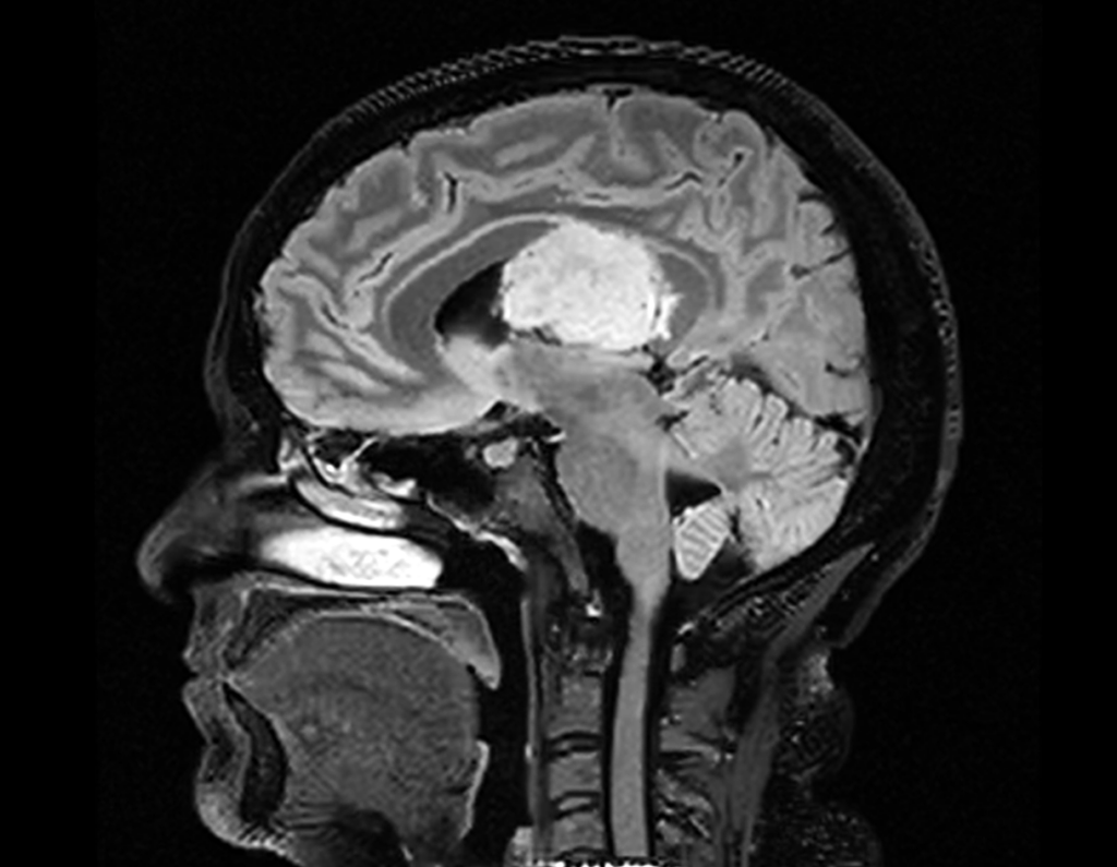
Figure 1. Sagittal FLAIR image showing a midline mass centered on the third ventricle, with a heterogeneous signal intensity and mass effect on adjacent structures, extending superiorly toward the corpus callosum and inferiorly toward the brainstem.
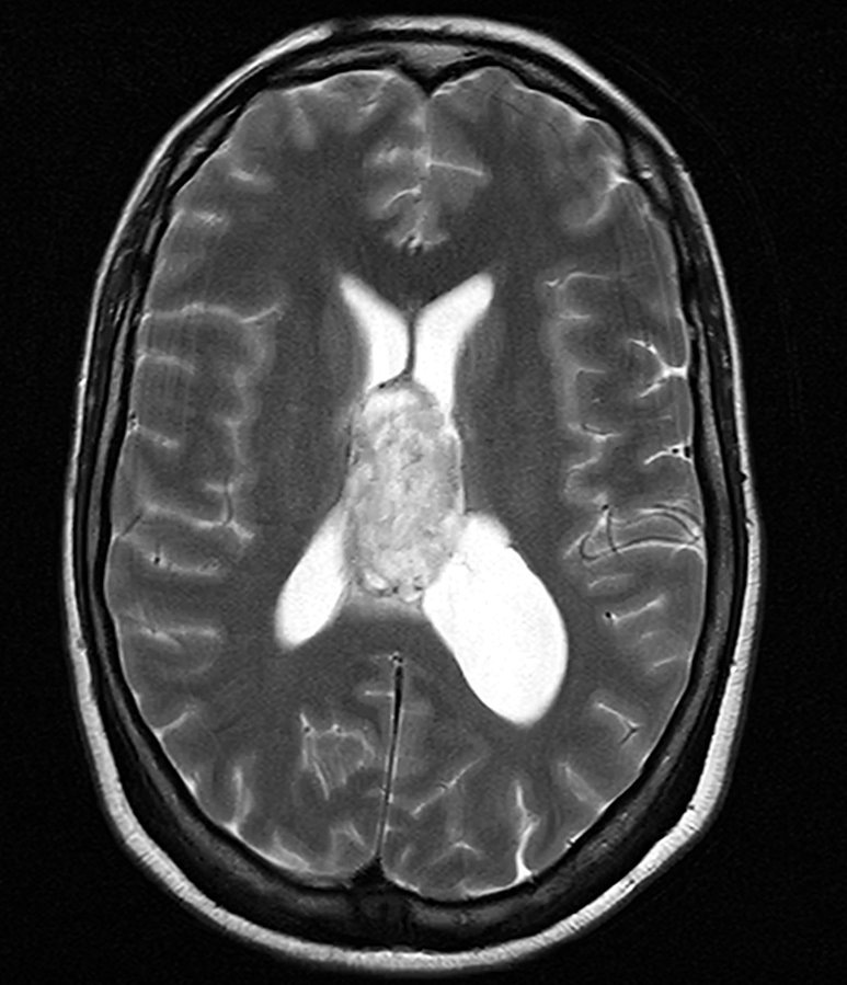
Figure 2. Axial T2-weighted image demonstrating a heterogeneously
hyperintense lesion centered on the the third ventricle, causing obstruction of the foramina of Monro and the dilation of the lateral ventricles, consistent with
obstructive hydrocephalus.
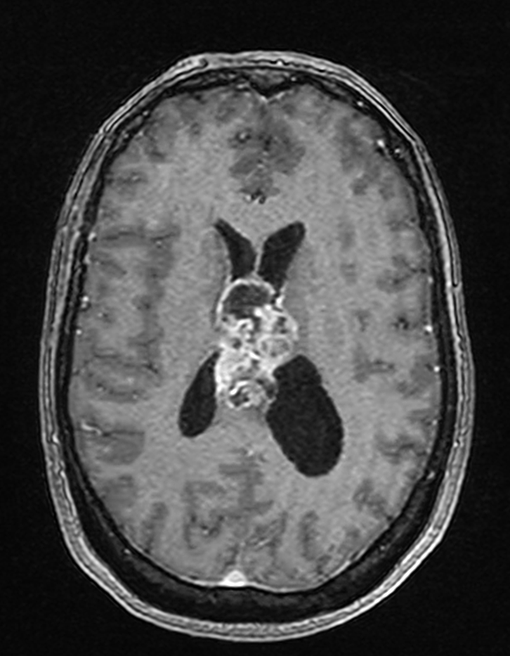
Figure 3. Axial post-contrast T1-weighted image showing irregular and heterogeneous peripheral enhancement of the lesion, with a central non-enhancing necrotic
component
Given the deep-seated location of the lesion and the high surgical risk, a stereotactic biopsy was performed to obtain histological confirmation. Histopathological analysis revealed a phenotype consistent with glioblastoma, characterized by frequent mitoses, palisading necrosis, and microvascular proliferation. Immunohistochemical analysis supported the diagnosis of an IDH-wildtype glioblastoma, classified as WHO (World Health Organization) grade 4 according to the 2021 classification. The methylation status of the MGMT promoter was methylated.
Following the multidisciplinary team meeting, a treatment plan including concomitant radio-chemotherapy according to the Stupp protocol was established. However, the patient experienced an unfavorable clinical course and died before the initiation of therapy.
3. Discussion
The case report concerns a patient with no prior neurological history who presented with a midline glioblastoma centered on the third ventricle and extending to the corpus callosum and trigone. This unusual localization illustrates the diagnostic and therapeutic complexity of such rare glioblastoma
presentations. According to the 2021 WHO classification, glioblastoma is the most frequent and most aggressive primary glial tumor in adults, with a median overall survival of only 12–15 months despite multimodal treatment (1, 2). Within the same WHO framework, diffuse midline glioma, H3K27-altered, is now recognized as a distinct molecular entity, defined by the presence of a histone H3K27M mutation and typically arising in pediatric or young adult patients. However, not all midline tumors harbor this alteration. Some, such as the present case, correspond to conventional glioblastoma, IDH-wildtype, that exceptionally occur in midline structures but lack H3K27 alteration.
Recognizing this distinction is essential for accurate diagnosis and appropriate interpretation within the current molecular classification (3).
While GBM typically arises within the cerebral hemispheres, particularly the frontal and temporal lobes, involvement of midline structures such as the septum pellucidum, corpus callosum, and third ventricle has been only rarely described (4,5,6,7). In this context, the precise anatomical origin of our patient’s tumor remains uncertain. Although the lesion appeared centered on the third ventricle, its extension toward the corpus callosum and the trigone raises the possibility of a juxtaventricular parenchymal origin, most likely from the septum pellucidum or the callosal body. True intraventricular glioblastomas are exceptionally rare, since glial tumors typically arise from the brain parenchyma rather than the ventricular lining (4,5). In most published reports, so-called “third ventricular glioblastomas” were in fact parenchymal tumors with secondary invasion of the
ventricular cavity (6,7). This distinction is not only of theoretical relevance, but also of practical importance for differential diagnosis, as intraventricular masses in this region more commonly correspond to ependymomas, central neurocytomas, germ cell tumors, or primary CNS lymphomas (8). In our case, the biphasic MRI pattern, the close relationship with both lateral ventricles, and the absence of thalamic or hypothalamic involvement suggested a midline parenchymal tumor with ventricular extension rather than a tumor arising purely from the third ventricle itself.
From a histopathological and molecular standpoint, the diagnosis was further characterized as follows. Histopathological examination confirmed a high-grade glial neoplasm consistent with glioblastoma. Immunohistochemical analysis demonstrated an IDH-wildtype (negative IDH1 R132H) profile. H3K27M immunostaining was not performed due to limited tissue availability; nevertheless,
the overall histopathological and radiological characteristics did not suggest a diffuse midline glioma, H3K27-altered. MGMT promoter methylation was positive, a molecular alteration that may influence therapeutic responsiveness but does not modify the classification or the inherently poor prognosis of glioblastoma, IDH-wildtype.
The standard treatment of glioblastoma relies on maximal safe resection, which has been clearly associated with improved overall survival. However, in deep-seated midline locations, particularly when in close proximity to critical structures such as the internal cerebral veins or the hypothalamus, such an approach is rarely feasible, and stereotactic biopsy often remains the only initial diagnostic
and therapeutic option (9). A neurosurgical consultation was also requested to discuss the possibility of a preventive ventricular shunt in view of ventricular dilatation; this option was not retained in the absence of clinical signs of intracranial hypertension, in accordance with the available literature.
Indeed, several studies have reported that shunting should only be considered in cases of symptomatic hydrocephalus, since its prophylactic use has not been demonstrated to be beneficial and carries a non-negligible risk of mechanical or infectious complications (10).
The standard adjuvant treatment is based on the Stupp protocol, which combines conformal radiotherapy delivering 60 Gy in 30 fractions with concomitant temozolomide, followed by adjuvant temozolomide (11). The patient presented with an excellent performance status (WHO 0), which justified this standard chemoradiation approach. Hypofractionated radiotherapy regimens, such as
40 Gy in 15 fractions or 25 Gy in 5 fractions, are generally reserved for elderly patients or those with poor functional status. (12,13).
Modern irradiation techniques (IMRT/VMAT) are preferred as they ensure optimal target coverage while sparing organs at risk. In our case, IMRT dosimetric planning was performed, but irradiation could not be initiated due to rapid clinical deterioration, highlighting the particularly poor prognosis of this presentation, with median survival generally not exceeding 12 to 15 months despite multimodal management (14).
We summarized clinical presentation, treatment details and outcomes for other similar 5 cases that were previously published (Table 1).
Table 1. Reported cases of third ventricular glioblastoma in the literature and comparison with our observation
| Lee & Manzano, 1997 (3) | 59 years old / Male | Third ventricle | Septum pellucidum, fornix with extension into the ventricle | Subtotal resection | Subtotal resection | Total brain irradiation followed by chemotherapy | Recurrence at 5 months Death at 7 months |
| Hariri et al., 2015 (4) | 62 years old /woman | Roof of the third ventricle | Juxtaventricular (septum pellucidum/fornix) | Subtotal resection | Subtotal resection | Radiochemotherapy refused → palliative care | Alive at postoperative day 71, severely impaired cognitive status |
| Yilmaz et al., 2016 (5) | 36 years old / woman, | Isolated third ventricle | Probable parenchymal origin (debated) | Stereotactic biopsy + double VP shunt | Stereotactic biopsy + double VP shunt | Radiochemotherapy | Alive and in good clinical condition at 6 months post-op, with resolution of hydrocephalus and radiological progression toward tumor necrosis. |
| Gacic et al., 2021 (6) | 22 years old /male | Third ventricle and diencephalon | Juxtaventricular (diencephalothalamic). | Maximum tumor resection | Maximum tumor resection | Radiotherapy + temozolomide | Recurrence at 4 months Death < 1 year |
| Notre cas (2024) | 42 years old / woman | Median mass centered on the third ventricle, extension of the corpus callosum and trigone | Probable juxtaventricular origin (septum pellucidum/corpus callosum) | Stereotactic biopsy | Stereotactic biopsy | Planned Stupp protocol | Early death before treatment |
Conclusion
This case illustrates the rarity of midline glioblastomas involving the third ventricle and highlights the difficulty in determining their exact origin. It emphasizes the surgical and therapeutic limitations associated with this deep and atypical localization, which account for the particularly dismal prognosis of such uncommon presentations.
Abbreviations
GBM – Glioblastoma
RT – Radiotherapy
TMZ – temozolomide
MRI – Magnetic Resonance Imaging
ADC – Apparent diffusion coefficient
NAA – N-acetyl-aspartate
WHO – World Health Organization
IMRT – Intensity-modulated radiotherapy
VMAT – Volumetric arc therapy by intensity modulation
Statements
Authors’ Contributions: NK and WH planned the analysis. WH, SK, and KS contributed to the interpretation of the case. NK took the lead in writing the manuscript. TB, ZA, and FZ F gave final approval. All authors provided critical feedback and contributed to the development of the research, analysis, and manuscript.
Consent for Publication: As the corresponding author, I confirm that the manuscript has been read and approved for submission by all listed authors.
Conflict of Interest: The authors declare that they have no competing interests.
Funding: No funding was received for the conduct of this study.
Ethics Approval and Consent to Participate: This manuscript does not contain any studies involving human participants or animals performed by the authors.
Written Informed Consent for Publication: Written informed consent was obtained from the patient for the publication of this case report
and the accompanying images.
References
-
Ostrom QT, Price M, Ballard C, Benedetti J, Neff C, Cioffi G, et al. CBTRUS Statistical Report: Primary Brain and Other Central Nervous System Tumors Diagnosed in the United States in 2017–2021. Neuro Oncol. 2024;26(Suppl 6):vi1-vi85. doi:10.1093/neuonc/noae145.
-
Louis DN, Perry A, Wesseling P, Brat DJ, Cree IA, Figarella-Branger D, et al. The 2021 WHO Classification of Tumors of the Central Nervous System: a summary. Lancet Oncol. 2021;22(11):1532-1546. doi:10.1016/S1470-2045(21)00475-8.
-
Brat DJ, Aldape K, Bridge JA, Canoll P, Colman H, et al. Molecular biomarker testing for the diagnosis of diffuse gliomas. Arch Pathol Lab Med. 2022;146(5):547–574. doi:10.5858/arpa.2021-0287-CP.
-
Lee TT, Manzano GR. Third ventricular glioblastoma multiforme: case report. Neurosurg Rev. 1997;20(4):291-294. doi:10.1007/BF01105903.
-
Hariri OR, Quadri SA, Farr S, Gupta R, Bieber AJ, Dyurgerova A, et al. Third Ventricular Glioblastoma Multiforme: Case Report and Literature Review. J Neurol Surg Rep. 2015;76(2):e227-e232. doi:10.1055/s-0035-1560048.
-
Yılmaz B, Ekşi MŞ, Demir MK, Akakın A, Toktaş ZO, Yapıcıer Ö, et al. Isolated third ventricle glioblastoma. SpringerPlus. 2016;5:115. doi:10.1186/s40064-016-1746-z.
-
Gacic A, Beculic H, Skomorac R, Efendic A. A rare case of third ventricular glioblastoma. Medeni Med J. 2021;36(2):167-171. doi:10.5222/MMJ.2021.45548.
-
Osborn AG, Hedlund GL, Salzman KL. Osborn’s Brain: Imaging, Pathology, and Anatomy. 2nd ed. Philadelphia: Elsevier; 2017. p. 730-750.
-
Brown TJ, Brennan MC, Li M, Church EW, Brandmeir NJ, Rakszawski KL, et al. Association of the extent of resection with survival in glioblastoma: A systematic review and meta-analysis. JAMA Oncol. 2016;2(11):1460-1469. doi:10.1001/jamaoncol.2016.1373.
-
Hong B, Polemikos M, Heissler HE, Hartmann C, Nakamura M, Krauss JK, et al. Challenges in cerebrospinal fluid shunting in patients with glioblastoma. Fluids Barriers CNS. 2018;15(1):16. doi:10.1186/s12987-018-0101-x.
-
Stupp R, Mason WP, van den Bent MJ, Weller M, Fisher B, Taphoorn MJB, et al. Radiotherapy plus concomitant and adjuvant temozolomide for glioblastoma. N Engl J Med. 2005;352(10):987-996. doi:10.1056/NEJMoa043330.
-
Perry JR, Laperrière N, O’Callaghan CJ, et al. Short-course radiation plus temozolomide in elderly patients with glioblastoma. N Engl J Med. 2017;376(11):1027–1037. doi:10.1056/NEJMoa1611977.
-
Takahashi K, Yoshida S, Natsume A, et al. A randomized phase III study of short-course radiotherapy combined with temozolomide in elderly patients with newly diagnosed glioblastoma (JCOG1910 AgedGlio-PIII). BMC Cancer. 2021;21:1105. doi:10.1186/s12885-021-08834-0.
-
Sipos D, Raposa BL, Freihat O, Simon M, Mekis N, Cornacchione P, et al. Glioblastoma: Clinical Presentation, Multidisciplinary Management, and Long-Term Outcomes. Cancers (Basel). 2025;17(1):146. doi:10.3390/cancers17010146.