


Author(s) :
Fatima-Ezzahra Alouan1, Nouhaila Tnifasse1, Marwa Biyoud1, Rabah Hamlaoui1, Thomas Ohnleiter1, Nicolas Bauer1
1 Radiation Oncology Unit, Mulhouse Sud Alsace Regional Hospital Group, Mulhouse, France
Corresponding author: Fatima-Ezzahra Alouan, Email: fatimaezzahra.alouan@usmba.ac.ma
Publication History: Received - 2 September 2025, Revised - 8 December 2025, Accepted - 30 December 2025, Published Online - 30 December 2025.
Copyright: © 2025 The author(s). Published by Casa Cărții de Știință.
User License: Creative Commons Attribution – NonCommercial (CC BY-NC)

 PDF
PDF
 Share
Share
Highlights
-
Case presentation of a 76-year-old male diagnosed with Adenoid Cystic Carcinoma that originated in the lacrimal gland and invaded the zygomatic bone and temporal fossa.
-
Rare pathologies pose diagnostic challenges and need comprehensive immunohistochemistry evaluation
-
Despite microscopically involved margins (R1 status) and extensive bone invasion, the patient achieved durable local control through a combination of radical surgical resection (with free flap reconstruction) and high-precision helical tomotherapy guided by MRI-CT fusion.
Abstract
Adenoid cystic carcinoma (ACC) is a rare malignant tumor that primarily affects the salivary glands, with orbital involvement being exceptionally uncommon. We report the case of a 76-year-old patient who presented with progressive left-sided proptosis and intermittent diplopia. Imaging, along with histopathological and immunohistochemical analyses, confirmed a trabecular-pattern ACC with perineural invasion and R1 resection status.
Due to the tumor’s proximity to critical structures and the presence of microscopically involved margins, the patient underwent orbital exenteration followed by high-precision postoperative radiotherapy using tomotherapy. This case underscores the importance of multidisciplinary coordination and individualized therapeutic planning in the management of orbital ACC. Advanced radiotherapy techniques provide effective local control with excellent tolerance in anatomically complex regions.
1. Introduction
Adenoid cystic carcinoma (ACC), also known as cylindroma, is a rare epithelial malignancy. This tumor shows a slow evolution but retains a strong potential for local invasion. It most commonly arises from the salivary glands, particularly the submandibular and minor accessory glands (1). Orbital involvement is exceptionally rare and typically originates from the lacrimal gland. When present, this localization poses significant diagnostic and therapeutic challenges due to the proximity of critical neurovascular structures. Neuroimaging, especially MRI and CT, plays a key role in identifying lacrimal gland lesions and assessing their extent and impact on adjacent tissues.
2. Clinical Case
We report the case of Mr. X, a 76-year-old patient with multiple comorbidities, including dyslipidemia, a history of duodenal ulcer, chronic obstructive pulmonary disease (COPD), deep vein thrombosis with pulmonary embolism, and prior left knee meniscectomy. The patient had a medical history of dyslipidemia, duodenal ulcer, COPD, phlebitis, pulmonary embolism, and left knee meniscectomy. His chronic medications included atorvastatin, bromazepam (Lexomil), cyamemazine (Tercian), alimemazine (Théralène), apixaban (Eliquis), ramipril, nebivolol, desloratadine, and the inhaled combination Ellipta (fluticasone/umeclidinium/vilanterol).
The initial presentation was atypical: the patient consulted an ophthalmologist for progressive left-sided exophthalmos associated with intermittent diplopia, evolving over a four-month period. On clinical examination, the general condition was preserved (WHO performance status 1), with stable hemodynamic parameters. Ophthalmologic evaluation of the left eye revealed grade III exophthalmos, global oculomotor paresis, reduced visual acuity (2/10), and blepharitis. Examination of the right eye revealed no pathological findings.
Given this unusual orbital presentation, several differential diagnoses were considered, including primary orbital tumor, metastasis, lymphoma, and lacrimal gland tumor. A multimodal imaging workup was initiated. Cranial and orbital CT revealed an intraorbital expansive process centered on the zygomatic bone, measuring 4.3 × 2 × 4 cm, with osteolysis of the lateral orbital wall and frontal bone, grade III exophthalmos, and displacement of the left optic nerve.
Orbital Magnetic Resonance Imaging (MRI) further clarified the locoregional extension: a well-defined soft-tissue mass with polycyclic contours involving the superolateral and inferolateral quadrants of the left orbit, with bone lysis of the orbital pillar and protrusion into the left temporal fossa. The optic nerve was medially displaced, without intracranial extension or meningeal/parenchymal involvement. The lesion showed heterogeneous enhancement and restricted diffusion, suggestive of an aggressive intraorbital tumor (Figure1).
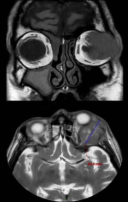
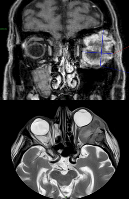
Figure 1. Initial MRI scans in coronal and sagittal views showed a left intraorbital pathological process invading the lateral orbital wall and the left temporal fossa. Grade III left-sided proptosis is evident.
Distant staging via thoraco-abdominopelvic CT revealed a suspicious thickening of the left bladder wall and heterogeneous prostatic hypertrophy, warranting further evaluation with PSA testing. No other secondary lesions were identified.
An intraorbital biopsy was performed. Histopathological analysis revealed a malignant epithelial proliferation arranged in sheets, with glandular structures composed of squamous, intermediate, and mucin-secreting cells within a sclerotic stroma disrupted by mucin lakes. Immunohistochemistry showed tumor positivity for AE1/AE3, CK7, CK5/6, P40, P63, and CD117, with a Ki-67 proliferation index of 44% and positive Alcian blue staining. These findings were consistent with a diagnosis of orbital mucoepidermoid carcinoma. Of note, HER2 expression and androgen receptor (AR) status were not assessed in the initial immunohistochemical panel.
The case was discussed in a multidisciplinary tumor board (MDT), which recommended surgical excision, although achieving clear margins was deemed unfeasible at the posterolateral orbital wall. Postoperative radiotherapy was planned.
The patient underwent complete tumor resection with reconstruction using a free anterolateral thigh (ALT) flap. Final histopathological examination confirmed an infiltrating salivary adenocarcinoma, most consistent with adenoid cystic carcinoma of trabecular architecture, measuring 4.5 cm, with extensive bone invasion (orbital roof and posterolateral wall), perineural spread, and involvement of the posterior superior surgical margins. According to the 8th edition of the TNM classification (2017), the tumor was staged as pT3e Nx PnI R1.
A second MDT confirmed the indication for postoperative radiotherapy, based on the R1 status and bone involvement.
The patient underwent individualized preparation, including the fabrication of a thermoformed immobilization mask and a planning CT scan with fine slices (2.5 mm), performed without contrast injection. Target volume and organ-at-risk (OAR) delineation were based on fusion and rigid registration with preoperative MRI (Figure2).
Treatment was delivered using helical tomotherapy with modulated 6 MV photon beams from a linear accelerator equipped with a multileaf collimator. A single-phase irradiation was performed using an integrated boost technique. Two target volumes were defined: the surgical bed received 52 Gy in 33 fractions of 1.6 Gy, delivered five times per week. and the R1 region received 66 Gy in 33 fractions of 2 Gy, on the same schedule (Figures 3 and 4).
The patient’s comorbidities and treatments were considered when selecting oncologic management. In particular, anticoagulant therapy (Eliquis) and the presence of COPD were considered during radiotherapy planning to minimize the risk of respiratory motion and bleeding. No contraindication to radiotherapy was identified.
Daily patient positioning was verified using onboard megavoltage CT (MV-CT), fused with the planning CT to ensure treatment accuracy. The total duration of radiotherapy was 50 days, with no interruptions exceeding 2 days.
Supportive care was provided throughout the course of radiotherapy. Treatment tolerance was excellent: grade 2 asthenia, grade 1 radiation dermatitis, and moderate headaches (VAS 2/10). No adverse events above grade 2 were reported, according to CTCAE v4 criteria.

Figure 2. Planning CT scan with contouring of clinical target volumes and critical structures for postoperative radiotherapy
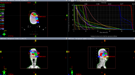
Figure 3. Postoperative radiotherapy dosimetric views showing a 66 Gy dose distribution to the PTV using intensity-modulated helical tomotherapy
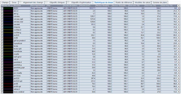
Figure 4. Dose–Volume Histogram (DVH) showing PTV coverage and dose distribution to organs at risk (OARs).
3. Discussion
Adenoid cystic carcinoma (ACC) is a rare malignant tumor that primarily arises in the salivary glands but may also involve other glandular structures such as the trachea, mammary glands, and, more exceptionally, the lacrimal gland (1-2). In its intraorbital form, ACC predominantly affects the lacrimal gland, located in the superolateral orbit, and accounts for approximately 2% to 16% of malignant orbital tumors in various series (3).
In France, the overall incidence of ACC across all sites is estimated at 1 to 2 cases per 100,000 inhabitants per year. It mainly affects adults aged 40 to 60, with a slight female predominance. The etiology of ACC remains poorly understood. No major risk factor has been clearly identified, and conventional environmental exposures such as tobacco or alcohol do not appear to be involved. A few cases have been linked to prior exposure to ionizing radiation, particularly following childhood cervicofacial radiotherapy (2). Genetically, alterations involving the MYB and NFIB genes have been identified, suggesting a specific molecular pathway in tumorigenesis (4).
Clinical manifestations of ACC vary depending on tumor location. In its intraorbital form, it typically presents as progressive proptosis, often accompanied by diplopia, orbital pain, and visual disturbances such as decreased acuity or blurred vision. Perineural invasion, a hallmark of this pathology, may lead to characteristic neuropathic pain: stabbing or burning sensations radiating to the temple, ear, or forehead (5,6).
Imaging plays a central role in the initial diagnostic workup. MRI is generally the first-line modality for characterizing the tumor mass, especially in suspected perineural involvement, due to its high tissue contrast and diffusion sequences (1,6). CT scanning provides a detailed evaluation of orbital bone structures and can detect irregular tumor margins or calcifications (5,8). However, neither MRI nor CT can definitively distinguish benign from malignant lesions.
Definitive diagnosis relies on histopathological examination obtained via surgical biopsy. Three histological subtypes are classically described in ACC: tubular, cribriform, and solid, the latter being associated with more aggressive behavior (3,4). In our case, the observed architecture was trabecular, a variant rarely described in the literature, which may coexist with classical forms. This trabecular organization reflects a degree of tumor differentiation, although its prognostic implications remain unclear.
As reported in the observation section, immunohistochemistry confirmed the carcinomatous nature of the lesion. Co-expression of epithelial markers (AE1/AE3, CK7, CK5/6) and differentiation factors (P40, P63, CD117) is consistent with a mixed phenotype, typical of mucoepidermoid carcinoma. A high Ki-67 proliferation index suggests significant mitotic activity, and Alcian blue positivity confirms the presence of extracellular mucin, reinforcing the diagnosis.
Taken together, these findings support an orbital localization of a mucoepidermoid carcinoma, a rare entity in this anatomical region. This localization poses specific prognostic and therapeutic challenges, particularly given the proximity of critical structures and the risk of local or metastatic spread.
In our case, HER2 and androgen receptor (AR) immunostaining were not performed, although both markers may be relevant in certain salivary gland carcinomas. AR expression is typically associated with salivary duct adenoid cystic carcinoma, where it suggests the potential efficacy of hormonal therapy, whereas its presence in lacrimal gland adenoid cystic carcinoma is rare. Similarly, HER2 amplification is uncommon in ACC of the lacrimal glands but, when present, may provide access to targeted anti-HER2 therapies. Although these markers are not routinely included in the diagnostic workup of conventional ACC, the presence of unusual histological features (such as the trabecular architecture observed here) may justify a broadened immunohistochemical panel to identify potential biomarkers and explore emerging therapeutic approaches. (7).
Staging should include thoracic imaging, typically computed tomography (CT), to assess for pulmonary metastases, which are common in this disease (1,9). In our case, the patient also underwent thoraco-abdominopelvic CT (TAP), allowing broader evaluation of both locoregional and distant extension.
18F-FDG PET-CT may be considered in selected cases, although its sensitivity in ACC is limited by low FDG uptake in tumor cells. This phenomenon is thought to be related to low expression of the glucose transporter GLUT-1 and physiological FDG absorption by salivary glands, which may confound interpretation (8).
Surgery remains the first-line treatment for non-metastatic ACC, with the primary goal of achieving complete resection with oncologically safe margins. This strategy is well supported in the literature, particularly for salivary gland tumors and head and neck locations (11). In orbital sites, surgical excision poses significant technical challenges due to the proximity of functional structures. Potential complications such as oculomotor disturbances, keratitis, or strabismus underscore the need for appropriate reconstruction. Free flap techniques, such as the anterolateral thigh (ALT) flap, have proven effective in restoring both morphology and function in these anatomically complex regions (12).
In our case, the decision to perform surgery with ALT flap reconstruction was driven by the need for optimal local control, despite positive margins (R1 status) and histologically confirmed bone invasion. This approach highlights the importance of a multidisciplinary therapeutic strategy, with tumor board (RCP) consensus guiding the decision to perform radical surgery followed by tailored postoperative radiotherapy.
Radiotherapy serves as a critical adjunct in ACC management, particularly in cases of positive surgical margins or microscopic residual disease. Several studies have shown that, despite a high rate of local recurrence in patients with involved margins (up to 83.3%), adjuvant radiotherapy achieves local control in a significant proportion of cases (13). ASCO guidelines recommend systematic postoperative radiotherapy (RTPO) for all surgically treated ACC patients, given the high risk of perineural spread and delayed recurrence (14).
In our experience, treatment was delivered via tomotherapy, with precise planning based on MRI-CT fusion. An integrated boost was administered to high-risk areas, with a total dose of 66 Gy to the tumor bed and 52 Gy to peripheral risk volumes. This approach provided optimal coverage of perineural pathways up to the skull base while respecting dosimetric constraints imposed by adjacent critical structures, including the globe, optic nerve, chiasm, and brainstem.
This technical protocol aligns with current recommendations for postoperative radiotherapy in ACC, emphasizing the value of advanced conformal techniques (IMRT, tomotherapy, proton therapy) to maximize local control while minimizing toxicity (13,14).
Treatment tolerance was excellent, with only mild side effects including transient ocular dryness and fatigue. No severe toxicity was reported, and treatment adherence was complete throughout the protocol. These outcomes confirm the feasibility and safety of high-precision radiotherapy in orbital-cranial ACC.
ACC is known for its slow but unpredictable progression, with a high risk of local recurrence and late metastatic dissemination, particularly to the lungs. This risk is especially pronounced in advanced stages (T3–T4), in cases of bone invasion or incomplete resection. Several studies have demonstrated that patients treated with conservative surgery, without orbital exenteration or postoperative radiotherapy, have significantly higher local recurrence rates (13,15).
In our case, the patient underwent orbital exenteration followed by targeted postoperative radiotherapy, justified by R1 status and histologically confirmed bone involvement. After 12 months of follow-up, no signs of local or metastatic recurrence were observed, reflecting the efficacy of the combined treatment approach. This outcome reinforces the value of an aggressive, multidisciplinary strategy in locally advanced ACC, particularly when deep orbital structures or bony walls are involved.
4. Conclusion
This case underscores the importance of accurate diagnosis and multidisciplinary coordination in the management of intraorbital ACC. Therapeutic adaptation to anatomical constraints, combining radical surgery with postoperative radiotherapy, enabled durable local control. Modern radiotherapy techniques, such as tomotherapy with MRI-CT fusion, are particularly relevant in complex anatomical sites, offering both precision and excellent tolerance.
Abbreviations
ACC – Adenoid Cystic Carcinoma
ALT – Anterolateral Thigh Flap
COPD – Chronic Obstructive Pulmonary Disease
CT – Computed Tomography
CTV – Clinical Target Volume
FDG – Fluorodeoxyglucose
GLUT-1 – Glucose Transporter Type 1
IMRT – Intensity-Modulated Radiation Therapy
MRI – Magnetic Resonance Imaging
MV-CT – Megavoltage Computed Tomography
WHO – World Health Organization
OAR – Organs At Risk
PET-CT – Positron Emission Tomography–Computed Tomography
PTV – Planning Target Volume
R1 – Microscopically Involved Surgical Margin
MDT – Multidisciplinary Team Meeting (Réunion de Concertation Pluridisciplinaire)
RTPO – Postoperative Radiotherapy
TAP – Thoraco-Abdominopelvic CT Scan
TNM – Tumor, Node, Metastasis Classification
VAS – Visual Analogue Scale
CTCAE – Common Terminology Criteria for Adverse Events
COPD – chronic obstructive pulmonary disease
Statements
Authors’ Contributions: All authors made significant contributions to the development of this case report. AFE coordinated the work, wrote the first draft of the manuscript, and supervised the entire process. TN, HW, and EKS were involved in the patient’s clinical management and data collection. SK and FFZ contributed to the analysis of clinical and imaging findings. AZ provided a critical revision of the manuscript. BT performed the final editing and approved the version for submission.
All authors have read and approved the final manuscript and agree to be accountable for all aspects of the work.
Consent for Publication: As the corresponding author, I confirm that the manuscript has been read and approved for submission by all listed authors.
Conflict of Interest: The authors declare no conflicts of interest.
Funding Sources: None.
Written Informed Consent for Publication: Written informed consent was obtained from the patient for the publication of this case report and all accompanying images.
Acknowledgements: We would like to sincerely thank the entire care team involved in the management of this patient. Their dedication and professionalism were essential to the quality of care provided.
We are also grateful to the radiology and pathology departments for their support and valuable input in understanding the case.
Above all, we extend our heartfelt thanks to the patient for their trust, generosity, and willingness to share their story for scientific purposes.
References
- Bouzidi A, Iferkhass S, Hansali ZE, Elmallaoui M, Laktaoui A. Left eye proptosis suggestive of orbital adenoid cystic carcinoma: a case report. Pan Afr Med J. 2015;22:168.
- Atallah S. Prospective cohort of 470 patients with adenoid cystic carcinoma of the head and neck included in the REFCOR database: prognostic analysis and evaluation of therapeutic strategies [doctoral thesis]. Paris-Saclay: Université Paris-Saclay; 2022. Available from: https://theses.hal.science/tel-04689999v1/file/108819_ATALLAH_2022_archivage.pdf
- El Azzouzi R, Dani B, Boulaadas M. Lacrimal gland tumors: Which surgical approach? [poster]. Presented at: CMF AOS Congress; 2021 Sep [cited 2023 Oct 27]. Available from: [suspicious link removed]
- Persson M, Andren Y, Mark J, Horlings HM, Persson F, Stenman G. Recurrent fusion of MYB and NFIB transcription factor genes in carcinomas of the breast and head and neck. Mol Oncol [Internet]. 2009;3(3):248–53 [cited 2023 Oct 27]. Available from: https://link.springer.com/content/pdf/10.1007/s12558-011-0148-y
- Hakam J, Belghmaidi S. Adenoid cystic carcinoma of the lacrimal gland: a case report. EM-Consulte. 2022; [Specific volume/issue/page if available].
- Achargui M. Unilateral exophthalmos revealing adenoid cystic carcinoma of the lacrimal gland. Presented at: CMF AOS Congress; 2022.
- De Laroche R, Bera G, Bertolus C, et al. Adenoid cystic carcinoma in PET/CT and PET/MRI: comparison of both modalities. Médecine Nucléaire [Internet]. 2016;40(3):118–25 [cited 2023 Oct 27]. Available from: https://www.em-consulte.com/article/1051854
- org. Disease profile: Adenoid cystic carcinoma [Internet]. 2021 [cited 2023 Oct 27]. Available from: https://www.radeos.org/maladie/fiche-carcinome-adenoide-kystique_468.html
- Medicover Hospitals. Adenoid Cystic Carcinoma – Overview and Symptoms [Internet]. [date unknown] [cited 2023 Oct 27]. Available from: https://www.medicoverhospitals.in/fr/diseases/adenoid-cystic-carcinoma/
- Spiro RH. Adenoid cystic carcinoma: factors influencing survival. Am J Surg. 1997;174(5):424–6.
- Mendenhall WM, Dagan R, Bryant CM, Amdur RJ, Werning JW. Perineural invasion and spread in head and neck cancer. Head Neck. 2012;34(7):1031–40.
- Garden AS, Weber RS, Morrison WH, et al. The influence of positive margins and nerve invasion in adenoid cystic carcinoma of the head and neck treated with surgery and radiation. Int J Radiat Oncol Biol Phys. 1995;32(3):619–26.
- Coca-Pelaz A, Rodrigo JP, Bradley PJ, et al. Adenoid cystic carcinoma of the head and neck—an update. Oral Oncol. 2015;51(7):652–61.
- van der Wal JE, Becking AG, Snow GB, van der Waal I. Distant metastases of adenoid cystic carcinoma of the salivary glands and the value of diagnostic examinations during follow-up. Head Neck. 2002;24(8):779–83.
- Fan C-Y, Melhem M, Hosal AS, Grandis JR, Barnes EL. Androgen receptor-positive mucoepidermoid carcinoma: case report and literature review. Int J Surg Pathol [Internet]. 2015;23(7):527–32 [cited 2023 Oct 27]. Available from: https://pubmed.ncbi.nlm.nih.gov/25559272/