

Author(s) :
Mara Răzniceanu1, Monica Buzemurgă1
1 Radiation Oncology Department, Regional Institute of Oncology, Iasi, Romania
Corresponding author: Mara Răzniceanu, Email: mara.razniceanu@gmail.com
Publication History: Received - May 1, 2025, Revised - June 18, 2025, Accepted - July 15, 2025, Published Online - August 2, 2025.
Copyright: © 2025 The author(s). Published by Casa Cărții de Știință.
User License: Creative Commons Attribution – NonCommercial (CC BY-NC)

 PDF
PDF
 Share
Share
Highlights
- Multimodal treatment for Merkel cell carcinoma (MCC), involves surgery, radiotherapy, and immunotherapy.
- A case of preoperative radiotherapy combined with early Avelumab led to a complete pathological response, while a traditional postoperative approach resulted in stable disease.
- These contrasting outcomes highlight the importance of flexible, patient-tailored strategies and the potential for a combined radioimmunotherapy approach to improve disease control in MCC.
Abstract
Merkel cell carcinoma (MCC) is a rare but aggressive cutaneous neuroendocrine malignancy with high rates of recurrence and metastasis. Optimal management requires a multidisciplinary approach, including surgery, radiotherapy, and, increasingly, immunotherapy with checkpoint inhibitors.
We present a comparative report of two male patients with axillary nodal involvement from MCC who underwent multimodal treatment. The first patient, a 73-year-old male with known primary cutaneous MCC of the forearm, underwent surgical lymphadenectomy followed by postoperative radiotherapy and immunotherapy with Avelumab between February and September 2021, achieving stable disease without progression. The second patient, a 66-year-old male with nodal MCC of unknown primary origin, initially progressed on cisplatin-etoposide chemotherapy. However, early initiation of Avelumab combined with preoperative radiotherapy, followed by surgical lymphadenectomy, resulted in significant tumor regression and a complete pathological response.
These cases underscore the crucial importance of integrating early systemic immunotherapy, meticulous radiotherapy planning, and personalized sequencing of multimodal therapies to optimize disease control. The contrasting outcomes emphasize the need for flexible, patient-tailored strategies in the management of Merkel cell carcinoma, particularly in the era of immunotherapy.
1. Introduction
Merkel cell carcinoma (MCC) is a rare, highly aggressive neuroendocrine skin cancer, typically affecting elderly and immunosuppressed individuals. It is associated with a high risk of locoregional recurrence, early nodal involvement, and distant metastasis. The incidence of MCC has been rising over the past decades, attributed in part to increased ultraviolet exposure, an aging population, and improved diagnostic awareness. Despite its rarity, MCC carries a disproportionately high mortality rate compared to other skin cancers, exceeding that of melanoma (1).
Traditional management of MCC relied heavily on surgical excision with wide margins, often followed by regional lymphadenectomy and radiotherapy. However, even after aggressive local treatment, recurrence rates remain significant, especially in patients with nodal involvement or immunosuppression. Conventional chemotherapy, although initially effective in some cases, has not been shown to provide durable responses or survival benefits, and its role has diminished in the era of immunotherapy (1, 2).
The introduction of immune checkpoint inhibitors, particularly agents targeting the PD-1/PD-L1 axis such as Avelumab, has revolutionized the therapeutic landscape of advanced MCC. Avelumab has demonstrated significant and durable responses even in chemotherapy-refractory settings, leading to its approval as a first-line systemic therapy in metastatic MCC (3).
Nevertheless, the optimal integration of immunotherapy with traditional modalities, such as surgery and radiotherapy, remains an area of active investigation. In particular, the timing and sequencing of these treatments may profoundly influence outcomes, but standardized protocols are lacking due to the rarity of the disease (4).
In this context, we present two clinical cases of MCC with axillary lymph node involvement managed through different therapeutic sequences, illustrating the impact of early immunotherapy integration and the strategic use of radiotherapy and surgery. These contrasting outcomes offer insights into tailoring multimodal approaches for MCC patients in the modern era.
2. Case presentations
2.1. Case 1
A 73-year-old male with a medical history of chronic obstructive pulmonary disease (COPD), hypertension, ankylosing spondylitis stage IV, and bilateral gonarthrosis was diagnosed with Merkel cell carcinoma of the left forearm in January 2020. Surgical excision confirmed cutaneous MCC staged pT2 Nx L1V0 Pn0 R1.
He presented in October 2020 with palpable masses in the left axilla. CT imaging revealed multiple enlarged axillary lymph nodes, the largest measuring 46 × 39 × 41 mm.
The patient underwent a left axillary lymphadenectomy, during which 25 lymph nodes were resected. Histopathological analysis confirmed metastatic Merkel cell carcinoma (MCC) in 17 of the nodes, with extensive involvement. Immunohistochemistry was positive for CK20 (in a dot-like pattern), Synaptophysin, and CD56, supporting the diagnosis of MCC.
Although a follow-up CT scan was scheduled, it was delayed due to the patient’s hospitalization for other medical conditions. When imaging was eventually performed in January 2021, it showed persistent lymph node enlargement and a postoperative seroma. A multidisciplinary tumor board recommended postoperative radiotherapy along with systemic immunotherapy.
Adjuvant external beam radiotherapy was administered between February and March 2021. Treatment consisted of Intensity-Modulated Radiation Therapy (IMRT), delivering 50 Gy in 25 fractions to the axillary, supraclavicular, and subclavicular lymph node regions, with a sequential boost of 10 Gy targeting the residual disease. (Figure 1)
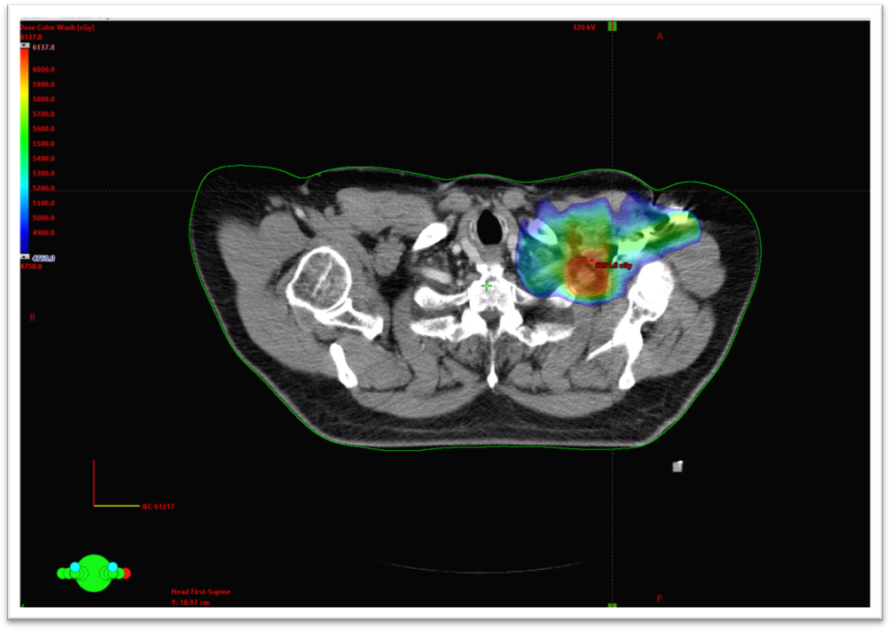
Figure 1 – Radiation dose coverage for 95% of the prescribed dose boost of the Planning target volume (PTV)
Avelumab 800 mg was administered every 2 weeks, starting on February 10, 2021, and continued until September 16, 2021. Throughout therapy, routine laboratory tests revealed no significant abnormalities in blood count, kidney function, or liver function. However, the patient did experience grade 4 diarrhea as a side effect of the immunotherapy.
At discharge and during the initial outpatient evaluations, the patient maintained a stable clinical status, with no radiologic evidence of disease progression. Active surveillance was recommended; however, the patient did not return for further examinations and was lost to follow-up.
2.2. Case 2
A 66-year-old male was diagnosed in February 2020 with right axillary lymph node metastases of Merkel cell carcinoma with unknown primary origin. He had a prior history of an excised nodular basal cell carcinoma with keratotic foci in 2016, but this was ruled out as a potential MCC source.
An initial PET/CT scan performed in May 2020 revealed increased metabolic activity in the axillary lymph node mass and identified a suspicious lesion on the forearm. The lymph node mass was inoperable. Despite receiving three cycles of first-line chemotherapy with cisplatin and etoposide, the disease progressed, with continued enlargement of the axillary mass. As a result, treatment was switched to Avelumab (800 mg every two weeks), beginning in May 2020. The lymph node lesion was inoperable at the time of diagnosis.
Preoperative radiotherapy, targeting the right axillary and supraclavicular regions with Volumetric Modulated Arc Therapy (VMAT), was planned for 66 Gy (50 Gy in 25 fractions to the right axillary, supraclavicular, and subclavicular lymph nodes, with a sequential boost of 16 Gy in 8 fractions to the axillary adenopathies). (Figure 2).

Figure 2 – Radiation dose coverage of 95% dose to PTV – axillary levels
During treatment, which consisted of both external beam radiotherapy and immunotherapy with avelumab, there was a significant reduction in the size of both the axillary mass and the forearm lesion, with the latter decreasing to approximately 1 × 1 cm from 2 × 2 cm in size. Due to concerns about the potential risk of brachial plexopathy as a late complication from high-dose radiation, the radiotherapy was discontinued at 50 Gy, rather than the initially planned 66 Gy, given the favorable response. The patient underwent a surgical re-evaluation in June 2020. It was decided to proceed with a right axillary lymphadenectomy approximately two months after the completion of radiotherapy, as well as surgical excision of the right forearm lesion, which had been marked preoperatively with tattoos on its medial, lateral, and distal margins.
At the 3-month imaging follow-up, we observed a significant reduction in the size of the right axillary lymphadenopathy. The most extensive lesion decreased from 23×36 mm to 14×25 mm. No pulmonary lesions or mediastinal lymphadenopathy were identified. (Figure 3)
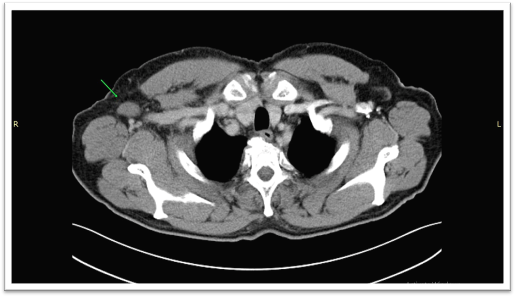
Figure 3 – CT scan – August 2020 – significant reduction in the size of the axillary lymphadenopathy.
The surgical intervention was initially postponed due to a COVID-19 infection. In April 2021, the CT scan revealed axillary lymphadenopathies with no significant morphological changes.
However, in May 2021, the patient successfully underwent a right axillary lymphadenectomy (levels I–III) and excision of the right forearm lesion, followed by skin grafting. Histopathological analysis revealed that the forearm lesion was a benign capillary hemangioma. Examination of the axillary lymphadenectomy specimen demonstrated a complete pathological response, with no residual tumor identified (ypT0N0).
A follow-up CT in November 2021 confirmed no residual disease.
As of May 2022, the patient remained clinically stable (ECOG 1) and continued Avelumab maintenance therapy (adjuvant setting) one year after the surgery, with no signs of disease recurrence.
3. Discussion
Case 1 illustrates the conventional sequence of surgery followed by adjuvant radiotherapy and immunotherapy. Surgery remains essential for locoregional control in MCC, but high nodal recurrence rates require adjuvant therapies. Postoperative radiotherapy is particularly beneficial in patients with high-risk features such as multiple positive nodes or extracapsular extension. Avelumab, an anti-PD-L1 immune checkpoint inhibitor, is emerging as a valuable systemic therapy in this context, with increasing evidence supporting its efficacy.
Table 1 – Comparative Overview of Two Merkel Cell Carcinoma Cases with Axillary Involvement
| Feature | Case 1 (Known Primary) | Case 2 (Unknown Primary) |
| Age | 73 | 66 |
| Primary Lesion | Left forearm MCC | No detectable primary |
| Initial Treatment | Surgery → Radiotherapy + Avelumab | Chemotherapy → Avelumab + Radiotherapy → Surgery |
| Radiotherapy Strategy | Postoperative | Preoperative |
| Radiotherapy Technique | IMRT | VMAT |
| Radiotherapy Dose | 50 Gy + boost 10 Gy | 50 Gy + boost 16 Gy
(delivered 50 Gy) |
| Surgery Timing | Before RT/Immnotherapy | After RT/Immunotherapy |
| Pathologic Response | – | ypT0N0 (complete response) |
| Avelumab Duration | 7 months | ≥2 years |
| Outcome | No disease progression | Complete remission |
In contrast, Case 2 demonstrates a non-traditional sequence, where early introduction of Avelumab after chemotherapy failure, in combination with preoperative radiotherapy, resulted in a complete pathological response (ypT0N0). These results suggest that integrating immunotherapy earlier, even in patients with bulky disease, may enhance tumor control. The preoperative use of radiotherapy could also contribute to immunomodulation, potentially increasing tumor antigen release and immune priming prior to surgery.
Several critical differences between the cases underscore the importance of individualized treatment strategies:
First, the value of multimodal therapy is evident, as surgical resection alone—even when extensive—often proves insufficient in MCC, as in Case 1, which experienced a recurrence. Postoperative radiotherapy improves locoregional control, especially in patients with high-risk features like multiple positive lymph nodes and extracapsular extension.
Second, the early use of immunotherapy plays a pivotal role. Case 2 illustrates the significant benefit of initiating Avelumab promptly after chemotherapy failure. The complete pathological response achieved suggests that checkpoint inhibitors can dramatically alter the disease trajectory, even in the presence of bulky nodal disease. (9, 11,12)
Third, the role and timing of radiotherapy remain central to MCC management. While postoperative radiotherapy, as employed in Case 1, serves to prevent recurrence, preoperative radiotherapy, as in Case 2, may increase tumor immunogenicity, reduce tumor burden prior to surgery, and stimulate systemic antitumor immune responses. Customizing radiotherapy—through dose modification and technique selection—based on individual patient risk factors, such as the need to avoid brachial plexopathy, is also essential. (5, 6, 9)
Fourth, the sequence of systemic and locoregional therapies may influence outcomes. In Case 2, initiating immunotherapy first, followed by radiotherapy and subsequently surgery, likely allowed for maximum immune-mediated tumor reduction prior to surgical resection. (9, 11)
Fifth, the management of treatment-related toxicity is critical. Both patients tolerated immunotherapy and radiotherapy well, suggesting that aggressive multimodal treatment is feasible even in elderly patients with comorbidities. Nonetheless, vigilant monitoring for late toxicities—particularly neurologic complications following axillary radiotherapy—remains essential.
Finally, the complete pathological response (ypT0N0) observed in Case 2 following combined Avelumab and radiotherapy supports the use of pathological complete response as a potential surrogate marker for favorable long-term outcomes in MCC, aligning with findings from other malignancies treated with neoadjuvant immunotherapy.
These observations highlight the evolving role of radioimmunotherapy in MCC and the importance of dynamic, patient-centered treatment planning.
4. Conclusion
These two clinical cases reflect real-world experience in managing advanced Merkel cell carcinoma, emphasizing the evolving role of radioimmunotherapy. The integration of Avelumab, an immune checkpoint inhibitor, with radiotherapy—whether in a preoperative or postoperative setting—was both effective and well-tolerated, even in elderly patients with comorbidities.
Our findings highlight that the radioimmunotherapy combination is not only feasible in clinical practice but may also improve disease control, as evidenced by the complete pathological response in one case and sustained disease stabilization in the other. Careful sequencing of surgery, radiotherapy, and immunotherapy allows for individualized treatment planning, maximizing therapeutic benefit while minimizing toxicity.
Overall, these cases support the growing promise of radioimmunotherapy in MCC and reinforce the need for flexible, multidisciplinary strategies tailored to each patient’s disease dynamics and treatment tolerance. Further studies are warranted to validate these observations and to define optimal treatment algorithms in this rare but aggressive malignancy.
ABBREVIATIONS
MCC – Merkel Cell Carcinoma
COPD – Chronic Obstructive Pulmonary Disease
CT – Computed Tomography
PET/CT – Positron Emission Tomography / Computed Tomography
RT – Radiotherapy
VMAT – Volumetric Modulated Arc Therapy
IMRT – Intensity-Modulated Radiation Therapy
ECOG – Eastern Cooperative Oncology Group
pCR – Pathological Complete Response
PD-1 – Programmed Death-1
PD-L1 – Programmed Death-Ligand 1
R1 – Microscopic Residual Tumor
LVI – Lymphovascular Invasion
IHC – Immunohistochemistry
STATEMENTS
Authors’ contributions: All authors equally contributed to writing the manuscript
Consent for publication: As the corresponding author, I confirm that the manuscript has been read and approved for submission by all named authors.
Conflict of interests: The authors declare no conflict of interest.
Funding Sources: None
Written informed consent for publication: Informed consent was obtained from the patients for the publication of this case report and any accompanying images.
REFERENCES
- Lugowska, I., Becker, J. C., Ascierto, P. A., Veness, M., Blom, A., Lebbe, C., … & ESMO Guidelines Committee. (2024). Merkel-cell carcinoma: ESMO–EURACAN Clinical Practice Guideline for diagnosis, treatment and follow-up. ESMO open, 9(5), 102977.
- Gauci, M. L., Aristei, C., Becker, J. C., Blom, A., Bataille, V., Dreno, B., … & European Association of Dermato-Oncology (EADO. (2022). Diagnosis and treatment of Merkel cell carcinoma: European consensus-based interdisciplinary guideline–Update 2022. European Journal of Cancer, 171, 203–231.
- Medina-Franco H, Urist MM, Fiveash J, et al.: Multimodality treatment of Merkel cell carcinoma: case series and literature review of 1024 cases. Ann Surg Oncol 8 (3): 204–8, 2001.
- Green, M. D., & Hayman, J. A. (2018). Radiotherapy in the multidisciplinary management of Merkel cell carcinoma. Journal of the National Comprehensive Cancer Network, 16(6), 776–781.
- Boyer, J. D., Zitelli, J. A., Brodland, D. G., & D’Angelo, G. (2002). Local control of primary Merkel cell carcinoma: review of 45 cases treated with Mohs micrographic surgery with and without adjuvant radiation. Journal of the American Academy of Dermatology, 47(6), 885–892.
- Wilson, L. D., & Gruber, S. B. (2004). Merkel cell carcinoma and the controversial role of adjuvant radiation therapy: clinical choices in the absence of statistical evidence. Journal of the American Academy of Dermatology, 50(3), 435–437.
- Morrison, W. H., Peters, L. J., Silva, E. G., Wendt, C. D., Ang, K. K., & Goepfert, H. (1990). The Essential Role of Radiation Therapy in Achieving Locoregional Control of Merkel Cell Carcinoma. International Journal of Radiation Oncology* Biology* Physics, 19(3), 583-591.
- Eng, T. Y., Boersma, M. G., Fuller, C. D., Cavanaugh, S. X., Valenzuela, F., & Herman, T. S. (2004). Treatment of Merkel cell carcinoma. American Journal of Clinical Oncology, 27(5), 510–515.
- Albores‐Saavedra, J., Batich, K., Chable‐Montero, F., Sagy, N., Schwartz, A. M., & Henson, D. E. (2010). Merkel cell carcinoma demographics, morphology, and survival based on 3870 cases: a population-based study. Journal of cutaneous pathology, 37(1), 20-27.
- Nghiem, P., MCKEE, P. H., & HAYNES, H. A. (1998). Merkel cell (cutaneous neuroendocrine) carcinoma. CA Cancer J Clin, 48, 6–29.
- Averbuch, I., Stoff, R., Miodovnik, M., Fennig, S., Bar‐Sela, G., Yakobson, A., … & Fenig, E. (2023). Avelumab for the treatment of locally advanced or metastatic Merkel cell carcinoma—A multicenter real‐world experience in Israel. Cancer Medicine, 12(11), 12065-12070.
- Dudzisz-Sledz, M., Sobczuk, P., Kozak, K., Switaj, T., Kosela-Paterczyk, H., Czarnecka, A. M., … & Rutkowski, P. (2022). Treatment of locally advanced Merkel cell carcinoma—A multi-center study. Cancers, 14(2), 422.