

Author(s) :
Alouan Fatima-Ezzahra1, Ouaddane Alami Salem1, Hassani Wissal1, El Khalfi Samia1, Soussy Kaoutar1, Farhane Fatima Zahra1, Alami Zineb1, Bouhafa Touria1
1 Radiotherapy Oncology Department, Hassan II, Fes, Morocco
Corresponding author: Alouan Fatima-Ezzahra, Email: fatimaezzahra.alouan@usmba.ac.ma
Publication History: Received - May 29, 2025, Revised - July 26, 2025, Accepted - July 31, 2025, Published Online - August 4, 2025.
Copyright: © 2025 The author(s). Published by Casa Cărții de Știință.
User License: Creative Commons Attribution – NonCommercial (CC BY-NC)

 PDF
PDF
 Share
Share
Highlights
- Pineoblastoma is a rare and aggressive brain tumor in children, making its therapeutic management particularly challenging.
- Radiotherapy is a key treatment option, especially when surgical removal is too risky due to the tumor’s location.
- The patient had a positive outcome with minimal side effects, demonstrating the potential of a non-invasive treatment approach.
Abstract
Pineoblastoma in children is a rare tumor of the central nervous system, with limited knowledge regarding its clinical features and outcomes. The optimal therapeutic strategy for pediatric pineoblastoma remains uncertain. This case presents an 11-year-old girl diagnosed with pineoblastoma. Due to potential impacts on quality of life and the available technical resources, tumor resection was not performed. Instead, the patient was treated exclusively with radiotherapy and remained free of recurrence for 36 months post-treatment.
This case underscores a minimally invasive approach to managing rare pineal tumors located near critical structures, demonstrating favorable responses and excellent neurological outcomes. Additionally, the radiographic and histopathological characteristics of pineoblastomas are reviewed, alongside a discussion of the various treatment options documented in the literature.
1. Introduction
Pineoblastoma in children is a rare tumor of the supratentorial region, classified among the primitive neuroectodermal tumors (PNETs). Representing only 3–8% of intracranial tumors in this age group in Western populations, it is associated with a poor prognosis due to its aggressive nature, making it the most malignant tumor of the pineal parenchyma.
We present a rare case of pineoblastoma in an 11-year-old child treated in our department. This case highlights the challenges of therapeutic management and emphasizes the crucial role of early histological diagnosis. Additionally, radiotherapy plays a key role in treatment, as the supratentorial location of the tumor presents significant surgical challenges.
2. Clinical case
The patient, A.S., an 11-year-old girl, has no notable personal or familial medical history. Symptoms began approximately 20 days prior with progressively worsening headaches, which later intensified with the onset of intracranial hypertension syndrome, accompanied by decreased visual acuity and diplopia. These symptoms led the family to seek consultation with a neurosurgeon in private practice.
During the initial physical examination, the child had a general condition classified as WHO 1, with stable hemodynamic and respiratory parameters. Her blood pressure was recorded at 123/82 mmHg. Neurological examination revealed that she was conscious, with a Glasgow Coma Scale score of 15. She was able to stand and walk without assistance.
Cranial nerve examination was normal, except for vertical diplopia associated with involvement of the fourth cranial nerve. Motor function was intact, with muscle strength rated at 5/5 in all four limbs. Regarding sensory function, the patient exhibited bilateral cervicobrachial neuralgia. No signs of cerebellar syndrome were observed, and deep tendon reflexes were present and symmetrical.
Magnetic resonance imaging (MRI) revealed a midline expansive lesion centered in the pineal region, measuring approximately 4 cm × 2.5 cm. The tissue signal was consistent with pineoblastoma. This mass pushed the third ventricle forward and exerted a compressive effect on the aqueduct of Sylvius, resulting in a discrete enlargement of the ventricular system accompanied by signs of transependymal resorption at the occipital horns. (Figure 1).


Figure 1. MRI aspect at diagnosis, the blue arrow showing the pineoblastoma (axial and sagittal view).
In the cervico-dorso-lumbar spine, the medulla showed normal caliber and a homogeneous signal.
The patient subsequently underwent a ventriculo-cisternostomy between the floor of the third ventricle (V3) and the interpeduncular cistern, as well as a stereotactic biopsy. Anatomopathological analysis of the biopsy revealed a tumor focus composed of round cells, whose immunohistochemical study revealed synaptophysin and CD56 positivity on tumor cells, with no PLAP expression despite the presence of a positive external control. The CD117 marker was more markedly expressed in tumoral areas, in favor of a diagnosis of pineoblastoma.
Biological tests, including tumor markers (beta-HCG, alpha-fetoprotein, carcinoembryonic antigen) and cerebrospinal fluid analysis, were normal.
The patient’s case was discussed at a neuro-oncology multidisciplinary consultation meeting, where it was decided to carry out exclusive radiotherapy. The patient underwent a centering CT scan in fine millimetric section, from the vertex to the union of the upper third and lower two-thirds of the thighs.
Organs at risk and target volumes were delineated following the fusion and co-registration of magnetic resonance imaging (MRI) scans. Due to the absence and unavailability of advanced techniques such as volumetric modulated arc therapy (VMAT) and tomotherapy in our department, Three-dimensional conformal radiotherapy (3D-CRT) was administered to the craniospinal axis with a total dose of 36 Gy, delivered in 20 daily fractions of 1.8 Gy. An additional dose of 18 Gy was delivered to the tumor using intensity-modulated radiotherapy (IMRT) with 6 MeV photon beams, in fractions of 2 Gy per session, five sessions per week. (Figure 2).
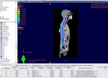

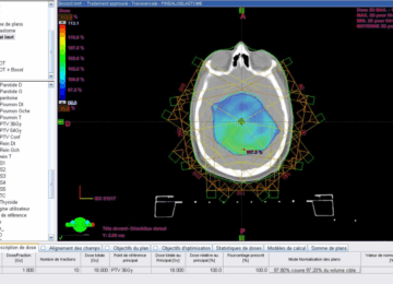

Figure 2: Dosimetric images of cranio-spinal of 36 Gy delivered by 3D conformal radiotherapy (3D-CRT), followed by a boost to the tumor itself up to 54 Gy using intensity-modulated radiotherapy (IMRT).
The total duration of treatment was 43 days, with no interruption of more than two days. Throughout, the patient was on corticosteroid therapy, accompanied by a salt-free diet and potassium and calcium supplementation.
Side effects included two patches of alopecia, CTACE grade I radiodermatitis, and secondary amenorrhea lasting one year, requiring a five-day hospital stay. After follow-up with an endocrinologist, the patient resumed a regular menstrual cycle. No side effects higher than grade 2 were reported.
Post-therapeutic evaluation by MRI, performed one month after the end of irradiation, showed a tumor residue of 9 mm. At six months, total regression of the pineal gland tumor residue was observed, leaving a cystic formation (Figure 3).


Figure 3: MRI images at 6 months after radiotherapy showing complete remission.
After a three-year follow-up, clinical examination showed that the patient was conscious and had no neurological deficits. Ophthalmological examination revealed visual acuity of 9/10. She had resumed a regular menstrual cycle and maintained stability on brain MRI, particularly concerning cystic formation. No other brain or spinal cord problems were detected.
The post-therapeutic follow-up of this patient is based on clinical and radiological monitoring every six months during the first five years, followed by annual evaluations thereafter. Its primary objective is to optimize the chances of long-term remission while enabling prompt management in the event of a relapse. This follow-up also contributes to the early detection of late-onset treatment-related complications, monitoring the patient’s overall progress, and assessing both functional and neurological prognosis in order to adjust care as needed.
3. Discussion
Pediatric pineoblastoma is a very rare tumor of the central nervous system. Classified as a supratentorial primitive neuroectodermal tumor, it develops in the pineal gland and tends to spread regionally along the neuraxis, while presenting a risk of local recurrence (1-2). Although it shares general similarities with other PNETs, its morphological and immunohistochemical features differ from those of infratentorial PNETs, such as medulloblastomas. Studies report a generally more favorable prognosis in children than in adults (3).
Magnetic resonance imaging (MRI) is the preferred modality for characterizing tumors of the pineal region. However, computed tomography (CT) can help detect tumor calcifications and rapidly assessing patients presenting with signs of intracranial hypertension.
In line with current SIOP-E imaging guidelines, a complete brain and spinal MRI is essential for all patients diagnosed with pineoblastoma. Whenever clinically feasible, spinal imaging should be performed prior to any surgical procedure to minimize interpretation challenges caused by postoperative anatomical changes (4).
On MRI, pineoblastoma is usually characterized by hyposignal or hypo- to iso-signal on T1-weighted sequences, and iso-signal or iso to hypersignal on T2-weighted sequences. The pineal region shows heterogeneous enhancement (5). In our patient’s case, several of these radiological features were observed.
On CT, this tumor often appears bulky, lobulated, and heterogeneous in its enhancement, with calcifications rarely observed (6). Moreover, patients with pineoblastoma generally present with more severe hydrocephalus than those with pineocytoma. These tumors are usually solid, but sometimes associated with cystic components, although the latter are more common in pineocytomas (7).
Histologically, pineoblastoma shares many similarities with other PNETs and is sometimes described as a supratentorial PNET tumor (2). It is a highly cellular tumor, composed of small, round, poorly differentiated cells organized in sheets or unstructured aggregates. The cells have round or oval, hyperchromatic nuclei and sparse cytoplasm, often arranged in Homer-Wright rosettes, which are interpreted as incomplete attempts at neuroblastic differentiation (2). Mitoses, rosettes, and areas of necrosis are common (8).
In addition, tumor cells show marked immunoreactivity towards specific neuronal markers, such as neurofilament, synaptophysin, chromogranin A, glial fibrillary protein, and S-100 protein. In the case studied, the majority of tumor cells were immunoreactive to synaptophysin.
The anatomical location and morphological characteristics of the lesion initially guide the pathological diagnosis of pineoblastoma. It is essential to distinguish pineoblastomas from other neoplasms of the pineal region, including pineocytomas, germ cell tumors, and glial tumors.
Beyond conventional histological evaluation, a multidisciplinary diagnostic approach incorporating histopathology, immunohistochemistry, and molecular biology has become standard. Advanced techniques such as DNA methylation profiling, next-generation sequencing (NGS), and Sanger sequencing allow for the identification of distinct molecular subtypes (PB-miRNA1/2, PB-MYC/FOXR2, PB-RB1), each with specific prognostic significance. The Heidelberg classifier, endorsed by the WHO, plays a critical role in refining tumor classification, particularly in diagnostically ambiguous cases. This integrated strategy improves diagnostic precision and supports personalized therapeutic decision-making in pediatric neuro-oncology. (9-10)
There is no established treatment strategy in the literature, mainly due to the extreme rarity of this disease. Only a few cases have been reported, with limited follow-up and modest results (11-12). However, some studies have shown that macroscopic resection of the tumor may play a key role in the treatment of pineoblastoma (13,14). Nevertheless, despite advances in surgical techniques and postoperative care, surgery in the pineal region remains complex. It carries significant risks: surgery-related mortality rates are between 4% and 7%, while serious complications can affect up to 10% of patients (15).
In recent years, significant progress has been made in the field of diagnosis. Upon confirmation of most non-germinal pineal tumors, surgical resection is required, with maximum safe resection as the primary surgical goal. Postoperative MRI should be performed within 48 hours of surgery to evaluate and document the extent of resection. The Chang staging system is used to assess metastatic disease, based on complete neuroaxial imaging and a lumbar puncture for CSF cytology on day 15 (16).
In our patient’s case, complete resection was not an option due to the tumor’s proximity to vital structures such as the midbrain and thalamus, as well as its intraventricular extension. In order to preserve quality of life, a non-invasive approach using radiotherapy was chosen, leading to complete tumor regression and excellent neurological results.
The efficacy of postoperative treatments remains uncertain in the literature. A few reports describe that radiotherapy led to tumor control and improved survival in pineoblastoma patients (17,18). However, these observations have not been statistically validated due to small sample sizes and a lack of homogeneity in the radiotherapy strategies and doses used.
Lee et al. (14) analyzed the factors influencing survival in 34 patients with pineoblastoma between 1969 and 1998. They observed that patients who received cranial irradiation ≥40 Gy had a median survival three times longer than those who received lower doses (29.8 months vs. 8.1 months). However, to date, no prospective study has confirmed the effectiveness of radiotherapy or determined the optimal radiobiological doses for treating pineoblastoma.
Recent findings indicate that localized radiotherapy, without intrathecal chemotherapy, may help suppress tumor progression following intensive systemic treatment. Although this strategy shows potential, especially in younger patients for whom intrathecal administration poses significant risks, its application remains investigational due to limited clinical data (19-20)
In the present case, the tumor was large and located near vital structures, leading to the decision to prioritize definitive radiotherapy to preserve the patient’s quality of life. Given the tendency of pineoblastomas to disseminate via cerebrospinal fluid, prophylactic craniospinal irradiation was administered at a dose of 36 Gy. Due to the relatively low sensitivity of this tumor to radiotherapy compared to germinomas or medulloblastomas (21,22) and considering its large volume, a local “boost” was delivered to the tumor site with an additional dose of 18 Gy in 2 Gy fractions. Adjuvant chemotherapy was also initiated. The treatment resulted in an almost complete regression of the tumor without inducing neurological deficits.
4. Conclusion
Pineoblastoma (PB) is the most common pineal parenchymal tumor in childhood (23), It is a highly aggressive tumor, classified as CNS WHO grade 4 (24), with a tendency to disseminate along the cranio-spinal axis (25,26), Although an optimal treatment for this condition has yet to be established, the present case successfully demonstrates that aggressive surgery can be avoided in patients whose tumors are located near critical structures. Given the risk of surgical complications, radiotherapy remains a viable and non-invasive treatment option. However, prospective studies including larger patient populations are needed to determine the efficacy and appropriate dosing of radiotherapy, to establish a standardized approach to the optimal management of pineoblastoma in children.
ABBREVIATIONS
3D-CRT – Three-dimensional conformal radiotherapy
CNS – Central nervous system
CT – Computed tomography
CTACE – Common Terminology Criteria for Adverse Events
CSF – Cerebrospinal fluid
DNA – Deoxyribonucleic acid
IMRT – Intensity-modulated radiotherapy
MeV – Megaelectronvolt
MRI – Magnetic resonance imaging
NGS – Next-generation sequencing
PNETs – Primitive neuroectodermal tumors
SIOP-E – International Society of Paediatric Oncology – Europe
VMAT – Volumetric modulated arc therapy
WHO – World Health Organization
STATEMENTS
Authors’ Contributions: All authors made significant contributions to the development of this case report. AF coordinated the work, wrote the first draft of the manuscript, and supervised the entire process. OA, HW, and ES were involved in the clinical management of the patient and data collection. SK and FF contributed to the analysis of clinical and imaging findings. AZ provided critical revision of the manuscript. BT performed the final editing and approved the version for submission. All the authors have read and approved the final manuscript and agree to be accountable for all aspects of the work.
Consent for Publication: As the corresponding author, I confirm that the manuscript has been read and approved for submission by all listed authors.
Conflict of Interest: The authors declare no conflicts of interest.
Funding Sources: None.
Written Informed Consent for Publication: Written informed consent was obtained from the patient for the publication of this case report and all accompanying images.
Acknowledgements: We would like to sincerely thank the entire care team involved in the management of this patient. Their dedication and professionalism were essential to the quality of care provided.
We are also grateful to the radiology and pathology departments for their support and valuable input in understanding the case.
Above all, we extend our heartfelt thanks to the patient for their trust, generosity, and willingness to share their story for scientific purposes.
REFERENCES
- Orphanet. Pineoblastoma [Internet]. Orphanet; [cited 2022 Nov 14]. Available from: https://www.orpha.net/consor/cgi-bin/OC_Exp.php?lng=fr&Expert=251909
- Herrick MK, Rubinstein LJ. The cytological differentiating potential of pineal parenchymal neoplasms (true pinealomas): a clinicopathological study of 28 tumors. Brain. 1979 Jun;102(2):289-320.
- Jakacki RI, Zeltzer PM, Boyett JM, Albright AL, Allen JC, Geyer JR, et al. Survival and prognostic factors following radiation and/or chemotherapy for primitive neuroectodermal tumors of the pineal region in infants and children: a report of the Children’s Cancer Group. J Clin Oncol. 1995 Jun;13(6):1377-83.
- Avula S, Peet A, Morana G, et al. European Society for Paediatric Oncology (SIOPE) MRI guidelines for imaging patients with central nervous system tumors. Child’s Nerv Syst. 2021 Aug;37(8):2497-508.
- Fujita A, Asada M, Saitoh M, Nakamura H, Kamikawa S, Kokunai T, et al. Pineoblastoma showing unusual ventricular extension in a young adult: case report. Neurol Med Chir (Tokyo). 1999 Aug;39(8):612-6.
- Chiechi MV, Smirniotopoulos JG, Mena H. Pineal parenchymal tumors: CT and MR features. J Comput Assist Tomogr. 1995 Aug;19(4):509-17.
- Sugiyama K, Arita K, Okamura T, Yamasaki F, Kajiwara Y, Ueda H, et al. Detection of a pineoblastoma with large central cyst in a young child. Child’s Nerv Syst. 2002 Apr 1;18(3):157-60.
- Schild SE, Scheithauer BW, Schomberg PJ, Hook CC, Kelly PJ, Frick L, et al. Pineal parenchymal tumors: clinical, pathologic, and therapeutic aspects. Cancer. 1993 Aug 1;72(3):870-80.
- World Health Organization. WHO classification of tumors: central nervous system tumors. 5th ed. Lyon (France): International Agency for Research on Cancer; 2021.
- Depania S, Vasiljevic A, Mynarek M, Dufour C, Pfaffe E, Maaz AU, et al. European clinical practice recommendations for the diagnosis and treatment of paediatric pineal tumors. EJC Paediatr Oncol. 2025;5:100217.
- Nozza P, Casciana ML, Rossi A, Cama A, Milanaccio C, Raso A, et al. Post-chemotherapy maturation of a pineoblastoma. Acta Neuropathol. 2010 May;119(5):651-3.
- Lutterbach J, Fauchon F, Schild SE, Chang SM, Pagenstecher A, Volk B, et al. Malignant pineal parenchymal tumors in adult patients: patterns of care and prognostic factors. Neurosurgery. 2002 Jul;51(1):44-55; discussion 55-6.
- Tate M, Sughrue ME, Rutkowski MJ, Kane AJ, Aranda D, McClinton L, et al. The long-term postsurgical prognosis of patients with pineoblastoma. Cancer. 2012 Jan 1;118(1):173-9.
- Lee JYK, Wakabayashi T, Yoshida J. Management and survival of pineoblastoma: an analysis of 34 adults from the Brain Tumor Registry of Japan. Neurol Med Chir (Tokyo). 2005;45(3):132-42
- Nomura K. Current status of treatment of brain tumors in Japan: report from the Brain Tumor Registry of Japan. No To Shinkei. 2001 May;53(5):444-52.
- Chang CH, Housepian EM, Herbert C Jr. An operative staging system and a megavoltage radiotherapeutic technique for cerebellar medulloblastomas. Radiology. 1969 Jun;93(6):1351-9.
- Chang SM, Lillis-Hearne PK, Larson DA, Wara WM, Bollen AW, Prados MD. Pineoblastoma in adults. Neurosurgery. 1995 Sep;37(3):383-90; discussion 390-1.
- Hinkes BG, von Hoff K, Deinlein F, Warmuth-Metz M, Soerensen N, Timmermann B, et al. Childhood pineoblastoma: experiences from the prospective multicenter trials HIT-SKK87, HIT-SKK92, and HIT91. J Neurooncol. 2007 Jan;81(2):217-23.
- Massimino M, Gandola L, Biassoni V, et al. Evolving therapeutic strategies for CNS-PNET. Pediatr Blood Cancer. 2013 Dec;60(12):2031-5.
- Dufour C, Foulon S, Geoffray A, et al. Prognostic relevance of clinical and molecular risk factors in children with high-risk medulloblastoma treated in the phase II trial PNET HR+ 5. Neuro Oncol. 2021 Jul;23(7):1163-72.
- Reddy AT, Janss AJ, Phillips PC, Weiss HL, Packer RJ. Outcome for children with supratentorial primitive neuroectodermal tumors treated with surgery, radiation, and chemotherapy. Cancer. 2000 May 1;88(9):2189-93.
- Claude L, Faure-Conter C, Frappaz D, Mottolese C, Carrie C. Radiation therapy in pediatric pineal tumors. Neurochirurgie. 2015 Apr 1;61(2):212-5.
- Lamba N, Groves A, Torre M, Yeo KK, Iorgulescu JB. The epidemiology of primary and metastatic brain tumors in infancy through childhood. J Neurooncol. 2022;156(2):419-29.
- Louis DN, Gerber K, von Hoff K, Resch A, et al. Treatment of children with central nervous system primitive neuroectodermal tumors/pineoblastomas in the prospective multicentric trial HIT 2000 using hyperfractionated radiation therapy followed by maintenance chemotherapy. Int J Radiat Oncol Biol Phys. 2014;89(4):863-71.
- Mynarek M, Pizer B, Dufour C, et al. Evaluation of age-dependent treatment strategies. In: Ohgaki H, Wiestler OD, et al. The 2007 WHO classification of tumors of the central nervous system. Acta Neuropathol. 2007;114(2):97-109.
- Neuro-oncology Working Group. Treatment outcomes for children and young adults with pineoblastoma: analysis of pooled European Society for Paediatric Oncology (SIOP-E) and US Head Start data. Neuro Oncol. 2017;19(4):576-85.