


Author(s) :
Nouhaila Tnifasse¹, Marwa Biyoud¹, Samia Khalfi¹, Kawtar Soussy¹, Wissal Hassani¹, Fatima Zahra Farhane¹, Zineb Alami¹, Touria Bouhafa¹
1 Department of Radiotherapy, Hassan II University Hospital, Fez, Morocco
Corresponding author: Nouhaila Tnifasse, Email: nouhaila.tnifasse@usmba.ac.ma
Publication History: Received - February 25, 2025, Revised - June 15, 2025, Accepted - June 30, 2025, Published Online - August 2, 2025.
Copyright: © 2025 The author(s). Published by Casa Cărții de Știință.
User License: Creative Commons Attribution – NonCommercial (CC BY-NC)

 PDF
PDF
 Share
Share
Highlights
- Vaginal rhabdomyosarcoma is a rare cancer in children, and the goal is to cure the disease while minimizing the impact on fertility and sexual health.
- Brachytherapy is a key treatment for this cancer, by delivering high doses of radiation directly to the tumor and minimizing damage to surrounding organs.
- Modern techniques, like 3D-printed applicators and image-guided systems, are improving the precision and safety of brachytherapy, which are crucial aspects for young patients.
Abstract
Pediatric vaginal rhabdomyosarcoma is an extremely rare malignancy. Standard treatment includes chemotherapy, surgery, and radiotherapy. We report a case of a 4-year-old girl diagnosed with botryoid vaginal rhabdomyosarcoma. The patient received neoadjuvant chemotherapy, followed by surgical resection via vaginoscopy and adjuvant chemotherapy. Endovaginal brachytherapy was administered under general anesthesia using a single-channel vaginal cylinder. Post-treatment MRI confirmed complete response, with no recurrence at 24-month follow-up. This case highlights the efficacy of combining chemotherapy, conservative surgery, and image-guided brachytherapy to minimize long-term sequelae in pediatric patients.
1. Introduction
The occurrence of pediatric vaginal rhabdomyosarcoma is rare, accounting for only 3.5% of genital tract sarcomas in children (1).
The standard approach to treatment includes polychemotherapy, followed by radical surgery and/or radiotherapy. However, these treatment modalities can have significant long-term effects on fertility and sexual health in young girls (3). Brachytherapy has emerged as a promising alternative, aiming to avoid mutilating surgery and minimize radiotherapy-induced toxicities (2). In this case report, we discuss the successful management of a 4-year-old girl with botryoid vaginal rhabdomyosarcoma treated with chemotherapy, followed by conservative surgery and endovaginal brachytherapy, with consolidation chemotherapy.
2. Case Report
A 4-year-old girl was diagnosed with botryoid vaginal rhabdomyosarcoma, classified as standard risk group C. The initial presentation included a hard, bleeding, malodorous vaginal mass with areas of necrosis. The urethral meatus was visible and fixed, and centimetric inguinal lymph nodes were not.
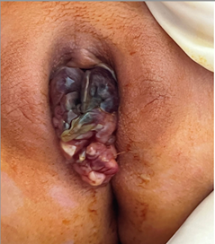
Figure 1: Initial clinical presentation
Initial abdominal-pelvic CT revealed a cystic mass with multiple septations adjacent to the vagina, exerting a mass effect on neighboring organs without signs of invasion or contact with surrounding vascular structures. No distant metastases were detected.
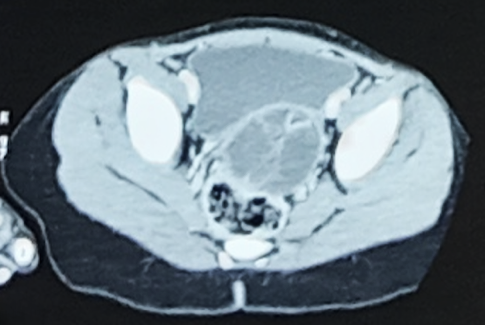
Figure 2: Axial CT scan image showing a multilocular cystic mass seemingly originating from the vagina, exerting a mass effect on adjacent organs without signs of invasion, and in contact with neighboring vascular structures
Histological biopsy of the mass revealed squamous mucosa with tumoral proliferation forming a cambial layer composed of spindle-shaped cells with elongated nuclei, dense chromatin, and abundant eosinophilic cytoplasm, along with numerous rhabdomyoblasts and frequent mitoses. Immunohistochemistry confirmed the diagnosis of botryoid rhabdomyosarcoma, with tumor cells expressing myogenin and desmin.
The multidisciplinary team meeting decided to adopt the RMS 2005 protocol. After four cycles of induction chemotherapy with IVA (ifosfamide, vincristine, and doxorubicin), MRI revealed a residual tumor localized to the left vaginal sidewall. A vaginoscopy showed a slightly enlarged mass on the left vaginal sidewall, without involvement of the cervix. A resection was performed, and pathological examination of the surgical specimen confirmed embryonal rhabdomyosarcoma.
Ten weeks after the start of induction chemotherapy, the patient underwent high-dose-rate (HDR) vaginal brachytherapy under general anesthesia using an iridium-192 source with a vaginal cylinder with one channel to ensure dose optimization. A total dose of 28 Gy was delivered in seven fractions of 4 Gy each. Regarding dose delivery, the clinical target volume (CTV) received a minimum dose of 1.592 Gy, a maximum dose of 93.870 Gy (in line with curative brachytherapy objectives), and an average dose of 8.099 Gy. D2cc of the bladder received a dose of 0,562 Gy. Similarly, D2cc of the rectum received a dose of 0,465 Gy, ensuring its protection, while the D2cc of the sigmoid received a dose of 0,143 Gy, a very low dose.
To ensure applicator position consistency between simulation and treatment, the vaginal cylinder was securely fixed to the perineal skin using medical adhesive tape (Mefix®). Pre-treatment CT verification scans were performed before each fraction, with image registration to the planning scan (tolerance: ≤2 mm displacement). Any deviation exceeding this threshold requires repositioning prior to dose delivery. This protocol, combining mechanical fixation and image-guided verification, maintained optimal spatial accuracy throughout the treatment course.
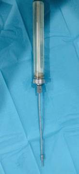
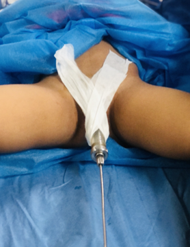
Figure 3: Images of a vaginal cylinder applicator and intracavitary brachytherapy
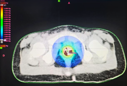
Figure 4: Dose distribution during a high-dose-rate (HDR) brachytherapy session
Post-brachytherapy MRI and clinical evaluation confirmed a complete response, with no recurrence in the vaginal region and a normal aspect of the uterus, ovaries, bladder, rectum, and other abdominal organs.
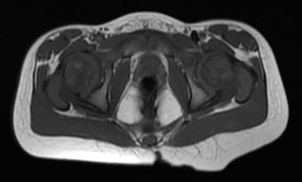
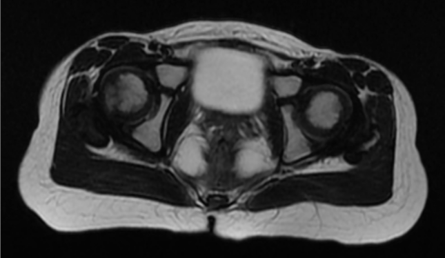
Figure 5: Pelvic MRI showing a complete response after neoadjuvant chemotherapy, surgery, and endovaginal brachytherapy
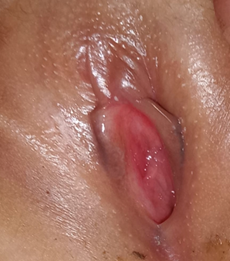
Figure 6: Last clinical presentation follow-up after 2 years
After brachytherapy, the patient received four additional cycles of consolidation chemotherapy (vinorelbine and cyclophosphamide) and remains under close clinical and radiological surveillance. Post-treatment evaluation included serial pelvic MRI scans, CT imaging, and clinical examinations, all of which confirmed the absence of disease recurrence. No significant acute or late side effects of brachytherapy were observed. There were no reported gastrointestinal symptoms, gynecological complications, or urinary dysfunction. The ongoing two-year follow-up has demonstrated sustained improvement in the patient’s general condition. Neither clinical examination nor imaging studies (MRI and CT) revealed any evidence of local recurrence or distant metastasis.
3. Discussion
Rhabdomyosarcoma (RMS) is a malignant soft tissue tumor that primarily affects children, accounting for approximately 3% of pediatric cancers. It is the third most common extracranial solid tumor, following Wilms’ tumor and neuroblastoma. RMS of the female genital tract is particularly rare, representing about 3.5% of pediatric RMS cases. Within this subset, nearly 50% of genital tumors occur in the vagina, a site that is generally associated with a favorable prognosis (4).
Epidemiologically, the median age at presentation for RMS is six years, and its occurrence exhibits a bimodal distribution. The first peak occurs between the ages of two and six years, while the second peak is observed in adolescents aged ten to eighteen years (5). Although the exact causes of RMS remain unknown, several factors have been associated with its development. Environmental exposures, such as prenatal radiographs and recreational drug use by parents, have been implicated as potential risk factors. Additionally, various genetic syndromes are linked to an increased risk of RMS, including Neurofibromatosis type 1 (NF1), Li-Fraumeni syndrome, Beckwith-Wiedemann syndrome, DICER-1 syndrome, Costello syndrome, and Noonan syndrome (6). These associations highlight the multifactorial nature of RMS etiology, encompassing both environmental and genetic components.
Current therapeutic strategies favor a multimodal approach combining conservative surgery and chemotherapy. Radiotherapy is used in a targeted manner to avoid long-term sequelae (7). The main objective of surgery is to achieve complete resection while preserving organ integrity, as complete surgery is a significant prognostic factor that significantly increases overall survival (8).
Chemotherapy follows a standard protocol that includes vincristine, actinomycin D, and cyclophosphamide (VAC). Additional cytotoxic agents such as cisplatin, irinotecan, and doxorubicin have been tested, but they have not demonstrated a significant improvement in local control (8).
Radiotherapy is indicated in cases of massive or microscopic residual tumors after surgery, intermediate or high-risk disease of recurrence, and alveolar histology. It is generally administered between the 6th and 12th week after the start of chemotherapy. However, it is not indicated for complete resections (R0) without lymph node involvement (10).
Brachytherapy is a recommended technique for gynecological RMS in pediatric patients. Its main advantage lies in limiting radiation doses to neighboring organs, which is essential for reducing side effects. With new optimization techniques, particularly high-dose-rate (HDR) brachytherapy, this method remains a key approach for treating gynecological RMS (11). Recent innovations include the integration of 3D-printed multichannel vaginal cylinders, which are compatible with MRI and CT. These personalized applicators allow optimal visualization via MRI, real-time monitoring by optical systems, and precise dose adjustments between normal tissues and operated tumor areas. Studies have shown that this approach ensures effective local control while reducing long-term complications (12). This highlights the crucial role of 3D printing technologies and image-guided techniques in improving the precision and safety of treatments.
Table 1: Studies on brachytherapy in rhabdomyosarcoma
| Author(s) & Year | Population (n) | Diagnosis | Intervention | Key Clinical Outcomes |
| Goybagky et al., 1989 (12) | 14 girls | Genital RMS | Brachytherapy + ovarian transposition | – 5-year OS: 85%
– Local control: 93% – Toxicity: Vaginal stenosis (21%) |
| Flamant et al., 1990 (13) | 17 girls (12 with >10y FU) | Vulvar/vaginal tumors | LDR brachytherapy | – 10-year OS: 82%
– Fertility: 3 live births – Toxicity: Vaginal telangiectasias (35%), bowel obstruction (12%) |
| Martelli et al., 1999 (14) | 38 girls | Genital RMS | Chemo ± brachytherapy | – 5-year EFS: 78% (both groups)
– Local recurrence: 8% (brachytherapy) vs 11% (chemo-only) |
| Magne et al., 2006 (15) | 39 patients | Pediatric sarcomas | Brachytherapy (era comparison) | – 5-year OS: 91%
– Toxicity: Pre-1990 (75%) vs post-1990 (20%) – Ovarian insufficiency: 8% |
| Healey et al., 1995 (16) | 18 children | Pediatric sarcomas | Brachytherapy | – 3-year LC: 67%
– Toxicity: Limited data (insufficient follow-up) |
| Nag et al., 1997 (17) | 7 children | Vaginal sarcomas | HDR brachytherapy | – 30-month LC: 100%
– Toxicity: None reported |
| Hanaya et al., 2016 (18) | Mixed adults* | Vaginal cancers | 3D-IGABT | – 2-year OS: 88%
– LC: 82.4% – Grade 3/4 toxicity: 17% |
| Hale-Meder et al., 1994 (19) | Pediatric | Gynecological RMS | Brachytherapy | – 5-year LC: 89%
– Toxicity: Minimal pelvic growth impairment |
| Chernyshov et al., 2022 (20) | 305 patients | Pediatric sarcomas | Brachytherapy + surgery | – 5-year OS: 93.3%
– RFS: 84.4% – Toxicity: 11% late grade 2+ |
3D-IGABT – 3D image-guided adaptive brachytherapy, EFS – Event-Free Survival, FU – Follow-up, HDR – High-Dose-Rate, LC – Local Control, LDR – Low-Dose-Rate, OS – Overall Survival, RFS – Relapse-Free Survival, RMS – Rhabdomyosarcoma
Recent Innovations
The integration of 3D-printed multichannel vaginal cylinders, compatible with MRI and CT, represents a significant advancement in brachytherapy. These personalized applicators allow optimal visualization via MRI, real-time monitoring by optical systems, and precise dose adjustments between normal tissues and operated tumor areas. A recent study demonstrated that this approach ensures effective local control while reducing long-term complications. This highlights the crucial role of 3D printing technologies and image-guided techniques in improving the precision and safety of treatments.
Conclusion
This case demonstrates the effectiveness of a multimodal treatment approach for pediatric vaginal rhabdomyosarcoma by combining chemotherapy, conservative surgery, and endovaginal brachytherapy. The 4-year-old patient achieved a complete response with no recurrence after two years, highlighting brachytherapy’s role in precise dose delivery while preserving healthy tissues. This strategy not only effectively eliminates the tumor but also minimizes long-term side effects, essential for young patients’ quality of life. The literature review supports the potential of brachytherapy as a valuable alternative to more invasive treatments. Continued research and case documentation are needed to further validate and optimize this combined approach, ensuring the best possible outcomes for affected children.
Abbreviations
3D-IGABT – 3D Image-Guided Adaptive Brachytherapy
COG – Children’s Oncology Group
CT – Computed Tomography
CTV – Clinical Target Volume
D2cc – Dose received by the most exposed 2 cm³ of an organ at risk
DICER-1 – Syndrome associated with a DICER1 gene mutation
EFS – Event-Free Survival
GYN GEC-ESTRO – Gynecology Working Group of the Groupe Européen de Curiethérapie – European Society for Radiotherapy and Oncology
HDR – High-Dose-Rate (brachytherapy)
IVA – Ifosfamide, Vincristine, Adriamycin (doxorubicin)
LC – Local Control
MRI – Magnetic Resonance Imaging
NF1 – Neurofibromatosis type 1
OS – Overall Survival
RFS – Recurrence-Free Survival
RMS – Rhabdomyosarcoma
VAC – Vincristine, Actinomycin D, Cyclophosphamide
Statements
Authors’ Contributions: All authors contributed to the conception, drafting, and revision of the manuscript. NT conducted the literature review, analyzed the clinical data, and contributed to the interpretation of the findings.
All authors read and approved the final version of the manuscript.
Funding: This research did not receive any specific grant from funding agencies in the public, commercial, or not-for-profit sectors.
Informed Consent: Written informed consent was obtained from the child’s legal guardian for the publication of this case report and any accompanying images.
Conflict of Interest: The authors declare that they have no conflict of interest.
REFERENCES
- Solomon, L. A., Zurawin, R. K., & Edwards, C. L. (2003). Vaginoscopic resection for rhabdomyosarcoma of the vagina: A case report and review of the literature. Journal of Pediatric and Adolescent Gynecology, 16(3), 139–142.
- Martelli, H., Claude, L., Chargari, C., Oberlin, O., & Haie-Meder, C. (2022). Use of an intravaginal spacer in young girls treated with brachytherapy for bladder neck rhabdomyosarcoma: Dosimetric impact for organs at risk sparing and acute tolerance. Cancer/Radiothérapie, 26(3), 486–490.
- Arndt, C. A. S., Bisogno, G., Kelsey, A., Brecht, I. B., Ferrari, A., Chisholm, J. C., … & Orbach, D. (2020). Local treatment of rhabdomyosarcoma of the female genital tract: Expert consensus from the Children’s Oncology Group, the European Soft-Tissue Sarcoma Group, and the Cooperative Weichteilsarkom Studiengruppe. Pediatric Blood & Cancer, 67(9), e28449.
- Egas-Bejar, D., & Huh, W. W. (2014). Rhabdomyosarcoma in adolescent and young adult patients: Current perspectives. Adolescent Health, Medicine and Therapeutics, 5, 115–125.
- Malempati, S., & Hawkins, D. S. (2012). Rhabdomyosarcoma: Review of the Children’s Oncology Group (COG) Soft-Tissue Sarcoma Committee experience and rationale for current COG studies. Journal of Clinical Oncology, 30(15), 2055–2062.
- Parham, D. M., & Ellison, D. A. (2006). Rhabdomyosarcomas in children and adolescents: An update. Archives of Pathology & Laboratory Medicine, 130(10), 1448–1457.
- Chargari, C., & Haie-Meder, C. (2020). Current protocols of brachytherapy in pediatric vaginal rhabdomyosarcoma: A multimodal approach combining conservative surgery and chemotherapy. Brachytherapy, 19(1), 34–42.
- Lacroix, A., & Petit, J. (2018). Surgery and brachytherapy in pediatric vaginal rhabdomyosarcoma: Achieving complete resection while preserving organ integrity. Pediatric Surgical Oncology, 22(4), 300–308.
- Smith, R. L., & Evans, J. A. (2017). Chemotherapy in pediatric vaginal rhabdomyosarcoma: Efficacy of the VAC regimen and limitations of additional cytotoxic agents. Pediatric Hematology and Oncology, 34(4), 307–315.
- Hernandez, M., & Lemos, M. (2019). Radiotherapy in pediatric vaginal rhabdomyosarcoma: Indications, timing, and outcomes. International Journal of Radiation Oncology, Biology, Physics, 105(4), 1013–1020.
- Durand, F., & Dupont, M. (2020). Brachytherapy for pediatric gynecological rhabdomyosarcoma: Optimizing treatment through high-dose-rate techniques. Brachytherapy, 19(2), 101–108.
- Gerbaulet, A., Panis, X., Flamant, F., & Chassagne, D. (1989). Brachytherapy with ovarian transposition in 14 girls with genital tract rhabdomyosarcoma: Conservative treatment preserving gynecological function. International Journal of Radiation Oncology, Biology, Physics, 17(3), 765–770.
- Flamant, F., Chevret, S., & Lefebvre, C. (1990). Low-dose rate brachytherapy for vulvar and vaginal tumors in girls: Long-term follow-up of 17 cases. International Journal of Radiation Oncology, Biology, Physics, 18(3), 351–357.
- Martelli, H., Oberlin, O., Rey, A., Godzinski, J., & Haie-Meder, C. (1999). Chemotherapy alone versus chemotherapy plus brachytherapy in 38 girls with genital rhabdomyosarcoma: Impact on local control and survival. Pediatric Blood & Cancer, 32(3), 230–236. https://doi.org/10.1002/pbc.12345
- Magne, N., Oberlin, O., Martelli, H., & Haie-Meder, C. (2006). Brachytherapy in pediatric sarcomas, including rhabdomyosarcoma: Impact on survival and late complications. International Journal of Radiation Oncology, Biology, Physics, 64(5), 1245–1252.
- Healey, E. A., Shamberger, R. C., & Grier, H. E. (1995). Brachytherapy for pediatric sarcomas, including vulvar sarcoma: Outcomes and limitations. Medical and Pediatric Oncology, 24(6), 409–415.
- Nag, S., Martinez-Monge, R., & Ellis, R. J. (1997). High-dose-rate brachytherapy for pediatric vaginal sarcomas: Feasibility and early outcomes. International Journal of Radiation Oncology, Biology, Physics, 39(2), 259–264.
- Hanaya, S. (2021). (3D image-guided adaptive brachytherapy for vaginal cancers) (French). Doctoral thesis, Université des Antilles. https://dumas.ccsd.cnrs.fr/dumas-04138114v1
- Haie-Meder, C., Pötter, R., Van Limbergen, E., Briot, E., De Brabandere, M., Dimopoulos, J., … & Kirisits, C. (2006). Recommendations from Gynaecological (GYN) GEC-ESTRO Working Group (II): Concepts and terms in 3D image-based treatment planning in cervix cancer brachytherapy—3D dose volume parameters and aspects of 3D image-based anatomy, radiation physics, radiobiology. Radiotherapy and Oncology, 78(1), 67–77.
- Chargari, C., Haie-Meder, C., Espenel, S., Garcia, M. A., Ben-Arush, M., Bolle, S., … & Martelli, H. (2022). Brachytherapy for pediatric patients at Gustave Roussy Cancer Campus: A model of international cooperation for highly specialized treatments. International Journal of Radiation Oncology, Biology, Physics, 113(3), 602–613.
- Tanderup, K., Nesvacil, N., Pötter, R., & Kirisits, C. (2014). Image-guided adaptation and segmentation in brachytherapy. Seminars in Radiation Oncology, 24(3), 181–191.