


Author(s) :
Bianca Elena Croitoru1, Edvina Elena Pirvu1
1Colțea Clinical Hospital, Medical Oncology Department, Bucharest, Romania
Corresponding author: Bianca Elena Croitoru , Email: croitorubianca7@yahoo.com
Publication History: Received - , Revised - , Accepted - , Published Online - 1 April 2023.
Copyright: © The author(s). Published by Casa Cărții de Știință.
User License: Creative Commons Attribution – NonCommercial (CC BY-NC)

 PDF
PDF
 Share
Share
Highlights
• Squamous cell carcinoma with sarcomatoid dedifferentiation represents a rare but highly aggressive variant of laryngeal cancer with poor locoregional control.
• Prior radiotherapy may contribute to sarcomatoid transformation, although the causal relationship remains controversial.
• Early recurrence, positive surgical margins, and limited response to systemic therapies underscore the need for vigilant surveillance and multimodal management.
Abstract
Sarcomatoid carcinoma is a rare and aggressive form of squamous cell carcinoma (SCC), a biphasic tumor that has been shown to be a dedifferentiated monoclonal form of classic squamous cell carcinoma. The factors that contribute to proliferation of SCC to sarcomatoid carcinoma are not precisely known but some studies revealed the importance of anterior radiation therapy. This case report describes a 62 year old man, heavy smoker and alcohol drinker diagnosed with laryngeal neoplasm. The pathological diagnosis was well-differentiated SCC of the piriform sinus. Surgery was performed and the pathology report revealed medium differentiated SCC, pT4aN1M0. The patient underwent external radiotherapy (RT) with 66Gy/tumour bed in 33 fractions. At 4 months post RT the clinical exam revealed tumor recurrence near the tracheal stoma. A second surgery was performed and the pathology diagnosed a recurrence of SCC with sarcomatoid-looking areas, with two surgical margins being invaded by the tumor. Postoperative computer tomography (CT) of the neck showed no evidence of macroscopic disease. The next treatment approach was chemotherapy with 4 cycles of carboplatin paclitaxel but soon after, an inoperable locoregional recurrence was diagnosed and he started treatment with Nivolumab followed by rapid locoregional progression. Squamous cell carcinoma with sarcomatoid dedifferentiation is a rare histology of laryngeal cancer, with an aggressive behavior that requires intense treatment and follow-up.
1. Introduction
The current incidence, prevalence and mortality of laryngeal cancer is estimated at 2.76 cases/year per 100,000 inhabitants, 14.33 cases/year per 100,000 inhabitants and 1.66 deaths/year per 100,000 inhabitants. More than 95% of laryngeal cancers are squamous cell carcinomas (1). Risk factors for laryngeal cancer are represented in particular by tobacco use, excessive alcohol ingestion, exposure to heat, gastro-esophageal reflux, anatomical malformations, Plummer-Vinson syndrome, chemicals, asbestos, nickel or ionizing radiations, along with some viral infections (e.g., human papilloma virus) (2).
2. Case presentation
A 62-year-old man, from an urban environment, with insignificant family history and a personal medical history from which we can note a single congenital kidney with chronic kidney disease stage II and arterial hypertension grade II, with occupational exposure to benzene, a 40 pack-year smoking history and history of alcohol consumption, presented on October 2020 to the ENT (Ear, Nose and Throat) department with dysphonia, hoarseness and anemia. Laryngoscopy detected significant right arytenoid edema with salivary stasis in the right piriform sinus, which completely covered the right vocal cord that could not be visualized. The CT scan revealed a tumor mass on the right piriform sinus and bilateral latero-cervical lymphadenopathy. The pathological diagnosis based on biopsy was well-differentiated squamous cell carcinoma of the piriform sinus, with invasive keratosis. The staging was completed with a CT of the chest, which didn’t reveal any metastasis. In march 2021 the patient underwent total subcricoid suprahyoid laryngectomy with bilateral jugulocarotid lymphadenectomy and right hemithyroidectomy. The pathology report showed medium differentiated squamous cell carcinoma, infiltrating the thyroid cartilage and the left aryepiglottic fold, pT4aN1M0, with negative microscopic margins (R0). Histological sections of the laryngectomy piece highlighted a malignant tumor proliferation composed of groups of epithelioid cells with moderate pleomorphism, prominent nucleoli and abundant eosinophilic cytoplasm (Fig 1).
A postoperative clinical exam showed no pathological changes. Surgery was followed in April-June 2021 by adjuvant radiotherapy when were administered 66Gy to the tumour bed (the upper edge at the level of the C3 vertebra, it includes the posterior part of the base up to the vertebral wall, and the lower up to the tracheostoma) and 50 Gy at the level of bilateral cervical ganglion areas Ib, IIa, IIb, III, IVa and the entire tracheostomy area. Radiotherapy was administered using a simultaneous integrated boost, with a dose of 2 Gy/fraction at the level of the volume irradiated with 66 Gy and 1,5 Gy per fraction at the level of the volume irradiated prophylactically, with good adherence and tolerance to treatment. Due to technical issues, treatment was postponed for 3 days during week 4.
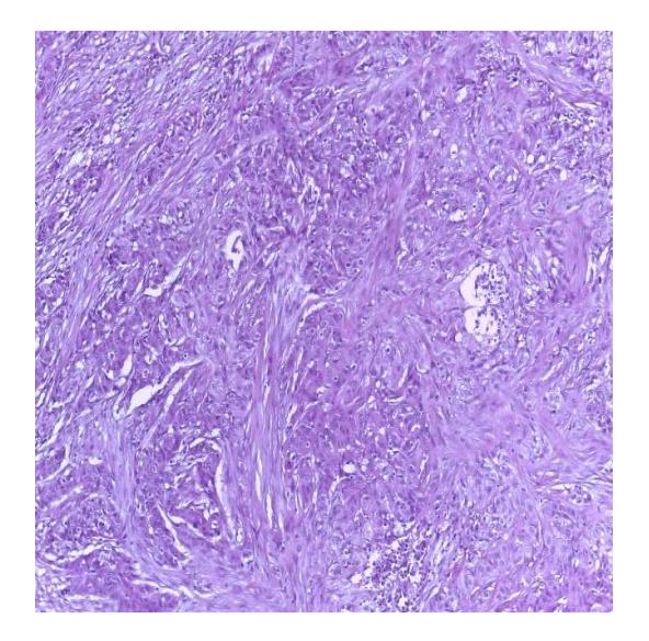
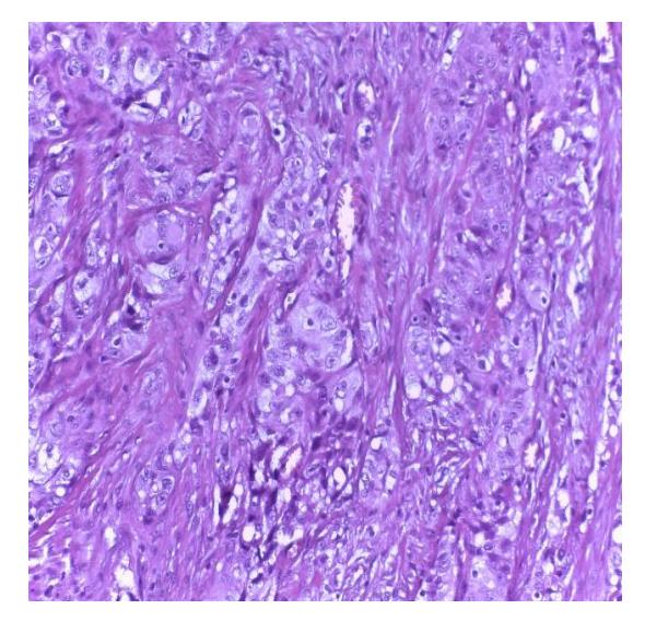
Figure. 1. The histological sections from the laryngectomy piece, with H&E staining with an x100 (A) and x200 (B) objective groups of epithelioid cells with moderate pleomorphism, prominent nucleoli and abundant eosinophilic cytoplasm
The first follow up exam 2 months post RT showed no evidence of disease, both on clinical exam and CT of the neck and thorax.
At 4 months post radiotherapy a neck CT showed a necrotic voluminous expansive mass developed at the level of the antero-inferior wall of the cervical region with a diameter of 35/33 mm, extending caudally to the proximity of the sternal handle. The ENT exam confirmed the tumor recurrence located inferior to the tracheal stoma. In October 2021 the patient underwent the second surgical intervention with en block peristomal tumor ablation with 3 tracheal rings, thyroid gland and remaining prelaryngeal musculature and restoration of the anterior cervical defect with a flap in the left large pectoral muscle. The pathology report revealed a tracheal stoma with a recurrence of squamous cell carcinoma with sarcomatoidlooking areas, with two of the surgical margins being microscopically invaded by the tumor. Histological sections of the excision piece of the tracheal stoma, in hematoxylin eosin staining, show malignant epithelial proliferation composed of spindle-shaped cells and rare groups of squamous epithelioid cells (arrow) suggestive for the diagnosis of keratinizing squamous focal carcinoma, with moderately differentiated appearance and sarcomatoid features (Fig 2). Immunohistochemistry confirmed squamous cell carcinoma with sarcomatoid dedifferentiation, that strongly stained for EMA, CK 5/6 and AE 1/3, and a high MIB-1 but negative for myoD1, SMA, desmin, and myf4 with a CPS level of 70. In November 2021, a postoperative CT of the neck showed no evidence of macroscopic disease.
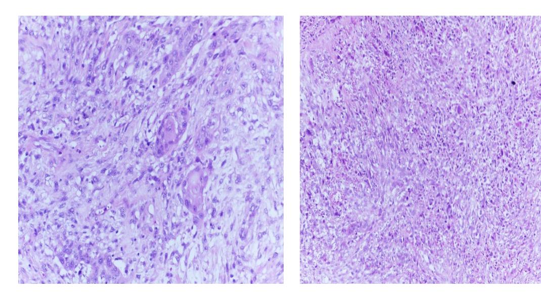
Figure 2. Histological sections from the excision piece of the tracheal stoma, with H&E staining with an x200 (A) and x40 (B) objective, show malignant epithelial proliferation composed of spindle cells and rare groups of epithelioid cells with a squamous appearance (arrow) suggestive of the diagnosis of carcinoma focal squamous keratinizing, moderately differentiated, with areas of sarcomatoid appearance;
Despite there being no evidence of residual tumor on the CT, the positivity of the surgical margins was a major risk factor for local recurrence. Reirradiation of the same anatomical region was not feasible, so 4 cycles of chemotherapy Carboplatin AUC6 / Paclitaxel 175mg/m2 were administered.
After the fourth cycle, the patient developed severe peripheral neuropathy and chemotherapy was stopped. In March 2022, the patient underwent plastic surgery with internal and external neurolysis of the common popliteal nerve, lateral popliteal and anterior tibialis, which resulted in partial recovery of neurological function and disappearance of neuropathic pain. In April 2022, an ENT exam described a continuation of local peristomal evolution on the right side, approximately 5/5 cm, with a necrotic tumor covered in detritus. An inoperable locoregional recurrence was diagnosed, and the patient immediately started treatment with Nivolumab 240 mg every 2 weeks. After three cycles, a rapid locoregional progression was visible clinically and confirmed on CT as a voluminous expansive tumor process, cauliflower-like, extended in the soft lower paracervical and straight supraclavicular parts with maximum diameters of 70/76/67 mm. It was tangent to the jugulo-carotidianne vascular structures (with preserved permeability and caliber). From June onwards, the patient started treatment with 5-Fluorouracil monotherapy 850mg/m²/day 1-4 (with reduced doses due to previous exposure to chemotherapy) with good tolerance. Unfortunately, two weeks after starting the treatment, the patient developed an infection of the tumor with sepsis and succumbed.
3. Discussion
First described by Virchow in 1863 as carcinosarcoma and further defined by Figi in 1933, [3] sarcomatoid carcinoma represents a rare, unusual, and aggressive form of SCC (<3%), a biphasic tumor that has been shown to be dedifferentiated monoclonal form of classic squamous cell carcinoma. It frequently recurs and metastasizes, affecting its prognosis [4,5,6]. The factors that contribute to transformation from SCC to sarcomatoid carcinoma are not precisely known but some studies revealed the importance of anterior radiation therapy on laryngeal squamous cell carcinoma (7,8). Case reports on this subject are very limited and treatment strategies are extrapolated from the classical recommendations for laryngeal cancer.
Laryngeal spindle cell carcinoma is a type of cancer that is more commonly seen in middle-aged to elderly men, and is strongly associated with the use of tobacco and alcohol (9). In most cases, this type of cancer is not caused by the human papillomavirus (HPV). While radiotherapy has not been identified as a causative factor, tumors that develop in patients who have received radiation treatment appear to be more aggressive in nature.(10)
There is a link in the literature between the development of Laryngeal spindle cell (sarcomatoid) carcinoma (LSCSC) and both radiation therapy and environmental factors (11). Thompson et al, in a clinicopathologic study of 187 cases of spindle cell carcinomas of the larynx found that while radiation therapy may lead to LSCSC in a small number of patients (observed in 9.1% of that clinical study and 7.7% in the literature review), the presence of a sarcomatoid pattern of growth in many patients without radiation exposure suggested that radiation was not necessarily a significant cause. (12) According to Thompson et al., 87% of their spindle cell carcinoma patients were reported to consume tobacco, while 48% were reported to consume alcohol, these factors could also influence the incidence of the disease. (13)
This histology should be treated as a carcinoma rather than a sarcoma. This is because primary sarcomas of the larynx are rare, accounting for only 0.3-1% of all laryngeal malignancies Sarcomas that do not produce matrix in the upper aerodigestive tract (UADT), such as in the larynx, are not common and tend to be deeply located. They do not typically cause a polypoid mass protruding from a mucosal surface, which is a characteristic feature of squamous cell carcinoma of the larynx (SCCL). Therefore, if a spindle cell neoplasm is found on a mucosal surface of the UADT and appears as an exophytic polypoid lesion or is detected in a more superficial location in the submucosa, it should be considered as SCCL. This holds true even if there is no differentiated squamous epithelial component present or if there is no reaction to epithelial markers.(14)
A retrospective study was conducted by Gamez at Mayo clinic that included patients with a median age at presentation of 73.5 years, most of whom (71%) had stage I or II glottic larynx cancer with polypoid or pedunculated tumors detected during the initial physical examination. The primary treatment for 27 patients (71%) was surgery alone, while 9 patients (25%) received adjuvant radiation therapy to the primary surgical site and/or elective neck, based on their initial stage or the presence of high-risk pathologic features. The median follow-up time for patients who survived was 45 months. According to the study, patients with early-stage laryngeal cancer could be effectively treated with a single modality therapy, while those with advanced-stage cancer may benefit from a multimodal treatment approach. (15)
Regarding treatment options, there is a general agreement in the literature that laryngeal spindle cell carcinomas are often resistant to radiotherapy and have a high likelihood of recurrence even after radiotherapy treatment. (16,17) This viewpoint is largely based on a literature review conducted by Lambert et al. in 1980, which reported recurrence of the cancer in 11 out of 14 patients across eight separate studies. (18) There is lack of studies to evaluate new systemic treatments for this rare disease.
One of the most comprehensive studies regarding this histology was designed by Dubal et al and included 312 cases of LSpCC (Laryngeal spindle cell carcinoma). Results showed 1-, 5-, and 10-year disease-specific survival (DSS) rates of 90.9%, 74.1%, and 57.9%; whereas 1-, 5-, and 10-year relative survival rates were 91.0%, 77.7%, and 64.5%, respectively.
A better prognosis was noted for tumors of the glottis with a 5-year DSS of 84.0% compared to 51.9% for nonglottic tumors (P < 0.0001) and a worse prognosis was observed for high-stage (III/IV) LpSCC with 5-year DSS 36.8% compared with lower stages (I, II), with a DSS of 91.8% (P < 0.0001). (19) Sarcomatoid carcinoma is a rare histology and patients are small in number, and underrepresented in clinical trials. Thus, the level of evidence regarding treatment options is limited. Due to their unfavorable evolution this type of tumors would benefit randomized clinical trials. Unfortunately, being such a rare disease, the clinical trials are not easy to set up.
4. Conclusion
Squamous cell carcinoma with sarcomatoid dedifferentiation is a rare histology of laryngeal cancer, with an aggressive behavior, that requires intense treatment and follow up. For the patients with squamous cell carcinoma of the larynx which have an in-field local relapse after radiotherapy, confirming sarcomatoid dedifferentiation has a well-defined prognostic value. Dedicated multicentric trials are needed for this rare type of laryngeal cancer in order to improve the clinical practice.
References:
- Rothman KJ, Cann CI, Flanders D, Fried MP. Epidemiology of laryngeal cancer. Epidemiol Rev. 1980;2:195-209. doi: 10.1093/oxfordjournals.epirev.a036223. PMID: 7000536.
- American Cancer Society. Cancer Facts and Figures 2019. Atlanta: American Cancer Society, 2019. Available online: https://www.cancer.org/content/ dam/cancer-org/research/cancer-facts-andstatistics/annual-cancer-facts-andfigures/2019/ cancer-facts-and-figures-2019.pdf
- Virchow R. Die krankhaften Geschwe`ulste. Berlin, Germany: A. Hirschwald; 1863:1 online resource. https://archive.org/details/diekrankhafteng06vircgoog. Accessed April 4, 2015
- Lewis JE, Olsen KD, Sebo TJ. Spindle cell carcinoma of the larynx: review of 26 cases including DNA content and immunohistochemistry. Hum Pathol. 1997;28:664–73.
- Viswanathan S, Rahman K, Pallavi S, Sachin J, Patil A, Chaturvedi P, D’Cruz A, Agarwal J, Kane SV. Sarcomatoid (spindle cell) carcinoma of the head and neck mucosal region: a clinicopathologic review of 103 cases from a tertiary referral cancer centre. Head Neck Pathol. 2010 Dec;4(4):265-75. doi: 10.1007/s12105-010-0204-4. Epub 2010 Aug 22. PMID: 20730609; PMCID: PMC2996502
- Figi FA. Sarcoma of the Larynx. Arch Otolaryngol Head Neck Surg 1933;18:21.
- Kim BY, Cho KR, Sohn JH, Kim JY. Sarcomatoid carcinoma after radiotherapy for early-stage oral squamous cell carcinoma: Case report. Medicine (Baltimore). 2019 Jul;98(27):e16003. doi: 10.1097/MD.0000000000016003. PMID: 31277093; PMCID: PMC6635294.
- Marchitto G, Marci V, Berrone M, et al. Early arising sarcoma after adjuvant radiotherapy for oral squamous cell carcinoma. J Oral Maxillofac Surg 2016;74:862e1-8.
- Miyahara H, Tsuruta Y, Yane K, Ogawa Y. Spindle cell carcinoma of the larynx. Auris Nasus Larynx 2004;31:177– 182.
- Roy S, Purgina B, Seethala RR. Spindle cell carcinoma of the larynx with rhabdomyoblastic heterologous element: a rare form of divergent differ entiation. Head Neck Pathol 2013;7:263–267.
- Batsakis JG, Rice DH, Howard DR. The pathology of head and neck tumors: spindle cell lesions (sarcomatoid carcinomas, nodular fasciitis, and fibrosarcoma) of the aerodigestive tracts, Part 14. Head Neck Surg 1982;4:499–513
- Spindle Cell (Sarcomatoid) Carcinomas of the Larynx A Clinicopathologic Study of 187 Cases Lester D. R. Thompson, M.D., Jacqueline A. Wieneke, M.D., Markku Miettinen, M.D., and Dennis K. Heffner, M.D.
- Sarcomatoid Carcinoma of the Larynx: An Aggressive Tumor Ipsita Dhal, Krishna Gopal Birmiwal1 , Bhagat Singh Lali, Aseem Mishra1 Departments of Pathology and 1 Head and Neck Oncology, Tata Memorial Centre, Homi Bhabha Cancer Hospital, Varanasi, Uttar Pradesh, India
- The Most Common Mistake in Laryngeal Pathology and How to Avoid it Amin Heidarian1 · Bruce M. Wenig
- Outcomes and Patterns of Failure of Sarcomatoid Carcinoma of the Larynx: The Mayo Clinic Experience Mauricio E. Gamez, MD ; Elizabeth Jeans, MAE; Michael L. Hinni, MD; Eric Moore, MD; Geoffrey Young, MD, PhD; Daniel Ma, MD; Lisa McGee, MD; Matthew R. Buras, MS; Samir H. Patel, MD
- Brodsky G. Carcino(pseudo)sarcoma of the larynx: the controversy continues. Otolaryngol Clin North Am 1984;17:185–197.
- Batsakis JG. “Pseudosarcoma” of the mucous membranes in the head and neck. J Laryngol Otol 1981;95:311–316
- Lambert PR, Ward PH, Berci G. Pseudosarcoma of the larynx: a comprehensive analysis. Arch Otolaryngol 1980;106:700–708.
- Laryngeal Spindle Cell Carcinoma: A Population-Based Analysis of Incidence and Survival Pariket M. Dubal, BA; Emily Marchiano, BA; David Kam, BS; Rahul Dutta, BS; Evelyne Kalyoussef, MD; Soly Baredes, MD, FACS; Jean Anderson Eloy, MD, FACS