


Author(s) :
Marwa Biyoud1, Fatima Ezzahra Alouane1, Nouhaila Tnifasse1, Thomas Ohnleiter1, Nicolas Bauer1, Rabah Hamlaoui1
1 Radiation Oncology Unit, Mulhouse Sud Alsace Regional Hospital Group, Mulhouse, France
Corresponding author: Marwa Biyoud, Email: marwa.biyoud@usmba.ac.ma
Publication History: Received - 15 October 2025, Revised - 18 December 2025, Accepted - 31 December 2025, Published Online - 31 December 2025.
Copyright: © 2025 The author(s). Published by Casa Cărții de Știință.
User License: Creative Commons Attribution – NonCommercial (CC BY-NC)

 PDF
PDF
 Share
Share
Highlights
- Re-irradiation require rigorous planning adapted to cumulative doses.
- Extended lower limb re-irradiation with good cutaneous tolerance is feasible.
- Multidisciplinary approach is needed, especially for elderly patients with multiple malignancies.
Abstract
CD30+ cutaneous T-cell lymphomas are rare lymphoid proliferations. These are characterized by radiosensitivity and can present as localized or recurrent lesions. Although rarely reported, re-irradiation is an interesting therapeutic option in cases of recurrence and localized cutaneous relapse. Modern irradiation techniques, such as tomotherapy, make this more feasible.
We report the case of an 84-year-old patient with multiple comorbidities and an incidental mediastinal adenosquamous carcinoma. He was followed for recurrent CD30+ cutaneous T-cell lymphoma of the right lower limb. The patient first received 3D conformal radiotherapy (30 Gy in 10 fractions) to the primary lesion. After recurrence, he underwent tomotherapy (30 Gy/10 fractions) to the recurrent lesion. Following further progression, a third re-irradiation (30 Gy/15 fractions) was delivered to the entire lower limb cutaneous tissue. This case shows that modern radiotherapy techniques, including tomotherapy, are safe and effective for locally recurrent cutaneous lymphomas and provide excellent local control.
1. Introduction
Cutaneous T-cell lymphomas (CTCL) represent a heterogeneous group of non-Hodgkin malignant lymphoid proliferations. They are characterized by a predominantly cutaneous tropism. Although they account for less than 5% of all non-Hodgkin lymphomas (1,2), they require a tailored approach to management. This is due to their specific clinical and evolutionary features.
Among CTCLs, CD30-positive forms, including both primary cutaneous anaplastic large cell lymphomas and lymphomatoid papulosis, are characterized by an often indolent course and excellent sensitivity and response to radiotherapy (3,4).
Clinically, patients with CTCL present with progressive skin lesions ranging from patches to tumors, typically evolving from macules. The indolent form of the disease may be present months before clinical presentation (5).
Radiotherapy thus plays a central role in the treatment of these lymphomas, whether with curative or palliative intent. It achieves high rates of complete response, even at low doses (5–7). However, local or multifocal recurrences remain frequent, sometimes necessitating re-irradiation. In such cases, tissue tolerance, particularly in elderly patients or those with multiple comorbidities, becomes a major concern (8–12).
The emergence of modern radiotherapy techniques, such as intensity-modulated radiotherapy (IMRT) and tomotherapy, has significantly improved treatment precision. These advances now allow re-irradiation of extensive volumes while preserving healthy tissue as much as possible (8,9,13–16).
Here, we report the case of an elderly patient with a recurrent CD30-positive cutaneous T-cell lymphoma who underwent re-irradiation of the right lower limb with tomotherapy, achieving excellent clinical and dosimetric tolerance. This case illustrates the safety and feasibility of a modern, advanced technical approach in the context of extensive cutaneous recurrence.
2. Clinical Case
We report the case of an 84-year-old male patient managed for recurrent cutaneous T-cell lymphoma (CTCL) localized to the right lower limb. His medical history included arterial hypertension, dyslipidemia, type 2 diabetes, and a chronic smoking history of 92 pack-years, having quit 22 years ago. Surgically, he had undergone an aorto-bi-iliac prosthesis, bilateral saphenous vein stripping, and angioplasty with stenting of the right external iliac artery.
The patient’s clinical history began in early 2023 with the appearance of a small millimetric cutaneous lesion on the right lower limb, with slow progression. Approximately one year after the gradual increase in size and onset of pruritus, the patient consulted a dermatologist in February 2024.
Dermatologic examination revealed a single cutaneous lesion measuring 2 cm in its largest diameter, pruritic, infiltrated, and ulcerated, located at the center of the middle anterior third of the right leg. Examination of the lymph node areas was unremarkable, and no other cutaneous lesions were identified. A skin biopsy was performed on the same day.
Histopathological analysis confirmed the diagnosis of CD30-positive, ALK-negative T-cell lymphoma with a clonal T-cell receptor (TCR) gamma rearrangement.
Two weeks after histological confirmation, on February 26, 2024, an initial 18F-FDG PET scan showed hypermetabolism strictly limited to the cutaneous lesion, without nodal or visceral involvement, classifying the disease as localized.
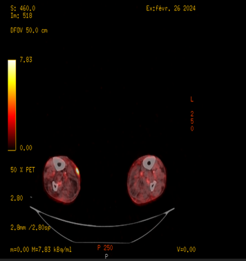
Figure 1: 18 FDG PET scan showing hyperfixation of the initial lesion (February 2024).
2.1. First Irradiation
A dosimetric CT simulation was performed on April 24, 2024, with 2.5-mm slices, without contrast, from the knee to the foot. Three-dimensional conformal radiotherapy (3D-CRT) was delivered using a TrueBeam 6 MV linear accelerator at a total dose of 30 Gy in 10 fractions of 3 Gy, five sessions per week, from April 29 to May 13, 2024, with two temporary interruptions due to machine maintenance. CTV 1 included macroscopic tumor volume (GTV 1) plus a 2 cm anatomoclinical margin. PTV 1 consisted of CTV + 5 mm isotropic margin
Planning utilized two anterior-posterior beams to ensure homogeneous coverage. Dosimetric analysis followed ICRU recommendations, with a maximum dose of 31.97 Gy and D95% of 29.03 Gy. Patient positioning was verified daily via portal imaging.
Clinical tolerance was excellent, with no radiodermatitis or acute toxicity (CTCAE v4). Three months after radiotherapy, the lesion had completely resolved, and the patient remained in complete cutaneous response for approximately 9 months.
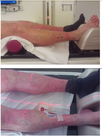
Figure 2. Simulation setup for the initial radiotherapy of the first lesion on the right leg.
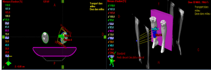

Figure 3. Dosimetric plan of the first radiotherapy on the initial lesion of the right leg.
2.2. Cutaneous Recurrence and Discovery of Lung Cancer
At the end of January 2025, the patient presented with a cutaneous recurrence consisting of a 2.5 cm ulcerated nodule in the right popliteal fossa, associated with multiple smaller nodules on the leg up to mid-thigh. Popliteal and inguinal lymph nodes were unaffected.
A PET scan on February 12, 2025, revealed intense hypermetabolism of the cutaneous lesions and hypermetabolic left hilar and subcarinal lymph nodes. Endobronchial ultrasound (EBUS) with biopsy confirmed a mediastinal nodal adenosquamous lung carcinoma harboring a KRAS G12C mutation, staged as cTx N2 M0 (stage IIIA). Clinically, the patient had general deterioration, weight loss, anorexia, and reduced autonomy (PS 2).
A comprehensive pre-therapeutic workup was performed, including laboratory analyses, vaccinations, echocardiography, thoraco-abdomino-pelvic and brain CT scans, and functional respiratory evaluation.
2.3. Second Irradiation (Popliteal Nodule)
On February 21, 2025, a multidisciplinary meeting recommended irradiation of the popliteal nodule. Tomotherapy was delivered from February 26 to March 11, 2025, at 30 Gy in 10 fractions of 3 Gy. CTV 2 was created by delineating the popliteal nodule and adding 2 cm anatomoclinical margin. PTV 2 consisted of CTV 2 to which we added a 5 mm isotropic margin.
Clinical regression of the nodule was achieved, although new cutaneous lesions appeared on the leg and thigh.
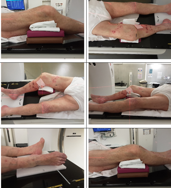
Figure 4. Simulation setup for the second irradiation at the right popliteal lesion.
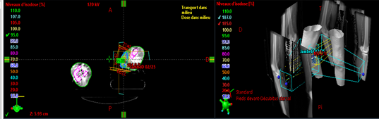

Figure 5. Dosimetric plan of radiotherapy delivered to the second right popliteal lesion.
2.4. Mediastinal Radiotherapy
For lung cancer, a 4D simulation was performed on May 16, 2025, to account for respiratory motion. Mediastinal radiotherapy targeting affected lymph nodes was delivered from May 26 to July 11, 2025, at 66 Gy in 33 fractions of 2 Gy, delivered 5 times per week. No aggressive chemotherapy was administered due to the low chemosensitivity of adenosquamous carcinoma, advanced age, and multiple comorbidities.
2.5. Extended Re-Irradiation of the Right Lower Limb
Due to cutaneous progression, extended re-irradiation of the right lower limb was performed. CT simulation was performed on June 27, 2025, with 2.5-mm slices from the pelvis to the feet. Tomotherapy IGRT 6 MV delivered 30 Gy in 15 fractions of 2 Gy, five sessions per week, from July 15 to August 2, 2025. CTV 3 ccovered the skin of the right lower limb, including all at-risk areas A 5 mm was used from CTV to PTV.
Fields were planned to fully cover the limb. Planning was optimized to spare normal tissues (skin, bone, muscle, vessels). Quality control included portal and absolute dosimetry, and daily positioning was verified via MV-CT. Dosimetric analysis showed a maximum dose of 33.76 Gy and a D95% of 29.04 Gy.
The re-irradiation achieved a good clinical response and excellent cutaneous tolerance, with no radiodermatitis or lymphedema. One month after treatment, dermatological examination showed significant regression of the lesions.
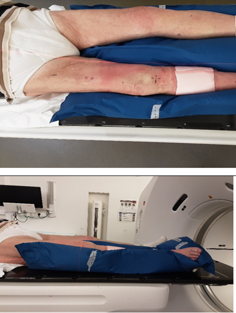
Figure 6. Simulation for total re-irradiation of the right lower limb.
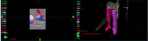

Figure 7. Dosimetric plan for the re-irradiation of the entire right lower limb.
2.6. Outcome and Follow-up
Two months after thoracic radiotherapy, thoraco-abdomino-pelvic and brain CT scans showed pulmonary and cerebral metastatic progression, leading to exclusive supportive care. The patient subsequently developed confusion and swallowing difficulties and died at the end of September 2025 due to lung cancer.
Table 1: Overview of radiotherapy treatments delivered to the patient.
| 1st Irradiation | 29 April – 13 May 2024 | Initial lesion, right leg | 3D-CRT (TrueBeam 6 MV) | 30 Gy | 10 × 3 Gy | CTV 1 : 2 cm anatomoclinical margin around tumor (GTV 1)
PTV 1: CTV + 5 mm isotropic |
Excellent, no radiodermatitis or acute toxicity |
| 2nd Irradiation | 26 February – 11 March 2025 | Right popliteal nodule | Tomotherapy | 30 Gy | 10 × 3 Gy | CTV 2 : popliteal nodule (GTV 2) + 2 cm anatomoclinical margin
PTV 2: CTV + 5 mm isotropic |
Good, no acute toxicity |
| Mediastinal Radiotherapy | 26 May – 11 July 2025 | Mediastinal lymph nodes (lung cancer) | 4D Tomotherapy | 66 Gy | 33 × 2 Gy | CTV: lymph nodes + margins according to respiratory motion PTV : CTV + 5 mm |
Moderate, no severe events reported |
| Extended Re-Irradiation | 15 July – 2 August 2025 | Right lower limb (skin) | Tomotherapy IGRT (MV-CT) | 30 Gy | 15 × 2 Gy | CTV 3: entire skin of right lower limb, including all at-risk areas PTV: CTV + 5 mm isotropic |
Excellent, good clinical response and cutaneous tolerance; planning optimized to respect previous cumulative doses |
Figure 8. Anatomical diagrams approximately illustrate the main therapeutic target volumes used for treatment
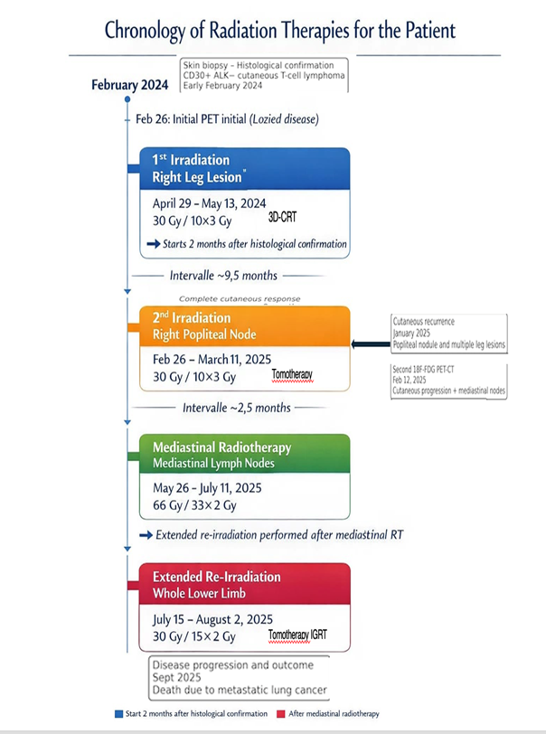
Figure 9. Illustration of the timeline of radiotherapy sequences performed for our patient.
3. Discussion
Cutaneous T-cell lymphomas represent a rare and varied group of malignant lymphoid proliferations with cutaneous tropism, accounting for less than 5% of all non-Hodgkin lymphomas (1,2). Among them, CD30+ forms are characterized by often localized progression, a potential for recurrence, and notable sensitivity to radiotherapy (3,4).
Radiotherapy is considered the standard treatment for localized forms, achieving complete response rates of over 90% at doses of 20-36 Gy (5–7). However, local or multifocal recurrence is not uncommon and may warrant re-irradiation (9). This raises important questions regarding tissue tolerance, technical feasibility, and cumulative dose, particularly in elderly or polymorbid patients (10–13).
Innovation in radiotherapy and advances in irradiation techniques, particularly three-dimensional intensity-modulated radiotherapy (IMRT), volumetric modulated arc therapy (VMAT), and tomotherapy, have led to improved dose homogeneity and reduced tissue sparing. Volumetric modulated arc therapy (VMAT) and tomotherapy have enabled greater dose homogeneity and optimized sparing of healthy tissue, opening the possibility of more effective and safer re-irradiation (9,10,14–17).
Tomotherapy combines helical intensity modulation and daily control using integrated imaging (MV-CT), ensuring millimeter-level positioning accuracy and a significant reduction in irradiation margins and toxicity risk (15–17).
The tolerable cumulative dose for skin tissue in re-irradiation remains debated, but several studies suggest that re-irradiation at moderate doses of 20-30 Gy can be performed without a major increase in toxicity. The prescribed surface dose needs to be homogeneous and doses to critical volumes to be strictly respected for avoiding significant toxicity (18,19,22).
In our case, extensive re-irradiation of the right lower limb using 30 Gy in 15 fractions was well tolerated, with no notable side effects, including radiodermatitis, lymphedema, or acute complications, confirming the clinical feasibility of this approach.
The concomitant presence of adenosquamous lung carcinoma highlights the rarity of multiple neoplasms in patients with CLL. These associations may be related to advanced age, immunosuppression, or cumulative carcinogenic exposure (1,2,20,21). The incidental discovery of this second primary cancer led to a personalized and individualized multidisciplinary management strategy, taking into account the constraints of dual cancer and the patient’s general condition.
Our observation has some limitations, notably its isolated, descriptive nature, which prevents us from drawing more generalizable conclusions or recommendations. Follow-up remains limited, so late side effects could not be assesed
However, the case illustrates the feasibility and safety of extensive skin re-irradiation using tomotherapy, even in frail, polymorbid elderly patients. It also highlights the value and benefits of rigorous dosimetric planning and daily monitoring using integrated imaging, ensuring both therapeutic efficacy and the preservation of surrounding healthy tissue. This approach could be considered and extended to similar cases, particularly when surgery or chemotherapy is contraindicated, and therapeutic options are limited.
Written informed consent was obtained from the patient for publication of this case report and any accompanying images.
4. Conclusion
Re-irradiation of recurrent cutaneous T-cell lymphomas remains a therapeutic challenge, particularly in elderly patients with multiple comorbidities. Technological advances, such as tomotherapy, now allow highly precise treatments with careful dosimetric planning.
The synchronous lung tumor further highlights the complexity of oncological management and the need for a multidisciplinary approach integrating clinical, technical, and ethical considerations.
Modern radiotherapy techniques, including tomotherapy, therefore represent an effective option for the re-irradiation of locally recurrent cutaneous T-cell lymphomas, contributing to improved local control in frail patients. However, further studies are needed to better understand long-term outcomes, refine treatment strategies, and establish standardized guidelines for this challenging clinical scenario.
Abbreviations
CTCL – Cutaneous T-cell lymphoma
PET scan – Positron Emission Tomography Scan
CD 30 – Cluster of Differentiation 30
CTCAE v4 – Common Terminology Criteria for Adverse Events Version 4
3D CRT – 3D conformal radiotherapy
IGRT – Image-Guided Radiation Therapy
ICRU – International Commission on Radiation Units and Measurements
EBUS – Endobronchial Ultrasound
KRAS – Kirsten Rat Sarcoma viral oncogene homolog
MV-CT – Megavoltage Computed Tomography
GTV – Gross Tumor Volume
CTV – Clinical Target Volume
PTV – Planning Target Volume
Statements
Authors’ Contributions: MB conceived and designed the study, coordinated the work, managed the patient, collected and analyzed the data, interpreted the clinical and dosimetric findings, drafted the manuscript, and performed all revisions following the peer-review process. FEA and NT were involved in patient surveillance and data collection. TO participated in patient management and treatment planning, and contributed to the technical aspects of radiotherapy. RH and NB provided supervision and critically reviewed the manuscript.
Consent for publication: All authors approved the final version of the manuscript and agree to be accountable for all aspects of the work.
Patient Consent: The patient provided written and verbal informed consent for the publication of this case report, in accordance with ethical guidelines and the journal’s recommendations.
Conflict of Interest: The authors declare no conflicts of interest.
Funding: No funding was received.
Acknowledgements: The authors thank the medical and radiotherapy staff for their support.
References
-
Willemze R, Jaffe ES, Burg G, Berti E, Kerl H, Meijer CJ, et al. WHO-EORTC classification for cutaneous lymphomas. Blood. 2005 May 15;105(10):3768-85.
-
Swerdlow SH, Campo E, Harris NL, Jaffe ES, Pileri SA, Stein H, et al., editors. WHO classification of tumours of haematopoietic and lymphoid tissues. 4th ed. Lyon: IARC; 2017.
-
Bekkenk MW, Geelen FA, van Voorst Vader PC, Heule F, Geerts ML, van Vloten WA, et al. Primary and secondary cutaneous CD30+ lymphoproliferative disorders: a report from the Dutch Cutaneous Lymphoma Group on the long-term follow-up data of 219 patients and guidelines for diagnosis and treatment. Blood. 2000 Jun 15;95(12):3653-61.
-
Kempf W, Pfaltz K, Vermeer MH, Cozzio A, Ortiz-Romero PL, Bagot M, et al. EORTC, ISCL, and USCLC consensus recommendations for the treatment of primary cutaneous CD30+ lymphoproliferative disorders: an update. Blood. 2011 Oct 13;118(15):4024-35.
-
Hoppe RT. Mycosis fungoides: radiation therapy. Dermatol Ther. 2003;16(4):347-54.
-
Specht L, Dabaja B, Illidge T, Wilson LD, Hoppe RT. Modern radiation therapy for primary cutaneous lymphomas: field and dose considerations from the International Lymphoma Radiation Oncology Group. Leuk Lymphoma. 2015;56(10):2771-8.
-
Neelis KJ, Schimmel EC, Vermeer MH, Senff NJ, Willemze R, Noordijk EM. Low-dose palliative radiotherapy for cutaneous T-cell lymphoma: outcomes in 48 patients. Int J Radiat Oncol Biol Phys. 2009 May 1;74(1):154-8.
-
Thomas TO, Agrawal P, Guitart J, Ghaferi J, Kuzel TM, Behdad A, et al. Outcome of patients with localized cutaneous T-cell lymphoma treated with radiation therapy. Int J Radiat Oncol Biol Phys. 2013 Dec 1;88(5):1054-60.
-
Doyen J, Servagi-Vernat S, Bensadoun RJ, Fenoglietto P, Thariat J, French Radiation Oncology Society for Cutaneous Lymphomas. Radiotherapy of cutaneous lymphomas: French recommendations. Cancer Radiother. 2018 Dec;22(8):764-72.
-
Hoppe RT, Harrison C. Reirradiation in the management of cutaneous lymphomas. J Am Acad Dermatol. 2014 Jun;70(6):1042-7.
-
Dabaja BS, Specht L, Neelis K, Willemze R, Illidge T, International Lymphoma Radiation Oncology Group. Radiation therapy for relapsed or refractory cutaneous lymphoma. Clin Lymphoma Myeloma Leuk. 2017 Aug;17(8):510-5.
-
Guenoun N, Doyen J, Bondiau PY, Benezery K, Hannoun-Levi JM. Challenges of re-irradiation in skin lymphomas: dose, tolerance and outcomes. Radiother Oncol. 2021 May;158:132-8.
-
Mavroidis P, Pearcey R, Vuong T, Lind BK, Brahme A. Dose constraints and reirradiation tolerance in skin and subcutaneous tissues. Phys Med Biol. 2019 Apr 15;64(8):085007.
-
Thomas TO, Agrawal P, Guitart J, Ghaferi J, Kuzel TM, Behdad A, et al. IMRT for extensive cutaneous lymphoma: dosimetric advantages and clinical outcomes. Int J Radiat Oncol Biol Phys. 2012 Mar 15;82(4):1529-36.
-
Amini A, Rostad BT, Ghaferi J, Thomas TO, Agrawal P, Guitart J, et al. Helical tomotherapy for total skin irradiation: technical considerations and early results. Radiat Oncol. 2015 Apr 17;10:93.
-
Vieillot S, Fenoglietto P, Lemanski C, Gourgou S, Quetier I, Doyen J, et al. Tomotherapy for cutaneous lymphomas: clinical outcomes and tolerance. Cancer Radiother. 2019 May;23(3):184-91.
-
Wong E, Tsang RW, Gospodarowicz MK, Wells W, Sun A, Hodgson DC, et al. Image-guided radiotherapy in cutaneous lymphoma: safety and feasibility. Br J Dermatol. 2020 Oct;183(4):735-42.
-
Modh A, McHargue CA, Lim HW, Siddiqui F. Single-fraction radiation therapy provides highly effective palliation for cutaneous T-cell lymphoma. J Am Acad Dermatol. 2014 Aug;71(2):333-9.
-
Chen TY, Specht L, Illidge T, International Lymphoma Radiation Oncology Group. Reirradiation in lymphoma: dose tolerance and outcomes. Ann Oncol. 2020 Jul;31(7):957-65.
-
Kim YH, Hoppe RT. Second malignancies in cutaneous T-cell lymphoma: risk factors and patterns. Arch Dermatol. 2003 Aug;139(8):948-52.
-
Jawed SI, Myskowski PL, Horwitz S, Moskowitz A, Querfeld C. Cutaneous T-cell lymphoma: risk of second primary malignancies. JAMA Dermatol. 2014 Dec;150(12):1308-15.
-
Specht L, Illidge T, International Lymphoma Radiation Oncology Group. Radiation tolerance of skin and subcutaneous tissue in lymphoma re-irradiation. Acta Oncol. 2018 Jul;57(7):905-12.
-
Scorsetti M, Alongi F, Castiglioni S, Clerici E, Fogliata A, Navarria P, et al. Advances in reirradiation techniques: IMRT, VMAT, and tomotherapy applications. Front Oncol. 2020 May 12;10:615.