

Author(s) :
Ana Băncilă1 Ioan-Alexandru Diaconu-Marinescu2
1 Radiotherapy Department, Neolife Clinic, Bucharest, Romania
2 Radiotherapy Department, „Prof. Dr. Alexandru Trestioreanu” Oncology Institute, Bucharest, Romania
Corresponding author: Ana Băncilă, Email: ana.bancila@neolife.ro
Publication History: Received - , Revised - , Accepted - , Published Online - 31 August 2026.
Copyright: © The author(s). Published by Casa Cărții de Știință.
User License: Creative Commons Attribution – NonCommercial (CC BY-NC)
 Share
Highlights
- Mycosis fungoides is the most common cutaneous T-cell lymphoma, and its high radiosensitivity makes radiotherapy a cornerstone of treatment.
- Moderate-dose localized radiotherapy achieves complete clinical and metabolic remission of a symptomatic tumoral-stage lesion with minimal toxicity.
- Careful immobilization, surface bolus and IMRT planning ensure reproducible setup and adequate dose coverage at challenging anatomical sites.
- Systemic progression despite local control highlights the need for ongoing multidisciplinary management in advanced disease.
Abstract
Background: Mycosis Fungoides (MF) is the most frequent subtype of cutaneous T-cell lymphoma. Radiotherapy (RT) is considered one of the most effective treatment modalities because of the high radiosensitivity of lymphoid cells, providing excellent local control even in advanced stages.
Case presentation: We report the case of a 74-year-old female patient with tumoral-stage Mycosis Fungoides presenting with a painful, ulcerated lesion on the plantar surface of the left foot. The patient was treated with intensity-modulated radiotherapy (IMRT) to a total dose of 30 Gy in 10 fractions, using a frog-leg position and thermoplastic immobilization with surface bolus to ensure reproducible setup and adequate dose coverage. Treatment was well tolerated and resulted in complete local response without significant acute toxicity. Follow-up Positron Emission Tomography–Computed Tomography (PET-CT) performed three months after treatment demonstrated complete metabolic remission of the irradiated lesion, although new lymph node involvement was detected.
Conclusion: This case confirms that localized radiotherapy using modern IMRT techniques can achieve durable local control and symptomatic relief in Mycosis Fungoides, while underlining the importance of multidisciplinary management for patients with systemic disease progression.
1. Introduction
Mycosis Fungoides (MF) is the most common subtype of primary cutaneous T-cell lymphoma (CTCL), representing nearly half of all cutaneous lymphomas. It is characterized by an indolent yet progressive clinical course that typically evolves through three stages — patch, plaque, and tumor — with potential for lymph node and visceral involvement in advanced disease.
The pathogenesis of MF involves the proliferation of mature, skin-homing CD4+ T-lymphocytes, leading to the formation of epidermotropic infiltrates. Diagnosis often requires a combination of histopathological, immunohistochemical, and molecular findings, and delays in recognition are frequent, particularly in the early phases of disease.
Management strategies depend on disease stage and may include skin-directed therapies (topical corticosteroids, phototherapy, or local radiotherapy) and systemic approaches (immunomodulatory or cytotoxic agents). Among these, radiotherapy remains one of the most effective modalities, as lymphoid cells are highly radiosensitive. Both localized RT and total skin electron beam therapy (TSEBT) have demonstrated excellent clinical outcomes, with response rates often exceeding 90%(1,2).
Recent international guidelines from the European Organisation for Research and Treatment of Cancer (EORTC) and the International Lymphoma Radiation Oncology Group (ILROG) emphasize the role of conformal RT techniques, such as IMRT or electron beam therapy, as standard of care for localized or symptomatic lesions(3,5).
In this report, we describe the case of a 74-year-old female patient with tumoral-stage Mycosis Fungoides treated successfully with localized IMRT, achieving a complete clinical response at the irradiated site. The case highlights technical considerations, radiotherapy planning strategy, and outcome in comparison with published data..
2. Case Report
History and Clinical Presentation
A 74-year-old female patient with a history of hypertension and paroxysmal atrial fibrillation was initially diagnosed in 2016 with anaplastic large-cell T-cell lymphoma, ALK-negative. She received six cycles of COEP chemotherapy, achieving complete remission confirmed by PET-CT, and remained disease-free until 2022.
In late 2024, the patient developed a progressive erythematous, infiltrative plaque located on the plantar surface of the left foot, approximately 6 × 5 cm in size, centrally ulcerated, pruritic, and infiltrative.
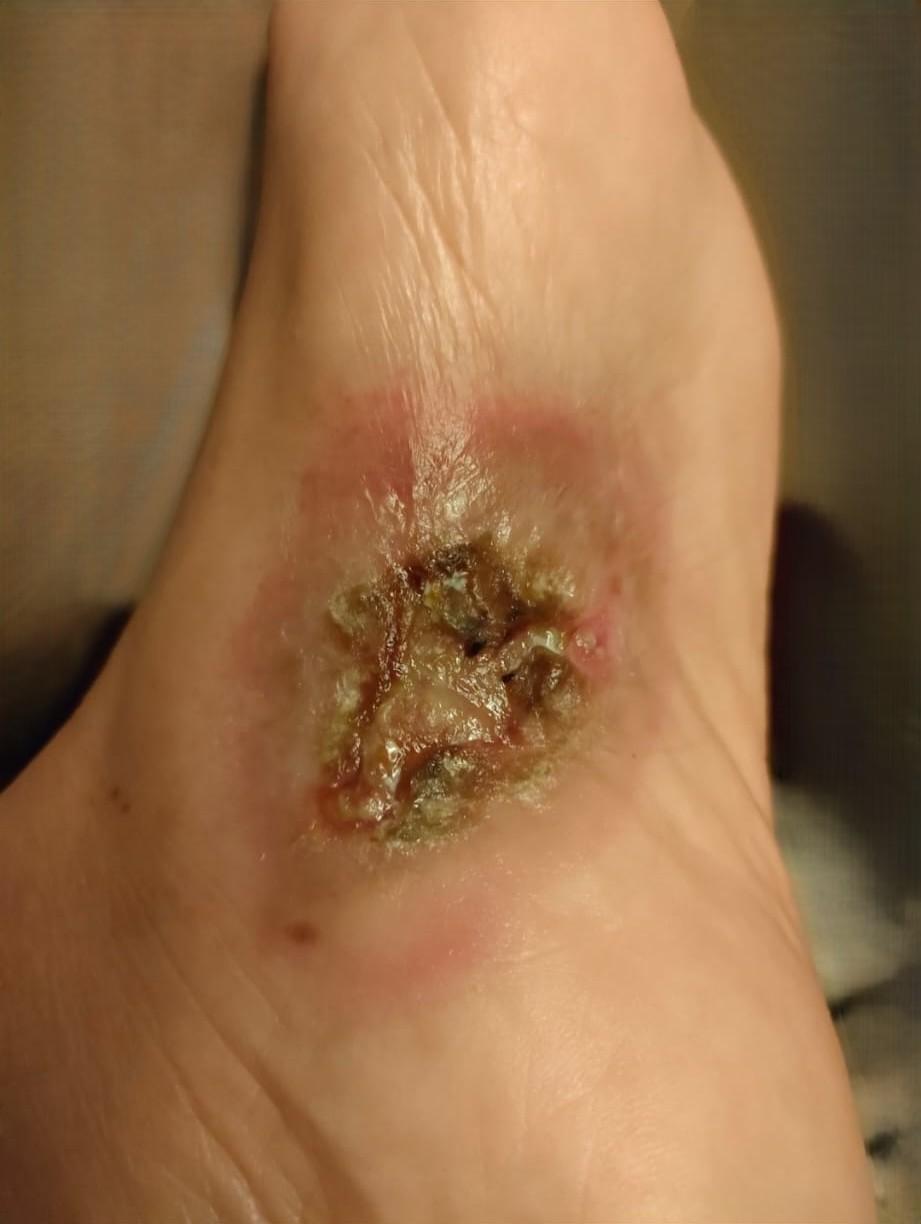 Figure 1 – Clinical aspect before radiotherapy
Investigations
Histopathological examination (January 2025) revealed diffuse dermal lymphoid proliferation of atypical medium-to-large T-cells. Immunohistochemistry showed a CD3+, CD5+, CD4+ phenotype, negative for CD30, CD25, and ALK-1, with a Ki-67 index of 20–25%.
A second opinion obtained in April 2025 confirmed epidermotropism and features consistent with the tumoral stage of Mycosis Fungoides(6). PET-CT (March 2025) identified a metabolically active lesion in the left plantar region Maximum Standardized Uptake Value – (SUVmax ≈ 7.1) with no evidence of systemic disease.
Treatment Approach
The multidisciplinary tumor board recommended definitive local radiotherapy, as surgical excision was not feasible.
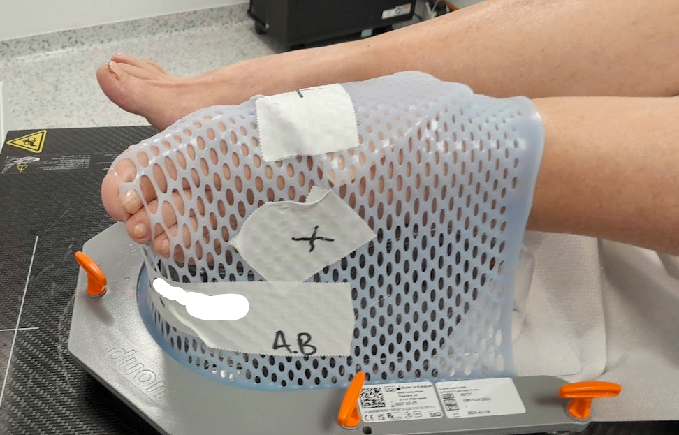
The patient was positioned in supine (dorsal decubitus) position, with the affected limb placed in a “frog-leg” configuration to optimize access to the plantar surface and ensure reproducible daily setup.
A thermoplastic immobilization mask was customized to stabilize the foot and lower leg, minimizing motion during irradiation. A 5 mm tissue-equivalent bolus was applied over the lesion for all fractions to ensure uniform dose distribution and adequate surface coverage.
Computed Tomograph (CT) simulation was performed with a 5 mm slice thickness for accurate target delineation according to ICRU Reports 50 and 62.
The Gross Tumor Volume (GTV) was defined as the macroscopic visible lesion on the plantar surface, measuring approximately measuring approximately 6 × 5 cm in greatest clinical dimensions, including the centrally ulcerated area identified on clinical examination and CT imaging.
The Clinical Target Volume (CTV) was generated by expanding the GTV by 0.5 cm in all directions to account for potential microscopic disease extension within the surrounding soft tissue.
The Planning Target Volume (PTV) was subsequently created by adding an additional 0.5 cm isotropic margin to the CTV to account for setup uncertainties and potential intra-fraction motion during daily treatment delivery.
All volume delineations followed the standardized methodology recommended by ICRU Report 50 and its Supplement 62.
Treatment planning and delivery were performed using intensity-modulated radiotherapy (IMRT) on a TrueBeam linear accelerator equipped with an Agility multileaf collimator (MLC).
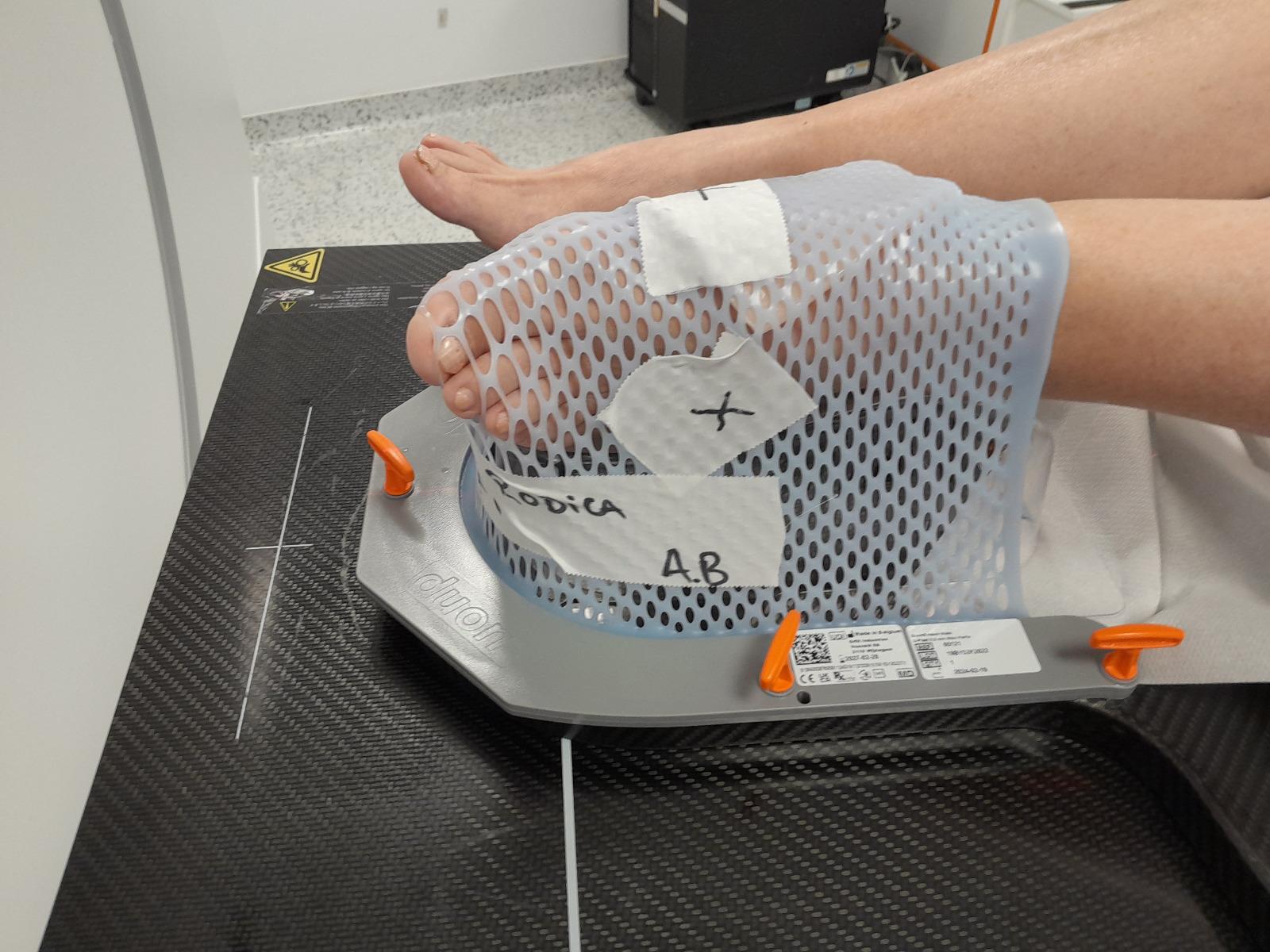
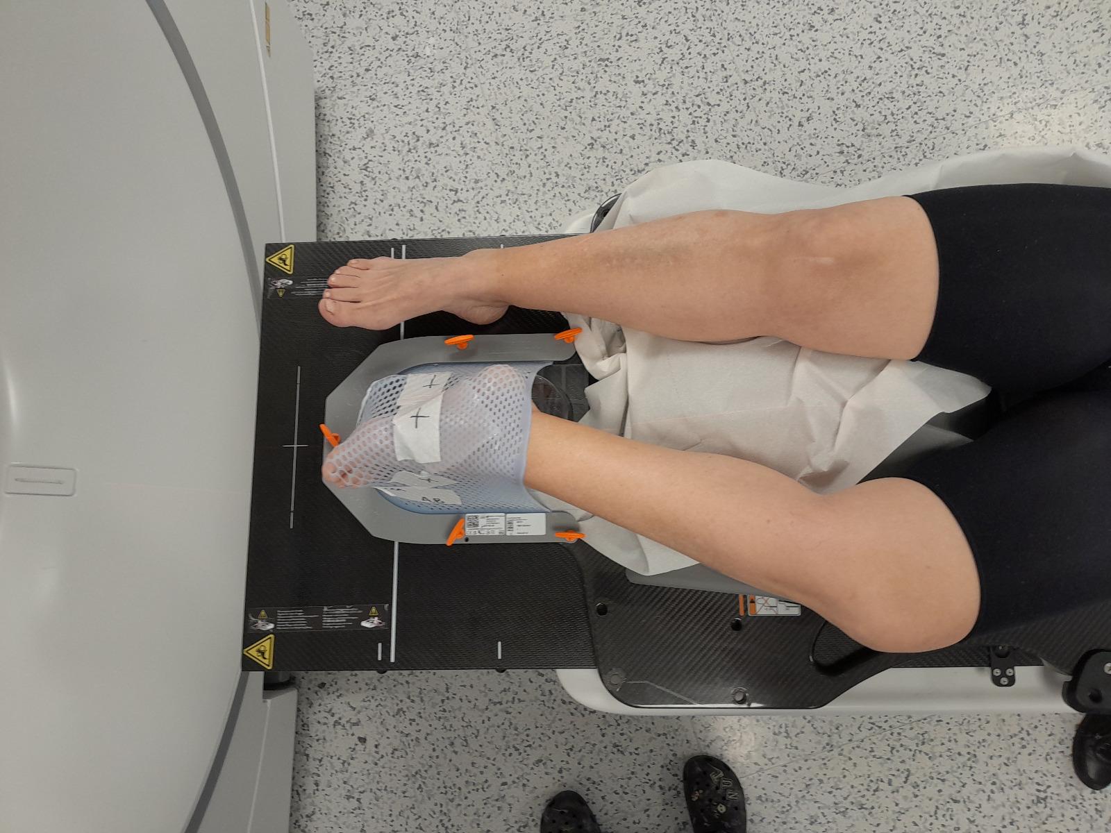
Figure 2A and 2B – Patient positioning and immobilization during IMRT
The plan was normalized such that 95% of the PTV received at least 100% of the prescribed dose (30 Gy in 10 daily fractions of 3 Gy each). The 80% isodose line, corresponding to approximately 24 Gy, encompassed the entire CTV, ensuring an optimal balance between target coverage and normal tissue sparing, as confirmed by the dose–volume histogram (DVH) (Figure 3). The treatment was well tolerated without significant acute toxicity or interruptions.
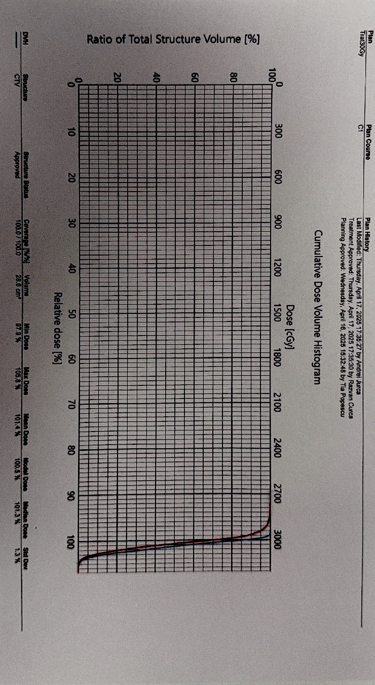
Figure 3 – Dose-volume histogram (DVH) of the treatment plan
Follow-Up
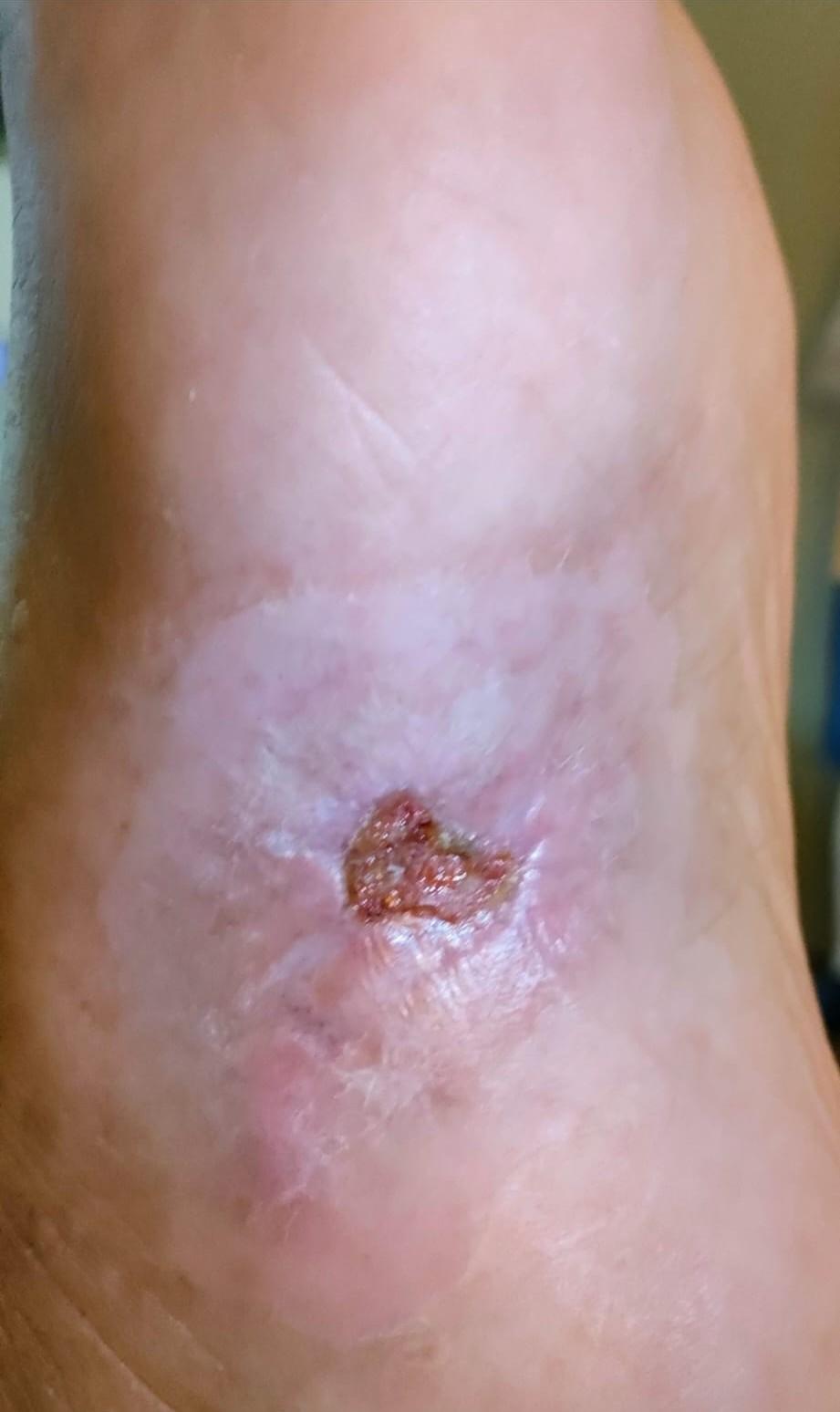
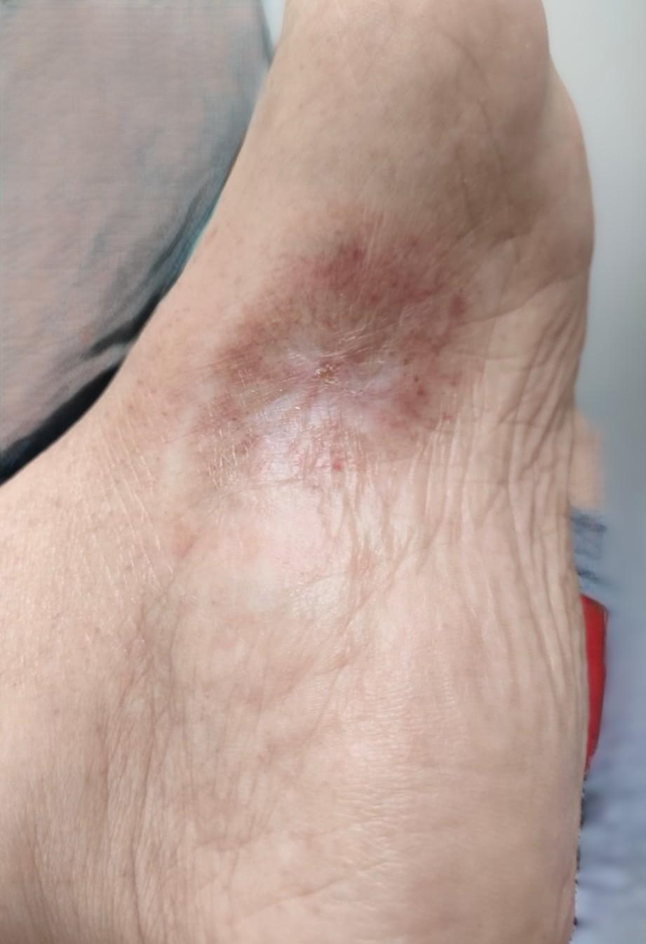
At treatment completion in May 2025, the patient reported marked symptomatic relief, with no significant acute toxicity. During June and July 2025 follow-ups, the irradiated lesion demonstrated progressive regression with residual erythematous scarring. By August 2025, complete epithelialization was achieved, with no residual ulceration or pain.
Follow-up PET-CT performed in August 2025 confirmed complete metabolic remission of the irradiated plantar lesion but identified new metabolically active iliac and inguinal-femoral lymph nodes (SUVmax ≈ 3.3), suggesting systemic progression.
The patient remains under hematology follow-up for further systemic management.
Figure 4A–B – Sequential evolution of the irradiated plantar lesion from May to August follow-up
3. Discussion
This case demonstrates the efficacy of radiotherapy in achieving local control in Mycosis Fungoides (MF), consistent with previous reports showing high response rates due to the radiosensitivity of malignant T-cells. Both conventional total skin electron beam therapy (TSEBT) regimens (30–36 Gy) and lower-dose regimens (10–12 Gy) have been shown to produce excellent local control, with lower toxicity allowing for repeat irradiation when needed.
In this patient, local IMRT with 30 Gy achieved complete remission of the irradiated lesion. However, the subsequent systemic progression underscores the importance of multidisciplinary management and the need for systemic therapies in advanced-stage disease.
Comparison with Published Literature
Our treatment results are consistent with multiple studies evaluating localized radiotherapy and TSEBT in Mycosis Fungoides.
Hoppe et al. (2015) reported complete response (CR) rates exceeding 90% with doses between 12 and 36 Gy(1), while Morris et al. (2017) demonstrated similarly high efficacy using lower-dose regimens (10–12 Gy)(2), with excellent tolerance and feasibility for retreatment.
Neelis et al. (2009) observed durable local control in over 85% of cases after 30–36 Gy(4), and Specht et al. (2015), in the ILROG recommendations, endorsed 24–36 Gy for localized lesions(3).
More recently, Higgins et al. (2025) reported dose–response data in Mycosis Fungoides with large cell transformation, further supporting the use of moderate-dose regimens for achieving durable local control(11).
These findings support the concept that moderate-dose radiotherapy (~30 Gy) achieves durable local control with minimal toxicity.
Our case confirms that an IMRT-based approach using a 0.5 cm PTV margin provides precise dose conformity and reproducible outcomes comparable to those reported in international literature.
Table 1. Summary of selected studies on radiotherapy in Mycosis Fungoides
| Author (Year) | Technique / Dose | Patients (n) | Response Rate | Main Findings |
|---|---|---|---|---|
| Hoppe et al. (2015) | Low-dose TSEBT (12–36 Gy) | 102 | 93% CR | Excellent local control; mild toxicity |
| Morris et al. (2017) | Low-dose TSEBT (10–12 Gy) | 105 | 88% CR | Comparable outcomes; suitable for retreatment |
| Neelis et al. (2009) | Local RT (30–36 Gy) | 43 | 86% CR | Durable local control; minimal late toxicity |
| Specht et al. (2015) | ILROG guidelines | – | – | 24–36 Gy recommended for localized MF |
| Higgins et al. (2025) | Local RT / TSEBT 8–40 Gy EQD2 | 49 (155 lesions) | ORR 94% (CR 60%, PR 35%) | Clear dose–response; EQD2 >12 Gy → 100% ORR, >36 Gy → 100% CR; excellent local control in LCT MF |
| Present case (2025) | IMRT 30 Gy / 10 fr | 1 | 100% CR (local) | Complete local remission; mild acute effects |
CR = Complete Response; TSEBT = Total Skin Electron Beam Therapy; MF = Mycosis Fungoides ORR=Overall response rate PR=Partial Response
These observations reinforce the established role of radiotherapy as a cornerstone in the management of Mycosis Fungoides. The choice of technique (IMRT vs. electron beams) and dose fractionation may be adapted according to lesion depth, site, and patient comorbidities, with equivalent efficacy when conformal coverage is achieved.
Guidelines from National Comprehensive Cancer Network (NCCN) and EORTC recommend radiotherapy for localized lesions, palliation, or TSEBT in disseminated disease.(5,7,10) Our findings align with these standards, confirming radiotherapy as both safe and effective, particularly when integrated into a multidisciplinary treatment plan.
4. Conclusion
Radiotherapy remains a cornerstone in the management of Mycosis Fungoides, offering excellent local control and symptomatic relief due to the intrinsic radiosensitivity of malignant T-cells.
In this case, localized IMRT delivered to a total dose of 30 Gy in 10 fractions achieved complete clinical and metabolic remission of the plantar lesion, with excellent tolerance and no significant acute toxicity. The use of image-guided setup, surface bolus, and thermoplastic immobilization ensured optimal dose conformity and reproducibility throughout treatment.
These results are consistent with published data supporting moderate-dose RT (24–36 Gy) as an effective and safe strategy for local disease control.(1–4,11) However, the detection of new nodal lesions during follow-up underscores the need for systemic assessment and ongoing multidisciplinary management.(5,7,10)
Overall, this experience highlights the crucial role of modern radiotherapy techniques such as IMRT in achieving durable local responses while maintaining favorable toxicity profiles, contributing to improved quality of life for patients with Mycosis Fungoides.
Statements
*Authors’ contributions: Ana Băncilă and Ioan-Alexandru Diaconu-Marinescu conceived and planned the case management and radiotherapy treatment. Ioan-Alexandru Diaconu-Marinescu collected the clinical and imaging data and drafted the initial version of the manuscript. Ana Băncilă contributed to the interpretation of the radiotherapy planning details and critically revised the manuscript for important intellectual content. Both authors approved the final version of the manuscript and agree to be accountable for all aspects of the work.
Consent for publication: As the corresponding author, I confirm that the manuscript has been read and approved for submission by all named authors.
Conflict of interests: The authors declare no conflict of interest.
Funding Sources: None.
Statement of Ethics: This case report does not contain any experimental studies on humans or animals performed by the authors. The case was managed according to routine clinical practice, in line with institutional and international ethical standards.
Written informed consent for publication: Written informed consent was obtained from the patient for publication of this case report and any accompanying images.
Acknowledgments: The authors would like to thank the radiotherapy technologists and medical physicists involved in the treatment planning and delivery, as well as the hematology team for their contribution to the multidisciplinary management of this patient.
Abbreviations
CR – Complete Response
CT – Computed Tomograph
CTV – Clinical Target Volume
DVH – Dose–volume Histogram
EORTC – European Organisation for Research and Treatment of Cancer
GTV – Gross Tumor Volume
ILROG – International Lymphoma Radiation Oncology Group
IMRT – Intensity-Modulated Radiotherapy
MF – Mycosis Fungoides
NCCN – National Comprehensive Cancer Network
OAR – Organs At Risk
ORR – Overall Response Rate
PET-CT – Positron Emission Tomography–Computed Tomography
PR – Partial Response
PTV – Planning Target Volume
RT – Radiotherapy
SUVmax – Maximum Standardized Uptake Value
TSEBT – Total Skin Electron Beam Therapy
References
- Hoppe RT et al. Low-dose total skin electron beam therapy in mycosis fungoides: results from a prospective trial. Int J Radiat Oncol Biol Phys. 2015;92(1):183-190.
- Morris S et al. Results of Low-Dose Total Skin Electron Beam Radiation in Patients With Mycosis Fungoides. Int J Radiat Oncol Biol Phys. 2017;99(3):627-633.
- Specht L et al. Modern radiation therapy for primary cutaneous lymphomas: ILROG guidelines. Int J Radiat Oncol Biol Phys. 2015;92(1):32-39.
- Neelis KJ et al. Low-dose total skin electron beam therapy for patients with MF. Int J Radiat Oncol Biol Phys. 2009;74(5):1547-1551.
- Rolf B et al. EORTC consensus recommendations for the treatment of MF/Sézary syndrome – Update 2023. Eur J Cancer. 2023;185:1-12.
- Willemze R et al. The 2018 update of the WHO-EORTC classification for cutaneous lymphomas. Blood. 2019;133(16):1703-1714.
- Trautinger F et al. EORTC consensus recommendations for the treatment of MF/Sézary syndrome. Eur J Cancer. 2006;42(8):1014-1030.
- Scarisbrick JJ et al. The PROCLIPI international registry of early-stage MF identifies diagnostic delay. Br J Dermatol. 2019;181(2):350-357.
- Kohn M et al. Total skin electron beam therapy for patients with MF: review of responses and toxicity. Cancer. 2014;120(20):3400-3411.
- NCCN Clinical Practice Guidelines in Oncology: Primary Cutaneous Lymphomas, v2.2024.
- Higgins M, Johnstone P, Prince HM, Lade S, Fahey MT, McCormack C, van der Weyden C, Bhabha F, Buelens O, Campbell BA. Radiotherapy dose–response of mycosis fungoides with large cell transformation. Radiother Oncol. 2025 Nov 19:111292.