

Author(s) :
Najam Uddin1, Muhammad Iqbal1, Umar Hussain1
1Atomic Energy Medical Centre (AEMC), Jinnah Post Graduate Medical Centre (JPMC), Karachi, Pakistan
Corresponding author: Najam Uddin, Email: najam_uddin78@hotmail.com
Publication History: Received - , Revised - , Accepted - 30 September 2026, Published Online - 06 October 2026.
Copyright: © 2026 The author(s). Published by Casa Cărții de Știință.
User License: Creative Commons Attribution – NonCommercial (CC BY-NC)
 Share
Share
Highlights
- Complete response after definitive chemoradiation is an important early marker of outcome in inoperable oral cavity squamous cell carcinoma.
- Larger nodal volume independently predicts a lower likelihood of complete response, supporting volumetric assessment of nodal disease.
- Bone involvement and nodal fixation are also associated with reduced response, while histological grade shows no predictive value.
- Overall disease burden drives treatment response, and larger prospective studies are needed to refine predictive models.
Abstract
Background: Complete response (CR) following definitive chemoradiation with curative intent is an important predictor of survival and local control in oral cavity squamous cell carcinoma (OCSCC). Although primary tumor volume and nodal volume are recognized predictors of treatment response, the prognostic significance of other clinicopathological factors remains less well defined. This study evaluated the independent predictors of complete response following definitive chemoradiation.
Material and Methods: We treated 114 patients with OCSCC with definitive concurrent chemoradiation consisting of 70 Gy radiotherapy with cisplatin and followed them for one year. Pre- and post-treatment assessments included primary tumor volume, largest nodal volume, histological grade, nodal fixation, and bone involvement. We evaluated treatment response according to the Response Evaluation Criteria in Solid Tumors (RECIST version 1.1). We used multivariable binary logistic regression in SPSS to identify independent predictors of complete response.
Results: The mean age of the study cohort was 41 years, with a predominance of male patients. The mean primary tumor volume and largest nodal volume were 3.63 cm³ and 2.37 cm³, respectively. Complete response (CR) was achieved in 64 patients (56.1%), whereas 50 patients (43.9%) had a non-complete response. In multivariable analysis, increasing largest nodal volume was independently associated with reduced odds of complete response (B = –0.441, p = 0.006, OR = 0.643, 95% CI: 0.470–0.880). Tumor volume showed a non-significant trend toward reduced odds of complete response (B = –0.282, p = 0.087, OR = 0.754, 95% CI: 0.546–1.042). Bone involvement and nodal fixation were also independently associated with reduced odds of complete response, while histological grade was not a significant predictor. The multivariable model performed well (Omnibus test χ² = 34.909, p < 0.001; Hosmer–Lemeshow test χ² = 5.271, p = 0.728), with an overall classification accuracy of 77.2% (sensitivity 79.7% and specificity 74.0%).
Conclusion: Larger primary tumor volume showed a non-significant trend toward reduced odds of complete response. In contrast, larger nodal volume was independently associated with a lower likelihood of achieving complete response following definitive chemoradiation. Bone involvement and nodal fixation were also associated with reduced odds of complete response. These findings highlight the importance of disease burden in predicting treatment response and support further prospective studies to refine prognostic models.
1. Introduction
Oral cavity squamous cell carcinoma (OCSCC) is a major global public health challenge. More than 350,000 new cases and approximately 170,000 deaths are reported annually (1). Despite advances in diagnosis and treatment, the overall 5-year survival rate remains approximately 50% (2). Tobacco use in various forms and betel nut chewing are among the most important etiological factors and contribute substantially to the high incidence and mortality of OCSCC, particularly in South and Southeast Asia (3,4). In Pakistan and other South Asian countries, oral cancer is the second most common malignancy and is one of the leading causes of cancer-related mortality among men, accounting for approximately 13% of all cancer deaths (5).
OCSCC often exhibits aggressive biological behavior and frequently presents as locally advanced disease because of delayed diagnosis, widespread use of tobacco products, and socioeconomic barriers to healthcare. Consequently, many patients are not suitable candidates for upfront surgical resection, the current standard treatment for resectable disease, and instead require definitive chemoradiation. This group includes patients with surgically unresectable tumors, those who are medically inoperable because of significant comorbidities and patients who decline surgical treatment. Although surgery remains the preferred treatment for early-stage disease, some patients with stage I or II tumors may also refuse surgery and undergo definitive chemoradiation.
Definitive concurrent chemoradiation has become the standard nonsurgical treatment for patients with unresectable or medically inoperable locally advanced OCSCC. The conventional regimen consists of external beam radiotherapy to a total dose of 70 Gy combined with either weekly or three-weekly cisplatin chemotherapy. However, treatment outcomes remain variable, with reported complete response (CR) rates ranging from 40% to 70% (6,7). Several clinicopathological and biological factors have been investigated as potential predictors of treatment response. These include primary tumor volume, nodal volume (8), histological grade, perineural invasion, lymphovascular invasion, and molecular biomarkers.
Previous studies have consistently demonstrated that patients with smaller primary tumors and lower nodal volumes are more likely to achieve a complete response following definitive chemoradiation (9,10). Therefore, the independent prognostic value of factors such as histological grade, bone involvement, and nodal fixation remains less well established. Furthermore, data regarding predictors of treatment response in patients with inoperable OCSCC are limited, particularly from South Asian populations, highlighting the need for additional prospective studies (11,12).
The present study aimed to evaluate the association of primary tumor volume, largest nodal volume, histological grade, nodal fixation, and bone involvement with complete response following definitive chemoradiation in patients with inoperable or unresectable OCSCC. Although tumor burden is recognized as an important prognostic factor, the independent contribution of tumor and nodal volume to complete response after definitive chemoradiation remains incompletely defined. Our study provides prospective, region-specific data and evaluates these variables simultaneously using a multivariable logistic regression model to better define their independent predictive value.
2. Materials and Methods
a. Study Design
This is a prospective observational cohort study conducted at the Atomic Energy Medical Centre, Karachi (AEMCK), Pakistan, from January 2024 to January 2025. The study was approved by the institute atomic energy medical center, JPMC, Karachi, Pakistan, in November 2023.
Inclusion criteria
Patients were included in the study if they met the following criteria: biopsy-proven, medically inoperable or unresectable oral squamous cell carcinoma, or patients who refused surgery. All patients were assessed based on biopsy findings, imaging, prior endoscopy reports, laboratory results, and other relevant medical records.
Exclusion criteria
Patients with distant metastasis, recurrent disease, prior head and neck radiotherapy, those who had received induction chemotherapy, those with second primary tumors, or those in a post-operative setting were excluded. Patients with extensive locally advanced disease deemed unsuitable for curative treatment were also excluded. Additionally, patients with incomplete clinical records or those who did not complete the planned treatment and follow-up required for response evaluation were excluded from the final analyzed cohort.
Procedure
A contrast-enhanced simulation computed tomography (CT) scan of the face, neck, and chest with a slice thickness of 0.3 cm was obtained using a Canon 84-slice CT scanner. The bone window was used to evaluate medullary bone involvement. Lymph node fixation was assessed clinically by two radiation oncologists using standard palpation. The primary tumor and largest lymph node volumes for each patient were determined using software-based auto-generated measurements (in cm³) on the contouring station by an experienced radiation oncologist. Treatment response in the tumor and largest nodal volume was assessed according to RECIST version 1.1 criteria. A contrast-enhanced CT scan was repeated 6–8 weeks after treatment completion for response evaluation. A complete history, physical examination, and assessment of performance status and fitness for study participation, along with relevant investigations and workup, were completed for all patients. All cases were discussed at a multidisciplinary tumor board. Tumor size and largest nodal size were defined according to the AJCC 8th edition TNM staging system. Tumor stage grouping was not used as a primary stratification variable in this study. Instead, quantitative assessment of tumor volume and largest nodal volume (cm³) was used to characterize disease burden. Tumor and largest nodal responses were assessed separately for each patient.
Chemoradiation
Radiotherapy was delivered using external beam radiotherapy (3D-CRT) on a linear accelerator with treatment planning performed using the Monaco (Elekta) system. The gross tumor volume (GTV) was defined as gross disease identified on contrast-enhanced CT imaging and clinical examination. The GTV plus a 1 cm margin received 70 Gy in 35 fractions. The high-risk clinical target volume (CTV) comprised the region of any involved node(s) and received a total dose of 66 Gy. Elective nodal regions (levels I–III in node-negative disease, and uninvolved nodal levels in node-positive disease) received 50 Gy. Neck levels IV–V were also treated to 50 Gy in patients with T3–T4 disease. A planning target volume (PTV) margin of 3–5 mm was applied to all CTVs to account for setup uncertainties and patient motion. Radiotherapy was delivered in once-daily fractions of 2 Gy five days per week, ensuring at least 95% coverage of the prescribed dose to the PTV while respecting dose constraints to organs at risk, including the spinal cord, brainstem, parotid glands, and mandible. Concurrent chemotherapy consisted of intravenous cisplatin administered either weekly at 40 mg/m² or every three weeks at 100 mg/m² with appropriate hydration, premedication and monitoring. All patients received standard supportive care including nutritional support, pain management, oral hygiene counseling and management of treatment-related toxicities. Written informed consent was obtained from all patients prior to treatment.
Logistic regression
a. Descriptive statistics
Treatment response (0 = non-CR, 1 = CR), nodal fixation (0 = mobile, 1 = fixed), and bone involvement (0 = absent, 1 = present) were recorded as binary categorical variables, whereas histological grade was classified as Grade 1, Grade 2, or Grade 3. Categorical variables were summarized as frequencies and percentages. For descriptive and bivariate analyses, tumor volume was categorized as 1–2, 3–4 and 5–6 cm³ and largest nodal volume was categorized as 0–1 and 2–6 cm³ for which associations with complete response were evaluated using Pearson’s chi-square test. In the multivariable logistic regression analysis (Table 3), tumor volume and largest nodal volume were entered in their original continuous form (cm³).
Inferential Statistics
Inferential statistics were performed to evaluate factors associated with complete response (CR). The independent variables included tumor volume, largest nodal volume, bone involvement, nodal fixation, and histological grade. Tumor volume and largest nodal volume were analyzed as continuous variables (cm³), while bone involvement and nodal fixation were analyzed as binary variables and histological grade was entered as a categorical variable, with Grade 1 as the reference category. All clinically relevant variables were entered into a multivariable binary logistic regression model using the enter method. Bone involvement, nodal fixation, and histological grade were entered into the logistic regression as categorical variables. Odds ratios (ORs), 95% confidence intervals (CIs), regression coefficients, standard errors, Wald statistics, and p-values were reported. Model performance was assessed using the Omnibus test of model coefficients, Cox & Snell R², Nagelkerke R² and the Hosmer-Lemeshow goodness-of-fit test. Classification accuracy, sensitivity and specificity were also calculated. A two-sided p-value <0.05 was considered statistically significant.
3. Results
A total of 114 patients were included in the study. Of these, 98 patients (86%) had surgically unresectable disease, 3 (2.6%) were medically inoperable, and 13 (11.4%) declined surgery. The mean age of the cohort was 41 ± 9.08 years. The cohort comprised 83 (72.8%) males and 31 (27.2%) females yielding a male-to-female ratio of 2.6:1. The majority of patients had moderately differentiated (grade 2) tumors (50, 43.9%). Bone involvement was present in 68 patients (59.6%) and nodal fixation was observed in 60 patients (52.6%). The mean tumor volume was 3.63 cm³ and the mean nodal volume was 2.37 cm³. Most patients had a tumor volume of 3–4 cm³ (47, 41%) and the most common nodal volume category was 2–6 cm³ (83, 73%). A complete response was observed in 64 patients (56.1%), while no complete response was observed in 50 patients (43.9%), as shown in Table 1.
Table 1: Baseline Demographics. Clinical and pathological variables distribution in the Study Population (n = 114)
| Variable | Category | Value | % |
|---|---|---|---|
| Demographic Characteristics | |||
| Age (years) | Mean ± SD | 41 ± 9.08 | – |
| Gender | Male | 83 | 72.8% |
| Female | 31 | 27.2% | |
| Clinical and Pathological Characteristics | |||
| Tumor volume (cm³) | 1-2 cm³ | 32 | 28.0 |
| 3-4 cm³ | 47 | 41.0 | |
| 5-6 cm³ | 35 | 31.0 | |
| Largest Nodal Volume (cm³) | 0–1 | 31 | 27.0 |
| 2–6 | 83 | 73.0 | |
| Bone Involvement | Absent | 46 | 40.4 |
| Present | 68 | 59.6 | |
| Nodal Fixation | Mobile | 54 | 47.4 |
| Fixed | 60 | 52.6 | |
| Histological Grade | Grade 1 | 25 | 21.9 |
| Grade 2 | 50 | 43.9 | |
| Grade 3 | 39 | 34.2 | |
| Treatment Outcome | |||
| Treatment Response | CR | 64 | 56.1 |
| Non-CR | 50 | 43.9 |
CR – Complete Response
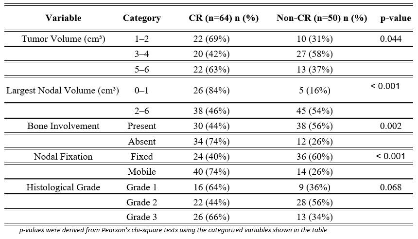
Patients were categorized into complete response (CR) and non-complete response (non-CR) groups. Baseline clinical variables were compared between the two groups using the Chi-square test. On bivariate analysis using the Pearson chi-square test, tumor volume category (χ² = 6.232, p = 0.044), largest nodal volume category (χ² = 13.297, p < 0.001), bone involvement (χ² = 9.893, p = 0.002) and nodal fixation (χ² = 13.401, p < 0.001) were significantly associated with complete response however histological grade was not significantly associated with complete response (χ² = 5.375, p = 0.068) as shown in Table 2.
Table 2: Comparison of Clinical Variables Between Complete Response and Non-Complete Response Groups
| Variable | Category | CR value (n=64) | CR % | Non-CR value (n=50) | Non-CR % | p-value |
|---|---|---|---|---|---|---|
| Tumor Volume (cm³) | 1–2 | 22 | 69 | 10 | 31 | 0.044 |
| 3–4 | 20 | 42 | 27 | 58 | ||
| 5–6 | 22 | 63 | 13 | 37 | ||
| Largest Nodal Volume (cm³) | 0–1 | 26 | 84 | 5 | 16 | < 0.001 |
| 2–6 | 38 | 46 | 45 | 54 | ||
| Bone Involvement | Present | 30 | 44 | 38 | 56 | 0.002 |
| Absent | 34 | 74 | 12 | 26 | ||
| Nodal Fixation | Fixed | 24 | 40 | 36 | 60 | < 0.001 |
| Mobile | 40 | 74 | 14 | 26 | ||
| Histological Grade | Grade 1 | 16 | 64 | 9 | 36 | 0.068 |
| Grade 2 | 22 | 44 | 28 | 56 | ||
| Grade 3 | 26 | 66 | 13 | 34 |
p-values were derived from Pearson’s chi-square tests using the categorized variables shown in the table
The mean tumor volume and mean largest nodal volume were 3.63 cm³ and 2.37 cm³, respectively. Complete response rates differed significantly across tumor-volume categories (p = 0.044), although the pattern was not monotonic. Patients with a largest nodal volume of 0–1 cm³ had a higher CR rate than those with a largest nodal volume of 2–6 cm³ (84% vs 46%, p < 0.001). The largest nodal volume in the cohort (n = 114) had a mean of 2.37 cm³ (SD 1.61) and a median of 2.0 cm³.
Multivariate Logistic Regression Analysis
Larger nodal volume was significantly associated with reduced odds of complete response (B = −0.441, Wald χ² = 7.59, p = 0.006, OR = 0.643, 95% CI: 0.47–0.88). When analyzed as a continuous variable in the multivariable model, tumor volume showed a trend toward reduced odds of complete response but did not reach statistical significance (B = −0.282, Wald χ² = 2.93, p = 0.087, OR = 0.754, 95% CI: 0.54–1.04) despite the significant association observed for categorized tumor volume in the bivariate analysis (p = 0.044) as shown in Tables 2 and 3.
Table 3: Logistic regression analysis of factors associated with complete response.
| Predictor | B (Coefficient) | SE | Wald χ² | p-value | Odds Ratio (Exp(B)) | 95% CI for OR |
|---|---|---|---|---|---|---|
| Nodal volume (cm3) | -0.441 | 0.160 | 7.59 | 0.006 | 0.643 | 0.47 – 0.88 |
| Bone involvement | -1.231 | 0.487 | 6.38 | 0.012 | 0.292 | 0.11 – 0.75 |
| Nodal fixation | -1.027 | 0.476 | 4.64 | 0.031 | 0.358 | 0.14 – 0.91 |
| Tumor volume (cm3) | -0.282 | 0.165 | 2.93 | 0.087 | 0.754 | 0.54 – 1.04 |
| Histological grade | — | — | 3.124 | 0.210 | – | — |
| Constant | 4.206 | 1.130 | 13.858 | <0.001 | 67.105 | — |
Complete response was coded as 1 and non-complete response as 0. Reference categories were absence of bone involvement, mobile nodes, and histological Grade 1. All predictors had df = 1 except histological grade (df = 2).
Model Performance
The Omnibus test of model coefficients (χ² = 34.90, p < 0.001) indicated that the model was statistically significant. Cox & Snell R² = 0.264 and Nagelkerke R² = 0.353, suggesting that the included variables explained approximately 26–35% of the variability in CR. The Hosmer–Lemeshow test showed χ² = 5.27 and p = 0.728, confirming adequate model calibration. The model correctly predicted treatment response in 77.2% of cases overall (sensitivity 79.7%, specificity 74.0%).
4. Discussion
Several studies have evaluated response to definitive chemoradiation in oral cavity squamous cell carcinoma (OCSCC). Our study adds to this body of evidence by investigating clinical predictors of complete response (CR).
Early tumor response following definitive treatment is a validated surrogate for long-term outcomes and pooled analyses and meta-analyses have demonstrated that CR is a meaningful early marker of therapeutic benefit (13). The treatment outcome of chemoradiation has been inconclusive, as several studies have reported a wide range of 5-year overall survival (OS) rates, ranging from 29% to 76% (14). A recent tertiary center cohort study from the region reported that CR after definitive chemoradiation was associated with improved progression-free outcomes in inoperable OCSCC (7).
Patients achieving CR can be managed with surveillance, whereas those with incomplete response may require early salvage surgery or intensified local therapy (15). Higher CR rates are also associated with improved overall survival attributable to effective eradication of resistant tumor clones (16). The complete response rate in our study (56.1%) is comparable to previously reported findings, including the study by Mishra et al., who reported a CR rate of approximately 50% (7). Several biological factors contribute to variability in treatment response, including tumor burden, hypoxia, and adverse molecular characteristics that may drive radioresistance (17).
Tumor extent can be described using different parameters including tumor size, tumor volume, and depth of invasion (DOI). These parameters are interrelated but not equivalent. Tumor size represents a linear measurement used in TNM staging and is a relatively coarse metric subject to interobserver variability. In contrast, volumetric tumor measurement captures true three-dimensional tumor burden and has been shown to correlate more closely with radiotherapy outcomes. DOI has also emerged as an important prognostic factor in recent studies, particularly in predicting local recurrence and survival (18,19). However, DOI is typically derived from histopathological assessment and was not available in our cohort. Recent studies from 2024 and 2025 have highlighted the prognostic significance of DOI, suggesting that its incorporation into future studies may improve predictive modeling (20,21).
In the bivariate analysis, categorized tumor volume was significantly associated with complete response (p = 0.044), although the pattern across categories was not monotonic. When tumor volume was analyzed as a continuous variable in the multivariable logistic regression model, it showed a trend toward reduced odds of complete response but did not reach statistical significance (p = 0.087). The difference in statistical significance reflects both the categorization used in the bivariate analysis and adjustment for other clinicopathological variables in the multivariable model. Prior studies have consistently demonstrated the prognostic value of tumor volume and larger sample sizes may further strengthen this association (22). Similarly, the predictive role of tumor volume has been shown to be robust across multiple treatment modalities, not limited to radiotherapy alone (23). Nodal volume emerged as a strong independent predictor of treatment response in our study. We observed that each incremental increase in nodal volume was associated with a 36% reduction in the odds of achieving CR (p = 0.006). This finding is consistent with existing literature and demonstrates that greater nodal tumor burden is associated with poorer treatment response, reduced locoregional control, and worse survival outcomes following chemoradiation (24,25). Volumetric assessment of nodal disease may therefore provide superior prognostic value compared to conventional diameter-based staging.
In the multivariable model, nodal fixation was associated with reduced odds of complete response consistent with the bivariate analysis in Table 2 and clinical expectation (fixed versus mobile nodes: B = –1.027, p = 0.031, OR = 0.358, 95% CI: 0.141–0.911). Similarly, bone involvement present versus absent was associated with reduced odds of complete response (B = –1.231, p = 0.012, OR = 0.292, 95% CI: 0.112–0.759). Nodal fixation and largest nodal volume are biologically related as fixed nodes tend to have a larger volumetric burden. Collinearity was assessed using linear regression collinearity diagnostics, and variance inflation factors were <2.1 for all predictors, indicating no severe multicollinearity, although some shared variance remains. Therefore, the independent effect of fixation should be interpreted cautiously.
Histological grade did not emerge as a significant predictor of complete response in our cohort. This observation is consistent with several recent studies (2021–2025) reporting limited independent prognostic value of tumor differentiation in predicting treatment response. However, the modest sample size of our study may have limited the ability to detect subtle differences between grade categories.
Bone involvement was associated with reduced odds of complete response in multivariable analysis. This suggests that tumor invasion into adjacent bone structures may influence treatment response, potentially reflecting increased tumor burden and an altered tumor microenvironment. Bone invasion has been associated with aggressive tumor biology, including hypoxia and reduced radiosensitivity, which may impact treatment outcomes.
However, the prognostic significance of bone involvement in OCSCC remains controversial. A multicenter study in 2024 reported no independent prognostic effect after multivariable adjustment (26), and a recent review highlighted variability in the definition and assessment of bone invasion, distinguishing cortical from medullary involvement (27). In our cohort, the observed association may also be influenced by interactions with nodal disease and treatment-related factors. Therefore, this finding should be interpreted with caution, and further prospective studies with standardized imaging and pathological assessment are warranted.
Despite its strengths, this study has several limitations. Its prospective observational single-center design limits generalizability, as patient characteristics, treatment protocols, and follow-up practices at our institution may not fully represent broader populations. The modest sample size and subgroup distribution may have reduced statistical power. Additionally, tumor and nodal volumes were derived from CT imaging and may be subject to measurement variability due to differences in slice thickness and contouring practices. Bone involvement and nodal fixation were assessed clinically and radiologically rather than histopathologically, introducing potential misclassification bias.
Furthermore, response assessment using RECIST version 1.1 criteria, although widely accepted, may not fully capture treatment response in head and neck cancers due to post-radiation changes such as fibrosis and inflammation. Treatment heterogeneity, particularly variation in cisplatin scheduling, may also have influenced outcomes.
Additionally, some correlation between largest nodal volume and nodal fixation exists, which may inflate variance estimates, although collinearity diagnostics showed variance inflation factors <2.1, indicating no severe multicollinearity. Finally, this study focused on short-term response, and long-term outcomes such as overall survival and progression-free survival were not assessed. These shortcomings collectively underscore the need for larger prospective multi-institutional studies incorporating standardized imaging, pathological assessment, and molecular profiling to validate and expand upon our findings.
5. Conclusion
In this prospective observational cohort study of oral cavity squamous cell carcinoma treated with definitive chemoradiation, larger nodal volume was independently associated with reduced odds of complete response. In contrast, tumor volume demonstrated a non-significant trend (p = 0.087). Bone involvement and nodal fixation were also independently associated with reduced odds of complete response, while histological grade was not a significant predictor. These findings highlight the importance of overall disease burden in determining treatment response and support the use of volumetric assessment in clinical evaluation. Further, larger prospective studies are needed to validate and refine prognostic models in this setting.
Abbreviations
AEMCK — Atomic Energy Medical Centre
CTV — Clinical target volume
CR — Complete response
CT — Computed tomography
DOI — Depth of invasion
OCSCC — Oral cavity squamous cell carcinoma
RECIST — Response criteria for solid tumors
Statements
Authors’ Contributions:
Concept, acquisition, analysis, interpretation of data, write-up, and review: NU. Data collection, methods and write-up: MI; methods, write-up, analysis and review: UHH.
Consent for publication:
As the corresponding author, I confirm that the manuscript has been read and approved for submission by all named authors.
Funding:
None.
Conflicts of Interest:
The authors have no conflicts of interest to declare.
Statement of Ethics:
This study was approved by an Institutional Ethics Committee
References
- Sung H, Ferlay J, Siegel RL, et al. Global cancer statistics 2020: GLOBOCAN estimates of incidence and mortality worldwide for 36 cancers in 185 countries. CA Cancer J Clin. 2021; 71:209-249.
- González-Moles MÁ, Aguilar-Ruiz M, Ramos-García P. Challenges in the early diagnosis of oral cancer: evidence gaps and strategies for improvement: a scoping review of systematic reviews. Cancers (Basel). 2022; 14:4967.
- Kijowska J, Grzegorczyk J, Gliwa K, et al. Epidemiology, diagnostics, and therapy of oral cancer: update review. Cancers (Basel). 2024; 16:3156.
- Warnakulasuriya S, Filho AM. Oral cancer in the South and South-East Asia region, 2022: incidence and mortality. Oral Dis. 2025; 31:1398-1405.
- Khan MF, Hayhoe RP, Kabir R. Exploring the risk factors for oral cancer in Pakistan: a systematic literature review. Dent J (Basel). 2024;12(2):25. doi:10.3390/dj12020025.
- Lang K, Baur M, Held T, Shafie RE, Moratin J, Freudlsperger C, et al. Definitive radiotherapy for squamous cell carcinoma of the oral cavity: a single-institution experience. Radiol Oncol. 2021;55(4):467-473. doi:10.2478/raon-2021-0041.
- Mishra VK, Gandhi AK, Rastogi M, Verma R, Khurana R, Hadi R, et al. Retrospective analysis of clinical outcome of 100 inoperable oral cavity carcinoma treated with definitive concurrent chemoradiotherapy with or without induction chemotherapy. Ecancermedicalscience. 2023; 17:1630. doi:10.3332/ecancer.2023.1630.
- Romeo V, Cuocolo R, Ricciardi C, Ugga L, Cocozza S, Verde F, et al. Prediction of tumor grade and nodal status in oropharyngeal and oral cavity squamous-cell carcinoma using a radiomic approach. Anticancer Res. 2020;40(1):271-280. doi:10.21873/anticanres.13949.
- Spoerl S, Gerken M, Mamilos A, Fischer R, Wolf S, Nieberle F, et al. Lymph node ratio as a predictor for outcome in oral squamous cell carcinoma: a multicenter population-based cohort study. Clin Oral Investig. 2021;25(4):1705-1713. doi:10.1007/s00784-020-03471-6.
- Zhou Y, Hu L, Zhao Z. Correlation analysis of tumor size and survival rate in oral cancer based on the SEER database. Curr Probl Surg. 2025; 69:101783. doi: 10.1016/j.cpsurg.2025.101783.
- Trikha M, Patil VM, Noronha V, Menon NS, Singh AC, Dhanawat A, et al. Neoadjuvant chemotherapy followed by definitive chemoradiation in technically unresectable oral cavity squamous cancers. J Clin Oncol. 2023;41(16 Suppl): e18073. doi: 10.1200/JCO.2023.41.16_suppl.e18073.
- Janmunee N, Peerawong T, Rordlamool P, Bridthikitti J, Tangthongkum M, Kongkamol C, et al. Tumor volume as a prognostic factor on the median survival in locally advanced oral cancer treated with definitive chemoradiotherapy. Indian J Cancer. 2023;60(1):72-79. doi: 10.4103/ijc.ijc_86_20.
- Black CM, Keeping S, Mojebi A, Ramakrishnan K, Chirovsky D, Upadhyay N, et al. Correlation between early time-to-event outcomes and overall survival in patients with locally advanced head and neck squamous cell carcinoma receiving definitive chemoradiation therapy: systematic review and meta-analysis. Front Oncol. 2022; 12:868490. doi:10.3389/fonc.2022.868490.
- Gore SM, Crombie AK, Batstone MD, et al. Concurrent chemoradiotherapy compared with surgery and adjuvant radiotherapy for oral cavity squamous cell carcinoma. Head Neck. 2015;37(4):518-523. doi:10.1002/hed.23626.
- Rades D, Zwaan I, Idel C, Pries R, Bruchhage KL, Hakim SG, et al. A new prognostic instrument for predicting the probability of completion of cisplatin during chemoradiation for head and neck cancer. J Pers Med. 2023;13(7):1120. doi:10.3390/jpm13071120.
- Ning Y, Song Y, Li H, He Y, Liu S, Liu Y. High pathological tumor response associates with enhanced overall survival in HNSCC patients following neoadjuvant immunochemotherapy and surgery. World J Surg Oncol. 2025;23(1):205. doi:10.1186/s12957-025-03865-4.
- Lai J, Huang R, Huang J. Predicting head and neck cancer response to radiotherapy with a chemokine-based model. Sci Rep. 2025; 15:28450. doi:10.1038/s41598-025-13346-z.
- Terzidis E, Friborg J, Vogelius IR, Lelkaitis G, von Buchwald C, Olin AB, et al. Tumor volume definitions in head and neck squamous cell carcinoma: comparing PET/MRI and histopathology. Radiother Oncol. 2023; 180:109484. doi: 10.1016/j.radonc.2023.109484.
- Ahmed I, Krishnamurthy S, Vinchurkar K. Prognosticating gross tumor volume in head-and-neck cancer: redefining gross tumor volume beyond contouring. J Med Phys. 2023;48(1):68-73. doi: 10.4103/jmp.jmp_101_22.
- Hong WJ, Shieh LT, Yen CY, et al. The significance of the depth of invasion and tumor size in resected pathologic T4a gingivobuccal squamous cell carcinoma. Sci Rep. 2025; 15:14618. doi:10.1038/s41598-025-98222-6.
- Koh ES, Pandey A, Banuchi VE, Kuhel WI, Tassler A, Scognamiglio T, et al. Depth of invasion as an independent prognostic factor in early-stage oral cavity squamous cell carcinoma. Am J Otolaryngol. 2024;45(3):104269. doi: 10.1016/j.amjoto.2024.104269.
- Mao W, Zhang T, Li L, et al. Role of primary tumor volume and metastatic lymph node volume in response to curative effect of definitive radiotherapy for locally advanced head and neck cancer. Eur J Med Res. 2024; 29:98. doi:10.1186/s40001-024-01691-0.
- Hirai Y, Kurihara K, Sano D, Inamo M, Takahashi H, Ichikawa Y, et al. Pre-treatment tumor size and tumor growth rate as prognostic predictors for patients with recurrent/metastatic squamous cell carcinoma of the head and neck treated with nivolumab. In Vivo. 2023;37(6):2687-2695. doi:10.21873/invivo.13378.
- Bernasconi M, Bilic A, Kauke-Navarro M, Safi AF. Nodal tumor volume as a prognostic factor for oral squamous cell carcinoma: a systematic review. Front Oral Health. 2023; 4:1229931. doi:10.3389/froh.2023.1229931.
- Li P, Fang Q, Yuan J, Luo R. Lymph node metastasis burden identifies head and neck squamous cell carcinoma patients benefiting from adjuvant chemoradiation: a propensity score-matching study. Eur J Surg Oncol. 2024;50(7):108453. doi: 10.1016/j.ejso.2024.108453.
- Venkatesh B, Roy P, Bardia A, Mallick I, Chatterjee S, Arun P, et al. Challenges in the assessment of medullary bone invasion in oral cavity cancers and its prognostic significance. Head Neck Pathol. 2024;18(1):37. doi:10.1007/s12105-024-01642-5.
- Alfurhud AA. Prognosis, controversies and assessment of bone erosion or invasion of oral squamous cell carcinoma. Diagnostics (Basel). 2025;15(1):104. doi:10.3390/diagnostics15010104.