

Author(s) :
Marta Camacho Manzanares1, Eva Fernández Lizarbe1, Ana Lario Arribas2, Beatriz Mateos Muñoz3, José Antonio Domínguez Rullán1, Marina Alarza Cano1, Mercedes Martin Sánchez1, Patricia Martín Nieto1, Julen Azcona Martín1, Sonsoles Sancho García1
1Department of Radiation Oncology, Hospital Universitario Ramón y Cajal, Madrid, Spain.
2Department of Hematology, Hospital Universitario Ramón y Cajal, Madrid, Spain.
3Department of Gastroenterology, Hospital Universitario Ramón y Cajal, Madrid, Spain.
Corresponding author: ,
Publication History: Received - , Revised - , Accepted - 30 September 2026, Published Online - 06 October 2026.
Copyright: © 2026 The author(s). Published by Casa Cărții de Știință.
User License: Creative Commons Attribution – NonCommercial (CC BY-NC)
 Share
Share
Highlights
- Vanishing bile duct syndrome is a rare paraneoplastic manifestation of Hodgkin lymphoma that can progress to fatal liver failure.
- Liver dysfunction may preclude standard chemotherapy, leaving few treatment options for patients with this syndrome.
- Definitive involved-site radiotherapy achieves durable lymphoma remission and full recovery of liver function in this case.
- Radiotherapy may serve as a key treatment option when systemic therapy cannot be safely delivered, potentially avoiding liver transplantation.
Abstract
Background: Vanishing bile duct syndrome (VBDS) is a rare cholestatic disorder defined by a progressive loss of intrahepatic bile ducts. Although multifactorial, it may present as a paraneoplastic syndrome of Hodgkin´s lymphoma (HL), often associated with poor prognosis due to progressive hepatic failure.
Case presentation: We report the case of a 34-year-old woman presenting with jaundice, pruritus, and hyperbilirubinemia (>20 mg/dL). Liver biopsy demonstrated ductopenia without inflammation or tumor infiltration, confirming the diagnosis of VBDS. Imaging studies and a lymph node biopsy confirmed the stage II bulky classical HL with paraneoplastic VBDS. Despite corticosteroids, ursodeoxycholic acid, and dose-reduced chemotherapy, liver function deteriorated, precluding systemic therapy. Definitive involved-site radiotherapy was delivered at 40 Gy in 20 fractions using Volumetric Modulated Arc Therapy (VMAT). After the treatment, bilirubin levels normalized and complete metabolic remission was achieved.
Conclusion: This represents one of the few reported cases of HL-associated VBDS achieving a complete remission following radiotherapy as one of the main therapeutic modalities alongside chemotherapy, corticosteroids, and other treatment approaches. Local tumor control produced both durable lymphoma remission and full reversal of VBDS, suggesting that definitive radiotherapy may represent a therapeutic option when systemic therapy cannot be safely administered.
1. Introduction
Vanishing Bile Duct Syndrome (VBDS) is defined as a loss of interlobular intrahepatic bile ducts (1). Clinically, it may present as progressive cholestasis, accompanied by jaundice, pruritus, hepatic insufficiency, and if untreated, can ultimately progress to fulminant liver failure, resulting in death. Etiology remains unknown; however, it has been associated with autoimmune diseases, adverse drug reactions, viral infections, and paraneoplastic syndrome in certain malignancies such as Hodgkin lymphoma (2).
We describe a case of a patient with Hodgkin lymphoma whose initial clinical manifestation was VBDS. Treatment of Hodgkin lymphoma was considered an important component in the resolution of paraneoplastic VBDS; this multimodal approach included chemotherapy, corticosteroids, ursodeoxycholic acid, and radiotherapy.
2. Case Presentation
A 34-year-old woman with a history of hypertension treated with an antihypertensive presented to the Emergency Department. One week prior to presentation, she developed nausea, dizziness, scleral icterus, and acholic stools. Laboratory tests revealed hyperbilirubinemia of 13.28 mg/dL (direct bilirubin 9.42 mg/dL) along with elevated liver enzymes: aspartate aminotransferase (AST) 158 U/L, alanine aminotransferase (ALT) 398 U/L, gamma-glutamyl transpeptidase (GGT) 472 U/L. She was admitted for further evaluation, but no etiology was identified following a complete hepatic work-up (negative viral serology, autoantibodies, and metabolic liver disease screening). Abdominal ultrasound showed intrahepatic biliary ductal ectasia, which was not confirmed by Magnetic Resonance Cholangiopancreatography. Liver biopsy demonstrated marked bile duct loss without inflammatory activity or granulomas. Treatment with corticosteroids and ursodeoxycholic acid was initiated, showing mild initial improvement, but bilirubin levels worsened again after tapering corticosteroids. Given the progressive deterioration in liver failure, a pre-transplant assessment was initiated.
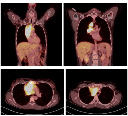
Contrast-enhanced CT scan showed a large prevascular mediastinal mass highly suggestive of a lymphoproliferative disorder, associated with multiple enlarged supradiaphragmatic lymph nodes (including prevascular, high right paratracheal, bilateral internal mammary, supraclavicular and ipsilateral retropectoral regions), causing compression of the superior vena cava. In this context, total bilirubin levels continued to rise (18.67 mg/dL), prompting referral to hematology. A fluorodeoxyglucose positron emission tomography–computed tomography (FDG PET-CT) scan confirmed these findings (see Figure 1), demonstrating increased metabolic activity in the mediastinal mass and involved lymph nodes, along with mild splenomegaly. CT-guided biopsy confirmed classical Hodgkin lymphoma (intermediate stage according to EORTC/LYSA criteria: stage II disease with bulky mediastinal involvement and B symptoms), associated with paraneoplastic bile duct loss.
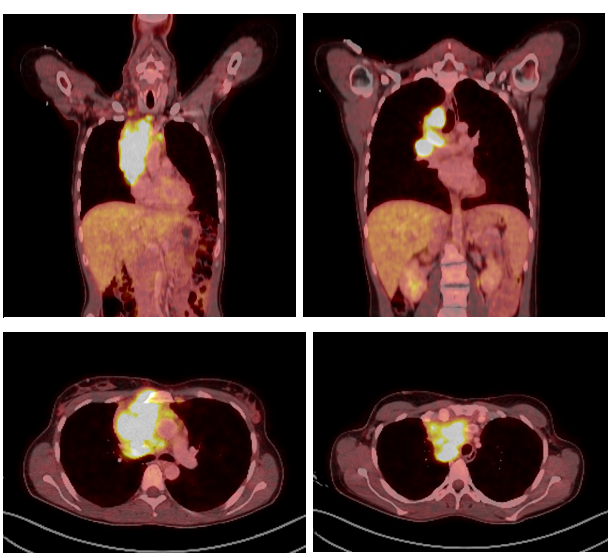
Figure 1: Diagnostic FDG PET-CT performed in August 2022: Large anterior mediastinal mass measuring approximately 7.6 × 7.5 × 10.5 cm (anteroposterior × transverse × craniocaudal), demonstrating markedly increased glucose metabolism (SUVmax 12,5), consistent with viable tumor tissue (bulky mass).
The patient initiated her first cycle of chemotherapy with ABVD (doxorubicin, bleomycin, vinblastine, and dacarbazine), administered at 50% of the standard dose due to impaired liver function. Treatment was delivered under inpatient monitoring, with continuation of corticosteroid therapy. Following a further decline in hepatic function, reflected by increasing bilirubin levels, the multidisciplinary tumor board recommended discontinuation of chemotherapy and a change in treatment strategy, including initiation of brentuximab vedotin (1.2 mg/kg), escalation of corticosteroid therapy, and consideration of radiotherapy (RT) (Figure 2).
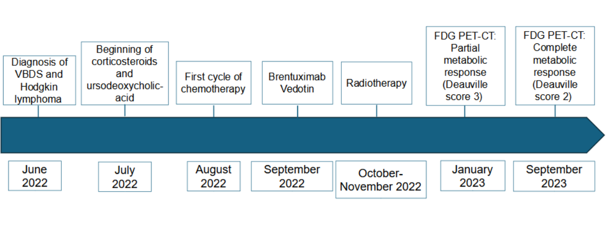
Figure 2: Treatment timeline.
To plan radiotherapy, a simulation CT scan was performed to acquire the necessary imaging for proper delineation of target structures and organs at risk. Immobilization was achieved using a thorax board, in the supine position with both arms raised, with 3 mm slice thickness. These CT images were then fused with prior PET-CT for accurate delineation of treatment volumes. The treatment volumes were defined in accordance with the International Lymphoma Radiation Oncology Group guidelines (ILROG) (3). Radiotherapy was administered between October 3rd and November 4th 2022, with a prescribed dose of 40Gy in 20 fractions of 2 Gy/day, to meet organ-at-risk constraints. Treatment was delivered using a linear accelerator (Infinity, Elekta), with 6 MV photon beams and two conformal volumetric arcs (see Figure 3). Daily low-dose CT scans (ConeBeam CT) were performed for patient positioning verification.
Prior to initiating radiotherapy, the patient experienced a worsening in liver function, with a peak total bilirubin of 21 mg/dL, leading to discontinuation of Brentuximab and continuation with RT alone. During treatment, she developed fatigue, mild dysphagia as acute toxicity, Additionally, the patient developed a mild respiratory infection and grade 1 radiation pneumonitis one week after RT, both of which resolved with oral antibiotics and corticosteroids.
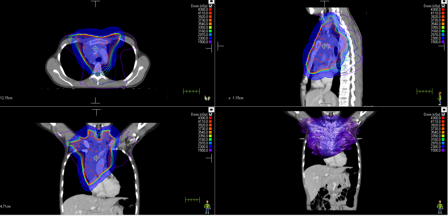
Figure 3: Radiotherapy treatment planning image using VMAT technique.
Two weeks after the end of treatment, the follow-up body CT scan showed a significant reduction in the size of the mediastinal mass and a slight decrease in total bilirubin (15.02 mg/dL). First re-evaluation FDG PET-CT (2 months post-radiotherapy) demonstrated marked metabolic improvement of the supradiaphragmatic lymphadenopathies and bulky mass, consistent with a metabolic response. Only minimal uptake was observed in a right internal mammary chain lymph node (Deauville score: 3), with no evidence of active macroscopic disease at other sites. The multidisciplinary tumor board decided to proceed with surveillance using FDG PET-CT every 3–4 months due to the mild uptake observed in the internal mammary chain, which was likely due to an inflammatory/reactive nature.
During surveillance, a complete metabolic response (Deauville score 2) was observed, with no evidence of radiotherapy-induced toxicity (see Figure 4). The patient remained clinically stable, and from February 2023 (3 months post-RT), total bilirubin levels began to decline significantly, reaching 0.66 mg/dL in April 2023, and remained stable through the most recent follow-up in May 2025 (total bilirubin: 1.11 mg/dL) (see Figure 3). Moreover, the patient maintains an active lifestyle, exercises daily, and is currently pregnant with her second child.
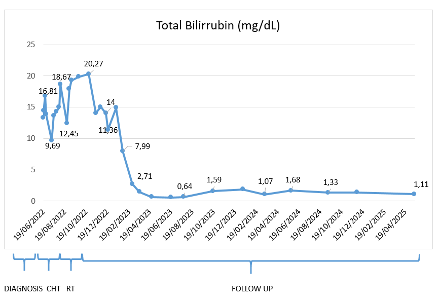
Figure 4: Graphic representation of the progression of total bilirubin levels from diagnosis in June 2022 through the most recent follow-up in April 2025 (European format).
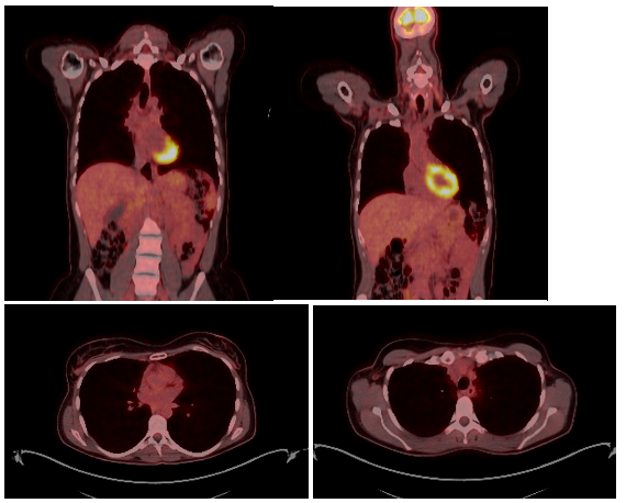
Figure 5: Image from the most recent FDG PET-CT performed on December 16, 2024, showing findings consistent with a sustained complete metabolic response.
3. Discussion
The presence of jaundice associated with Hodgkin´s lymphoma has been described in approximately 3-13% of cases (4). This hepatic alteration can be due to several causes, amongst which one could mention infiltration by tumor cells in the liver, viral infections, biliary obstruction secondary to nodal involvement, and in rarer cases, the paraneoplastic syndrome known as Vanishing Bile Duct Syndrome. Bouroncle et al. (5) in 1962 described cases of severe cholestatic jaundice in patients with Hodgkin lymphoma despite the absence of hepatic infiltration or mechanical biliary obstruction. Their observations suggested the existence of a lymphoma-related intrahepatic cholestatic process and provided some of the earliest evidence supporting a paraneoplastic mechanism of cholestasis in Hodgkin lymphoma.
A similar case to the one discussed in this paper is the one published by Knesl et al (6). A case of a 20-year-old female with early-stage Hodgkin lymphoma associated with VBDS, in which involved-field radiotherapy was used as a successful bridge to curative chemotherapy. Radiotherapy was initiated due to impaired liver function at diagnosis, as in this case. After hepatic function improvement, adjuvant chemotherapy was administered, achieving a complete metabolic response.
Scalabrini et al. (1), discussed a case of a female diagnosed with HL who also presented VBDS. The treatment for this patient consisted of an ursodeoxycholic acid and prednisone initially. Bilirubin levels continued to rise; therefore, chemotherapy was started. She received 6 cycles of ABVD. Six years later, the patient remains in complete response to HL with normal liver function. Wong et al. (4) describe the case of a 38-year-old patient presenting signs of severe cholestasis and diagnosed with VBDS associated with HL. After chemotherapy and autologous stem cell transplantation, intrahepatic bile duct regeneration was observed, suggesting that biliary damage in this context may be reversible.
The exact mechanism underlying the association between classical Hodgkin lymphoma (HL) and VBDS remains unclear; however, an immune-mediated process has been widely proposed. This syndrome is characterized by the progressive loss of interlobular bile ducts, likely resulting from sustained immune-mediated injury, particularly by T lymphocytes targeting the biliary epithelium, leading to cholangiocyte apoptosis (5).
One of the most frequently reported mechanisms is the release of toxic cytokines by lymphoma cells, which may directly damage the bile ducts (8). In addition, cytokine-driven upregulation of adhesion molecules and major histocompatibility complex expression has been described, potentially enhancing immune-mediated biliary injury (1). Liver biopsy typically demonstrates bile duct loss in the absence of significant tumor infiltration, supporting the concept that this condition is not due to direct hepatic involvement by lymphoma, biliary obstruction, or metastatic disease.
In the presented case, the patient started treatment with prednisone and ursodeoxycholic acid (Ursobilane) with limited improvement. The reason for initiating this treatment was that it has been shown to have a beneficial effect in diseases such as primary biliary cirrhosis and sclerosing cholangitis (1). Although it did not provide significant clinical benefit in this case, it has been described in many similar cases (8). Several published cases have reported treatment with full-dose chemotherapy, achieving not only a complete response of the lymphoma but also normalization of liver function (1). The patient received 1 cycle of ABVD with reduced doses of 50% of doxorubicin and vinblastine due to the hepatic dysfunction she presented, as these agents may exacerbate liver function and potentially require liver transplantation.
Other treatments, such as brentuximab, have also been reported as effective in treating HL (2). In this context, Min Fong et al. describe the case of a 35-year-old patient diagnosed with HL and severe liver failure secondary to VBDS, who received treatment with brentuximab vedotin along with other chemotherapy agents and prednisone sequentially, achieving significant improvement in the liver function and remission of the lymphoma (9).
| Author/year | Patient characteristics | Bilirubin level (mg/dL) | Treatment received | Role of radiotherapy | Liver transplantation | Clinical outcome | Limitations |
|---|---|---|---|---|---|---|---|
| Knesl et al., 2024 | 20-year-old female with pruritus, jaundice, and cholestatic hepatitis | 22 | Definitive involved-site radiotherapy and chemotherapy | Bridge therapy | No | Complete metabolic response and recovery of liver function | Single case report |
| Scalabrini et al., 2014 | 42-year-old woman with jaundice and fever | 18.3 | Ursodeoxycholic acid and prednisone initially, followed by 6 cycles of ABVD | No role for RT | No | Complete remission of HL and recovery of liver function | Limited information regarding the severity of liver dysfunction |
| Wong et al., 2013 | 38-year-old man with severe cholestasis and VBDS associated with HL | 14.8 | Chemotherapy followed by autologous stem cell transplantation | No role for RT | No | Intrahepatic bile duct regeneration and complete lymphoma remission | Single case report; intensive treatment approach limits extrapolation |
| Min Fong et al., 2019 | 39-year-old female with a 2-week history of malaise and back pain. | 23.6 | Sequential treatment with brentuximab vedotin, additional chemotherapy agents, and prednisone | No role for RT | No | Significant improvement in liver function and complete lymphoma remission | The contribution of each therapeutic component to the clinical response cannot be determined |
Table 1. Review cases of Hodgkin’s lymphoma-related vanishing bile duct syndrome.
In selected cases, liver transplantation may be considered as a salvage option in HL-associated VBDS with advanced liver failure, provided that adequate control of the underlying lymphoma has been achieved (7).
Although several authors assert that VBDS associated with HL is irreversible and that all patients ultimately require liver transplantation, in this presented case, lymphoma control was achieved with radiotherapy as one of the main therapeutic approaches, leading to the resolution of the VBDS. The dose and fractionation used in HL depend on the stage of the disease. In favorable early stages, i.e., stages I-II without risk factors, the treatment consists of 2 cycles of chemotherapy (ABVD) and adjuvant consolidative radiotherapy with a total dose of 20-30 Gy in 10 fractions of 1.8-2 Gy/day (10). In early stages with risk factors such as bulky disease, involvement of >3 nodal areas, and presence of B symptoms, treatment involves 4 cycles of chemotherapy and adjuvant radiotherapy up to 30 Gy in 15 fractions (2Gy/day) (10).
In the case of bulky disease, the dose may be increased to 36 Gy, as indicated by the ILROG 2020 guidelines (3). In more advanced stages (III-IV), the treatment consists of 6 cycles of chemotherapy, with adjuvant radiotherapy generally reserved for patients with bulky disease or persistent disease on PET-CT after chemotherapy, with doses ranging from 36-45 Gy (2Gy/day) (12).
In the present case, the patient presented an unfavorable stage II disease, for which a standard dose of 30 Gy in 2 Gy/fraction is typically administered. However, due to the inability to administer systemic treatment in this case, a more radical approach was taken, and the dose was increased to 40-46 Gy in 2 Gy/fraction, as it was the only oncological treatment that the patient was able to receive (13,14). Ultimately, a total of 40 Gy in 20 fractions was prescribed. The dose could not be escalated to 46 Gy due to the limitation of surrounding organs at risk, particularly the lungs.
To the best of our knowledge, this case constitutes one of the few reported cases of HL-associated VBDS achieving complete remission following radiotherapy, which, in combination with chemotherapy, corticosteroid therapy, and other therapeutic strategies, remains a key treatment modality. In most of the published cases to date, radiotherapy has played only a “bridge” role prior to chemotherapy (6).
Nevertheless, this report presents certain limitations. First, it describes a single case, which limits the generalizability of the findings. Second, the contribution of concomitant therapies, particularly corticosteroids and dose-reduced chemotherapy, cannot be completely excluded in the observed clinical improvement. Third, no mechanistic evidence is available to support the biological basis underlying the association between treatment and the resolution of VBDS. Finally, although radiotherapy may have contributed to the resolution of VBDS through improved lymphoma control, a definitive causality cannot be established from this case alone.
4. Conclusion
This case represents one of the few reported cases of complete resolution of VBDS, in which radiotherapy directed at mediastinal Hodgkin lymphoma constituted one of the principal therapeutic modalities, as part of a multimodal approach including the other treatment strategies described. It highlights the importance of a thorough diagnostic evaluation for the underlying causes of VBDS. It also points out its association with malignancies such as Hodgkin lymphoma, even though it is extremely rare.
Early and targeted treatment of the underlying malignancy may lead to reversal of VBDS. In this case, involved-site radiotherapy was used as one of the treatment approaches, achieving effective lymphoma control and complete recovery of liver function, thereby avoiding the need for liver transplantation. Although a direct causal relationship between radiotherapy and the VBDS resolution cannot be definitively established, the temporal association and the favorable clinical course suggest that radiotherapy may have played a key role in the multidisciplinary management of this patient.
Abbreviations
ABVD – Doxorubicin, bleomycin, vinblastine, and dacarbazine.
CHT – Chemotherapy
CTV – Clinical Target Volume
CT – Computed tomography
FDG PET-CT – fluorodeoxyglucose positron emission tomography–computed tomography
GTV – Gross Tumor Volume
HL – Hodgkin´s lymphoma
ILROG – International Lymphoma Radiation Oncology Group
PTV – Planning Target Volume
RT – Radiotherapy
VBDS – Vanishing bile duct syndrome
VMAT – Volumetric Modulated Arc Therapy
Statements
Authors’ contributions
MCM, EFL, ALA, BMM, JADR, MAC, SSG were involved in the differential diagnosis of VBDS, in the clinical care of the patient and in the data collection and report. All authors revised the manuscript and approved the final version for submission.
Consent for publication
The informed consent was obtained from the patient for publication of this case report and any accompanying images.
Conflict of interests
All authors declared no competing interests.
Funding Sources
None
Statement of Ethics
According to local institutional regulations, ethical approval was not necessary for this study. Written informed consent was obtained from the patient for the publication of this case report, including all accompanying images.
Acknowledgements
None
References
- Rota Scalabrini D, Caravelli D, Carnevale Schianca F, D’Ambrosio L, Tolomeo F, Boccone P, Manca A, De Rosa G, Nuzzo A, Aglietta M, Grignani G. Complete remission of paraneoplastic vanishing bile duct syndrome after the successful treatment of Hodgkin’s lymphoma: a case report and review of the literature. BMC Res Notes. 2014 Aug 14;7:529. doi: 10.1186/1756-0500-7-529. PMID: 25125318; PMCID: PMC4143581.
- Hallén K, Sangfelt P, Nilsson T, Nordgren H, Wanders A, Molin D. Vanishing bile duct-like syndrome in a patient with Hodgkin lymphoma – pathological development and restitution. Acta Oncol. 2014 Sep;53(9):1271-5. doi: 10.3109/0284186X.2014.897001. Epub 2014 Apr 3. PMID: 24697745.
- Involved Site Radiation Therapy in Adult Lymphomas: An Overview of International Lymphoma Radiation Oncology Group Guidelines. International Journal of Radiation Oncology, Biology, Physics. 2020. Wirth A, Mikhaeel NG, Aleman BMP, et al.
- Wong KM, Chang CS, Wu CC, Yin HL. Hodgkin’s lymphoma-related vanishing bile duct syndrome: a case report and literature review. Kaohsiung J Med Sci. 2013 Nov;29(11):636-41. doi: 10.1016/j.kjms.2013.05.002. Epub 2013 Aug 9. PMID: 24183359.
- Bouroncle Ba, Old Jw, Vazques Ag. Pathogenesis of Jaundice in Hodgkin’s Disease. Arch Intern Med. 1962;110(6):872–883. doi:10.1001/archinte.1962.03620240054009
- Knesl A, Mapp S, Tallis C, Lee YY. Tumour-directed radiotherapy as a successful bridge to curative chemotherapy in early-stage Hodgkin’s lymphoma-associated vanishing bile duct syndrome. BMJ Case Rep. 2024;17(5):e261879. doi:10.1136/bcr-2024-261879.
- Strazzabosco M, Fabris L, Spirli C. Pathophysiology of cholangiopathies. J Clin Gastroenterol. 2005 Apr;39(4 Suppl 2):S90-S102. doi: 10.1097/01.mcg.0000155549.29643.ad. PMID: 15758666.
- Bakhit M, McCarty TR, Park S, Njei B, Cho M, Karagozian R, Liapakis A. Vanishing bile duct syndrome in Hodgkin’s lymphoma: A case report and literature review. World J Gastroenterol. 2017 Jan 14;23(2):366-372. doi: 10.3748/wjg.v23.i2.366. PMID: 28127210; PMCID: PMC5236516.
- Fong M, Boyle S, Gutta N. Brentuximab vedotin in combination with sequential procarbazine, cyclophosphamide and prednisolone for the management of Hodgkin’s lymphoma-associated vanishing bile duct syndrome (VBDS) with severe obstructive liver failure. BMJ Case Rep. 2019 Feb 21;12(2):e227676. doi: 10.1136/bcr-2018-227676. PMID: 30796069; PMCID: PMC6388893.
- Rosenbrock J, Kaul H, Oertel M, et al. Involved-site Radiation Therapy is Equally Effective and Less Toxic Than Involved-field Radiation Therapy in Patients Receiving Combined Modality Treatment for Early-stage Unfavorable Hodgkin Lymphoma-An Analysis of the Randomized Phase 3 HD17 Trial of the German Hodgkin Study Group. Int J Radiat Oncol Biol Phys. 2024 Dec 1;120(5):1344-1352. doi: 10.1016/j.ijrobp.2024.04.015. Epub 2024 Apr 15. PMID: 38631539.
- Eich HT, Diehl V, Görgen H, et al. Intensified chemotherapy and dose-reduced involved-field radiotherapy in patients with early unfavorable Hodgkin’s lymphoma: final analysis of the German Hodgkin Study Group HD11 trial. J Clin Oncol. 2010 Sep 20;28(27):4199-206. doi: 10.1200/JCO.2010.29.8018. Epub 2010 Aug 16. PMID: 20713848.
- NCCN Clinical Practice Guidelines in Oncology (NCCN Guidelines®) – Hodgkin Lymphoma, v.2.2025 January 30, 2025; https://www.nccn.org
- Eichenauer DA, Engert A, André M, et al. Hodgkin lymphoma: ESMO Clinical Practice Guidelines for diagnosis, treatment and follow-up. Annals of Oncology. 2018; 29(Suppl 4): iv19–iv29. https://doi.org/10.1093/annonc/mdy115
- National Comprehensive Cancer Network (NCCN) Guidelines. Hodgkin Lymphoma. Version 1.2024. https://www.nccn.org