


Author(s) :
Lilia Bacalîm
1 University of Medicine and Pharmacy ″Nicolae Testemițanu″, Chișinău, Republic of Moldova
2 Breast Unit, Oncological Institute, Chișinău, Republic of Moldova
Corresponding author: Lilia Bacalîm, Email: lilia.bacalim@usmf.md
Publication History: Received - March 3, 2024, Revised - April 24, 2025, Accepted - July 26, 2025, Published Online - July 31, 2025.
Copyright: © 2025 The author(s). Published by Casa Cărții de Știință.
User License: Creative Commons Attribution – NonCommercial (CC BY-NC)

 PDF
PDF
 Share
Share
Highlights
- This retrospective study included non-metastatic patients with Paget’s breast diseases treated in the Republic of Moldova.
- Clinical symptoms of the disease frequently included eczema, skin peeling, and local itching, with most cases being diagnosed in an advanced stages.
- Most patients underwent a modified radical mastectomy, with the disease often associated with invasive ductal carcinoma and a high-grade G3 tumor differentiation.
Abstract
Background: Paget’s disease of the breast (PDB) is an unusual lesion that affects the nipple-areolar complex of the breast and is associated with heterogeneous clinical characteristics [1]. The incidence of PDB accounts for only 1-3% of total primary breast carcinomas and occurs more frequently in postmenopausal women [2]. In this study, we present the experience of our Oncology Department regarding the diagnosis and treatment characteristics of patients with PDB.
Methods: Data from consecutive patients with histologically proven Paget’s disease of the breast treated in the Oncology Institute of the Republic of Moldova from January 2019 to December 2023 were retrospectively collected. The study did not include metastatic cases.
Results: Twenty-two patients were included in the study. They were between 47 and 81 years old, and 16 (72.7%) were between 51 and 70 years old. Most patients were diagnosed with stage III (n=14; 63.6 %). The condition was predominantly detected through breast self-examination (BSE) (n=12; 54.5%) or breast cancer screening (n=7; 31.8%). All patients underwent an ultrasound of the breast with regional lymph nodes and mammography. The diagnosis was confirmed in 63.6% (n=14) through cytological examination by smear-imprint and core needle biopsies of the tumor in 63%. The predominant forms were Paget breast carcinoma with invasive ductal carcinoma (n=14; 63.6%), with a poorly differentiated grade G3 (n=17; 77.2%). Five cases (22.7%) had Luminal A, and fifteen cases (68.1%) had Luminal B histology. The surgical treatment involved modified radical mastectomy with axillary lymph node dissection in all cases. Adjuvant radiotherapy was performed in 31.8%, chemotherapy in 54.5%, and endocrine therapy in 63.6%.
Conclusion: In our study population, PDB was often diagnosed in advanced stages, being associated with poorly differentiated Luminal A and B invasive ductal carcinoma and was treated by radical mastectomy followed by chemotherapy and radiotherapy (according to indications).
1. Introduction
In 1874, Sir James Paget discovered a condition involving ulceration of the nipple that was associated with breast cancer. Subsequently, this condition is known as Paget’s disease of the breast. Paget’s disease of the breast mainly affects women aged 26 to 88, with the highest incidence among postmenopausal women around the age of sixty. Paget’s cancer is rarely found among adolescents and men (2). The association with invasive ductal carcinoma of the breast ranges from 67-100%, mainly associated with central and multifocal tumors located near the areola (3). PDB can also occur on ectopic breasts and accessory nipples.
The most commonly reported symptoms are bleeding, pain, and itching of the nipple-areolar complex. The appearance of skin changes in Paget’s disease of the breast can vary from an erythematous rash with fine scaling to a fissured, bleeding rash in more severe cases or erythema with associated transudate and deformation or retraction of the nipple. The usual clinical appearance of this type of cancer involves eczematous changes or ulcerations of the mammary-areolar complex, often accompanied by scaling, bleeding, crusts, or bloody discharge from the nipple, sometimes accompanied by pain, itching, tingling, or burning sensation for most patients. Also, hyperpigmented lesions similar to melanoma have been observed, with a superficial spread (4-6).
Screening ultrasound and mammography starting at the age of 40 can detect PBD. Ultrasound reveals a specific hypoechoic lesion with irregular contours affecting the nipple-areolar complex or the breast parenchyma, associated with invasive forms. Mammography can detect a subareolar mass, subareolar microcalcifications (either clustered or more diffuse), thickening of the skin, and/or retraction of the nipple. Cytological examination by smear-imprint and excisional biopsy is indicated in the early stages. In advanced cases, core needle biopsy serves as the primary diagnostic tool. Malignant Paget cells are characterized by abundant, clear cytoplasm that typically contains mucin and pleomorphic, hyperchromatic nuclei. These cells are organized in groups, resembling nests or glandular structures, and are preferably located in the basal layer of the epidermis. The number of cells can vary from a few to large quantities, sometimes completely replacing the epidermal cells.
The treatment depends on the stage of the condition, presence or absence of an underlying tumor, lymph node involvement, hormone receptors, HER2 receptor status, and overall general condition of the patient. Surgical options may include mastectomy or breast-conserving surgery (BCS) in case of a unifocal disease limited to the nipple-areolar complex. Chemotherapy treatment is prescribed for invasive forms of PDB with lymph node involvement, poorly differentiated, triple-negative, Luminal B HER2/neu negative, or in cases of distant metastases. Radiation therapy is recommended after (BCS) in the postoperative area and the axillary and subclavian regions in case of lymph node involvement at these levels. Other treatment options include endocrine therapy (ER and PR positive tumors) and targeted therapy (HER2/neu positive tumors).
Treatment must be personalized based on each patient’s individual needs and circumstances. Decisions regarding treatment options should be made in collaboration with a specialized medical team in breast cancer, including a breast surgeon, medical oncologist, and radiation oncologist.
Clinical characteristics are similar in women and men, but the prognosis is generally poorer in men. Five-year survival for patients without palpable mass was 92%, and ten-year survival was 82%. When breast mass is palpable, 38% survive five years, and 22% survive ten years (3, 6).
This study focuses on presenting our Oncology Department’s experience regarding PDB’s clinical, histological characteristics, and methods of treatment.
2. Materials and methods
We conducted a retrospective analysis of patients diagnosed with PDB between January 1, 2019, and December 31, 2023, admitted to the Department of Breast Surgery of the Oncological Institute of Chisinau, Republic of Moldova. Over these years, in our country, 5,863 patients were diagnosed with breast cancer, out of which 22 were diagnosed with PBD, which constitutes 0.37%. The criteria for inclusion were patients with histologically and immunohistochemically confirmed PDB without distant metastases. The exclusion criteria were cases without histological and immunohistochemical confirmation of the diagnosis, patients with other types of breast cancer than PDB, and those with stage IV. The staging criteria for PDB were as follows: stage 0 – lesion limited to the epidermis, without underlying ductal carcinoma in situ (DCIS) of the breast; stage I – associated with DCIS immediately beneath the nipple; stage II – associated with extensive DCIS; stage III – associated with invasive ductal carcinoma. Mammographic staging according to the BIRADS score was as follows: 1 – negative; 2 – benign; 3 – probably benign; 4 – suspicious for malignancy; 5 – highly suggestive of malignancy; 6 – known biopsy-proven malignancy. We presented results using descriptive statistics. There were no cases that we excluded from the study. We extracted clinical and pathological data from the records of patients undergoing treatment. The examination data were then processed and analyzed for comparison. The included patients signed an informed consent form for using their data for research.
3. Results
We identified 22 female patients who corresponded to our inclusion criteria. They were between 47 and 81 years old. The majority (16 patients, 72.7%) were aged 51-70 years. Most patients had Stage III disease (n=14; 63.6%), followed by Stage II (n=5; 22.7%). Most cases were diagnosed on the left breast (n=13; 59%) and were predominantly unifocal (n=20; 91%). Seventeen cases (77.2%) had positive lymph nodes. Seven patients (31.8%) had localized fibrocystic mastopathy that was not adequately treated. Five patients (22.7%) had a family history of cancer in first or second-degree relatives (Table 1).
Table 1. Patient’s characteristics with Paget’s breast disease
| Frequency (N=22) | Percentage | |
| Age | ||
| 47-50 | 3 | 13.6 |
| 51-60 | 9 | 40.9 |
| 61-70 | 7 | 31.8 |
| 71-80 | 2 | 9.1 |
| > 80 | 1 | 4.5 |
| Laterality | ||
| Left | 13 | 59 |
| Right | 9 | 41 |
| Unifocal | 20 | 91 |
| Multifocal | 2 | 9.1 |
| Lymph nodes | ||
| Negative | 5 | 22.7 |
| Positive | 17 | 77.2 |
| Clinical staging | ||
| Stage 0 | 1 | 4.5 |
| Stage I | 2 | 9.1 |
| Stage II | 5 | 22.7 |
| Stage III | 14 | 63.6 |
| Risk factors | ||
| Fibrocystic mastopathy | 7 | 31.8 |
| Ereditary factor | 5 | 22.7 |
| Methods for detection | ||
| Breast self-examination | 12 | 54 |
| Breast screening | 7 | 31.2 |
| Medical check-up | 2 | 9 |
| Ocassionaly | 1 | 4.5 |
| Radiological staging (BIRADS) | ||
| II | 8 | 36.6 |
| III | 3 | 13.6 |
| IV | 4 | 18.2 |
| V | 5 | 22.7 |
| VI | 2 | 9 |
Clinical, eczema, skin peeling, and local itching were present in 81% of the cases. Other nipple changes included nipple and areola hardening (45%) and nipple deformation (32%) (Figures 1 and 2).
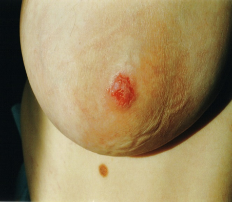
Figure 1. Two patients with Paget’s disease of the breast
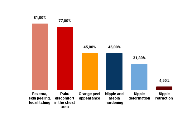
Figure 2. Paget’s disease clinical symptoms’ frequency
Twelve cases (54%) were diagnosed following BSE or noticing other abnormal aspects. Seven cases (31.2%) were diagnosed as a result of breast screening. Two cases (9%) were detected during a routine medical check-up, and in one case (4.5%), during examination for other conditions.
All patients underwent mammography and breast ultrasound.
The majority of patients (63.6%) were diagnosed by smear-impression. Fine-needle aspiration was used for 31.81%. Patients in whom cytological examination revealed cells with signs of atypia or carcinoma had preoperative tumor core biopsy (n=16;63%).
All patients had modified radical mastectomy.
Tumors’ largest dimensions varied from 5 to 57 mm. Most patients (n=12; 54.5%) had tumors smaller than 20 mm. In 8 cases (36.3%), the diameter of the tumor process varied from 20 to 40 mm. Only two patients had tumors sized between 40-60 mm, with the maximum size being 57x32x36 mm (Table 2).
Table 2. Postoperative characteristics of the tumor
| Frequency (N=22) | Percentage | |
| Tumor size (mm) | ||
| 12,3 (5-20) | 12 | 54.5 |
| 25,5 (20-40) | 8 | 36.3 |
| 55 (40-60) | 2 | 9.1 |
| Degree of differentiation | ||
| Gx | 2 | 9.1 |
| G1 | 1 | 4.5 |
| G2 | 2 | 9.1 |
| G3 | 17 | 77.2 |
| G4 | – | – |
| Histological types: Paget carcinoma | ||
| without DCIS | 1 | 4.5 |
| with DCIS | 2 | 9.1 |
| with extensive DCIS | 5 | 22.7 |
| with invasive ductal carcinoma | 14 | 63.6 |
| Immunohistochemical types | ||
| Luminal A | 5 | 22.7 |
| Luminal B Her2/neu – | 13 | 59.0 |
| Luminal B Her2/neu + | 2 | 9.1 |
| TNBC | 2 | 9.1 |
In 14 cases (63.6%), PDB was associated with invasive ductal carcinoma, and in 5 cases (22.7%), with extensive ductal carcinoma in situ (DCIS). Most patients had G3 tumors (n=17; 77.2%).
Twenty-two % had Luminal A tumors, and 68.1% had Luminal B tumors. TNBC was identified in two cases (9.1%).
Surgical treatment was the only treatment method in four cases. Five patients (31.81%) with positive postoperative lymph nodes underwent a mastectomy followed by radiation therapy. Adjuvant systemic therapy, such as chemotherapy and trastuzumab, was given in 12 cases (54.5%) for TNBC, Luminal B Her2/neu+, or Luminal B Her2/neu – with Ki67 over 21%. Endocrine treatment (Anastrozole) for five years was indicated for patients with luminal A and B tumors with positive hormonal receptors (63.6%).
4. Discussion
To our knowledge, this is the first study reporting a case series of PDB in the Republic of Moldova regarding epidemiological information, staging, histological and immunohistochemical results, and the treatment administered. The incidence of PDB in this study was slightly lower compared to other studies, representing 0.37% vs. 1-3% (3). In our study, the disease is predominantly encountered during the menopausal period, which aligns with previously published international data.
There are no precise data on etiology or triggering factors in PBD; however, precancerous conditions (31.2%) were frequent in our group.
More than 60% of the patients in our study group had stage III disease. Unfortunately, only one patient was diagnosed with stage 0. This high percentage of advanced-stage cases is probably explained by the fact that PDB clinically presents as eczema, dermatitis, or psoriasis, with patients avoiding referral to an oncologist. Based on a cohort study that included 72 patients, in about 33% of cases, a palpable mass is detected at the time of diagnosis, and in 54% of cases, the axillary lymph nodes on the same side are enlarged (9). In our group, a palpable mass was identified in 81% of the cases, and axillary lymph nodes in 77.2%. In another study, the main symptom that led patients to seek medical attention was eczema or nipple ulceration (98% of cases) (10). In our study, eczema, skin peeling, and local itching were reported by 81% of the patients.
Early diagnosis of the disease represents a favorable prognostic factor; sometimes, regular BSE can save lives. Like any tumor in the breast gland, breast self-examination represents the primary method of detecting formations worldwide. 54% of patients discovered cancer through breast self-examination (4, 9-10).
The rate of accurate diagnosis can improve through skin biopsy or core needle biopsy, performed in about 63% of the investigated patients. Due to technical limitations, we cannot perform sentinel lymph node biopsy (SLNB) in our country.
The stage of the disease influenced the therapeutic strategy, the levels of estrogen (ER) and progesterone (PR) receptors, HER2/neu expression, the degree of differentiation (G), tumor proliferation index (Ki67), vascular and perineural invasion, age, and menstrual status. PBD was associated with invasive breast carcinoma in 63.6% of the cases in our study group. Other reports found that over 80% of PDBs have HER2 overexpression or amplification (12,13). In our case, only 9.1% of the patients had HER2/neu+ tumors. Luminal tumors accounted for about 50%, consistent with other studies (14). According to the NCCN version 6.2024 guidelines, there is no category 1 data that specifically addresses the local management of PBD (11). All patients in our study group underwent modified radical mastectomy with complete axillary lymph node dissection (ALND). Breast reconstruction is still not performed.
Regarding radiotherapy treatment, in our country, only external radiotherapy is performed, as other radiotherapy methods are unavailable. Fifteen patients (68.18%) with postoperative metastasis in lymph nodes underwent a mastectomy followed by radiation therapy. Loco-regional irradiation included the ipsilateral chest wall and regional lymph nodes with conventional fractionation radiotherapy. External radiotherapy was performed with a Clinac linear accelerator and the Truebeam-Vitalbeam linear accelerator. Total dose = 50 Gy/25 fractions/2 Gy per fraction/5 weeks.
Similar to international data, in our country, chemotherapy treatment in PBD (without parenchyma involvement) after mastectomy is not indicated. In cases of Paget’s disease with breast parenchyma involvement, chemotherapy is recommended based on pTNM staging, histological results (degree of differentiation, perivascular and perineural invasion, resection margins), and the tumor’s immunohistochemical profile. Approximately 50% of patients received postoperative chemotherapy after mastectomy. Currently, there is no data that endocrine therapy, including tamoxifen or aromatase inhibitors, reduces the risk of recurrence of local disease in patients with PDB; about 63.6% of patients receive endocrine therapy, among these, six premenopausal patients were prescribed Tamoxifen 20 mg/day for 5 years, while in the remaining cases, Anastrozole 1 mg/day for 5 years was indicated (13,14). Hormonal therapy was initiated after the completion of chemotherapy and could be administered concurrently with anti-HER2/neu therapy.
The limitations of our study include the relatively small number of patients, the selection of only operable patients, the potential for missing or inaccurate data due to the retrospective design, and limitations in staging (only a low number of patients underwent CT/MRI investigation). In addition, we had limited possibilities to track and analyze the results obtained in other clinics.
5. Conclusion
Our retrospective clinical study is the first research in our country to publish data reflecting the experience accumulated over 5 years. In the study, BDB tends to be detected in advanced loco-regional stages, predominantly invasive ductal and Luminal B forms, and is treated by radical mastectomy followed by radiotherapy and chemotherapy (according to indications).
Abbreviations:
PDB – Paget disease of the breast
DCIS – ductal carcinoma in situ
BCS – breast-conserving conserving-surgery
BSE – breast self-examination
USG – ultrasound examination
HER2/neu – Human Epidermal Growth Factor Receptor 2
PR – Progesterone receptor
ER – Estrogen receptor
TNBC – Triple-negative breast cancer
CK 5 – basal cytokeratin
SLNB – sentinel lymph node biopsy
ALND – axillary lymph node dissection
Statements
Conflict of interests: None
Funding Sources: None
Statement of Ethics: Patient data was anonymized and the institutional Ethics Committee approved this retrospective study.
Informed consent: The patients provided written informed consent for using their clinical data for research purposes and for the pictures of their lesions to be published.
REFERENCES
- Scardina L, Di Leone A, Magno S, Franco A, Biondi E, Sanchez AM, D’Archi S, Gentile D, Fabi A, Masetti R, Franceschini G. Paget’s disease of the breast: Our 20 years’ experience. Front Oncol. 2022 November 22;12:995442. doi: 10.3389/fonc.2022.995442. PMID: 36483034; PMCID: PMC9722944
- Yasir M, Khan M, Lotfollahzadeh S. Mammary Paget Disease. (Updated 2023 June 3). In: StatPearls (Internet). Treasure Island (FL): StatPearls Publishing; 2023 Jan-. Available from: https://www.ncbi.nlm.nih.gov/books/NBK563228
- Dubar S, Boukrid M, Bouquet de Joliniere J, et al. Paget’s Breast Disease: A Case Report and Review of the Literature. Front Surg. 2017;4:51. Published 2017 October 23. doi:10.3389/fsurg.2017.00051
- Kar H, Altındağ SD, Etit D, et al. Clinicopathological features of mammary Paget’s disease: a single-center experience in Turkey. Turk J Med Sci. 2021;51(6):2994-3000. Published 2021 December 13. doi:10.3906/sag-2103-148
- Del Riego J, Pitarch M, Codina C, et al. Multimodality approach to the nipple-areolar complex: a pictorial review and diagnostic algorithm. Insights Imaging. 2020;11(1):89. Published 2020 August 5. doi:10.1186/s13244-020-00896-1
- Lyons, D., Wahab, R. A., Vijapura, C., & Mahoney, M. C. (2021). The nipple-areolar complex: a comprehensive imaging review. Clinical Radiology, 76(3), 172–184. https://doi.org/https://doi.org/10.1016/j.crad.2020.09.013
- Cai Y, Cheng Z, Nangong J, Zheng X, Yuan Z. Using immunohistochemistry to classify the molecular subtypes of Paget’s disease of the breast. Cancer Med. 2023;12(13):14104-14111. doi:10.1002/cam4.606
- Grewal IS, Rajagopal T. Studying the Presentation of Paget Disease of the Breast. Cureus. 2023 Jun 27;15(6):e41057. doi: 10.7759/cureus.41057. PMID: 37519599; PMCID: PMC10374979
- Song Q, Jin Y, Huang T, Zhang JH. Diagnosis and treatment of Paget’s disease of the breast: an analysis of 72 cases. Int J Clin Exp Med. 2015 October 15;8(10):19616-20. PMID: 26770622; PMCID: PMC4694522.
- Markarian S, Holmes DR. Mammary Paget’s Disease: An Update. Cancers (Basel). 2022;14(10):2422. Published 2022 May 13. doi:10.3390/cancers14102422
- https://www.nccn.org/professionals/physician_gls/pdf/breast.pdf
- Kothari AS, Beechey-Newman N, Hamed H, Fentiman IS, D’Arrigo C, Hanby AM, Ryder K. Paget disease of the nipple: a multifocal manifestation of higher-risk disease. Cancer. 2002 Jul 01;95(1):1-7. (PubMed)
- Markarian S, Holmes DR. Mammary Paget’s Disease: An Update. Cancers (Basel). 2022 May 13;14(10):2422. doi: 10.3390/cancers14102422. PMID: 35626023; PMCID: PMC9139704
- Gaurav A, Gupta V, Koul R, Dabas S, Sareen R, Geeta K, Arora V, Parikh PM, Aggarwal S. Practical consensus recommendations for Paget’s disease in breast cancer. South Asian J Cancer. 2018 Apr-Jun;7(2):83-86. doi: 10.4103/sajc.sajc_107_18. PMID: 29721469; PMCID: PMC5909301.