


Author(s) :
Artiom Mîrzătan1, Monica-Emilia Chirilă Claudiu Mihai Ciuciureanu3, Corneliu Prepelită Dumitru Sofroni1,4, Valentin Martalog1,4
1 University of Medicine and Pharmacy “Nicolae Testemiţanu”, Chişinău, Republic of Moldova
2 Clinical Development Department MVision Al. Helsinki, Finland
3 University of Medicine and Pharmacy “Carol Davila”, Bucharest, Romania
4 Oncology Institute Chisinău, Republic of Moldova
Corresponding author: Valentin Martalog, Email: valentin.martalog@usmf.md
Publication History: Received - , Revised - , Accepted - , Published Online - 1 October 2023.
Copyright: © The author(s). Published by Casa Cărții de Știință.
User License: Creative Commons Attribution – NonCommercial (CC BY-NC)

 PDF
PDF
 Share
Share
Highlights
-
Fluoroscopic-guided transthoracic needle biopsy (FGTNB) proved a practical, minimally invasive option for high-risk/comorbid patients: In a Moldovan tertiary cancer center (2019–2021), FGTNB enabled pathological confirmation when more invasive procedures or CT-guidance were not feasible.
-
Solid diagnostic yield for both lung and mediastinal lesions: Sensitivity was 79.6% for lung tumors (diagnosing malignancy but also TB and pulmonary fibrosis) and 83.3% for mediastinal tumors (predominantly lymphomas), with best technical suitability for upper-lobe lung lesions and anterior mediastinal masses.
-
Manageable complication rates, with higher pneumothorax risk in mediastinal biopsies: Lung biopsies had low pneumothorax (5.6%) and bleeding/hemoptysis (3.7%) rates, while mediastinal biopsies showed a notably higher pneumothorax rate (16.7%), supporting careful patient/lesion selection and post-procedure monitoring.
Abstract
Introduction: Pathological confirmation of a newly identified tumour is needed for choosing the adequate treatment. Patients may have a low performance status which makes them unsuitable for invasive diagnostic procedures. Percutaneous transthoracic biopsy is a minimally invasive method which can provide tissue samples for identification of lung or mediastinal tumours.
Material and method: Data from consecutive patients which underwent fluoroscopic-guided transthoracic puncture-biopsy (FGTPB) in a tertiary cancer center from Republic of Moldova from 2019 to 2021 were retrospectively collected.
Results: We identified 54 patients with lung tumors and 12 patients with mediastinal tumours. In the lung tumors group, median age was 57 years and in the mediastinal tumours group the median age was 27.5 years. Most of the lung tumors were situated in the superior lobes (79.6%) and had less than 5 cm (70.4%). The majority of mediastinal tumors were situated anteriorly (66.7%) and had more than 10 cm (58.3%). The sensitivity of transthoracic percutaneous biopsy was 79.6% in lung tumours and 83.3% in mediastinal tumours, identifying malignant or infectious pathology, or fibrosis. Lung biopsy had a low rate of pneumothorax (5.6%) and of bleeding (3.7%). Mediastinal tumor biopsy had a rate of pneumothorax of 16.7%.
Conclusion: Transthoracic puncture-biopsy made under fluoroscopic guidance is a safe and efficient alternative diagnostic procedure for unfit patients with lung or mediastinal tumours.
1. Introduction
Lung cancer is the most common cancer in the world and the leading cause of cancer death worldwide. Central lung cancer with airway obstruction is generally sampled bronchoscopically. Computed tomography (CT)-guided or Fluoroscopic-Guided Transthoracic Needle Biopsy is reserved for lesions difficult to visualize sonographically due to bony or paravertebral localization of the intervention. Lung biopsy is a reliable procedure performed to inform treatment strategy for patients with abnormal findings of the lung. Cytology of biopsy specimens has long been a major diagnostic modality for initial evaluation of patients with lung cancer (1,2) .
However, invasive diagnostic procedures can be challenging or impossible in patients with multiple underlying health conditions.
The prevailing lung biopsy techniques that are used today encompass the Percutaneous Transthoracic Lung Biopsy, Open Lung Biopsy, Video-Assisted Thoracic Surgery, Transbronchial Biopsy, and Cryo-biopsy (3). Mediastinal cancers, typically diagnosed in younger patients, often have diverse origins and prognostic factors, so a specific pathologic diagnosis is mandatory due to the array of possible differential diagnoses. Surgeons commonly employ thoracoscopy (including video-assisted thoracoscopic surgery) and mediastinoscopy to obtain tissue samples for histopathologic analysis. Various diagnostic possibilities also exist for transbronchial or transesophageal endoscopic techniques, with or without ultrasonic guidance.
Fluoroscopic-guided transthoracic needle biopsy (FGTNB) is a minimally invasive technique with a high degree of diagnostic accuracy, which offers the possibility of obtaining sufficient material for cytological and histological research. This method is particularly beneficial for patients with unresectable tumors, concurrent diseases that pose a significant risk for radical surgery, or those who refuse surgery, where the traditional histopathological diagnosis is unachievable (4).
When obtaining a biopsy in mediastinal tumors proves challenging or repeated biopsies yield negative results, there may be a calculated need to resort to parasternal mediastinotomy. When biopsy results for lung tumors remain inconclusive, a comprehensive discussion with the multidisciplinary team may warrant the performance of thoracotomy or median sternotomy. A separate study conducted in the Department of Thoracic and Abdominal Surgery in 2013 reported a false-negative rate for detection of malignancy by percutaneous biopsy of only 5%.
This study is focused on presenting the outcomes of FGTNB in diagnosing both lung and mediastinal tumors.
2. Material and Method
We enrolled patients with lung or mediastinal tumors admitted to the Department of Thoracic and Abdominal Surgery of the Oncological Institute of Chișinău, Republic of Moldova, from 2019-2021. The criteria for inclusion were the ineligibility for general anesthesia and the technical accessibility of the tumor. The superior-anterior part of the mediastinum or segment 3 of the upper lobe were considered as the most accesible locations. The exclusion criteria for the procedure were a performance status of 3 or 4, coagulation disorders, and an estimated life expectancy of less than two months. Tumours with endobronchial extension were primarily refferedd for bronchoscopy. FGTNB was performed in these patients only if the bronchiscopy was unavailable or unfeasible. We conducted a retrospective review of data from the patients’ clinical records.
A transthoracic needle biopsy guided by chest radioscopy was performed in these patients. The Bard Biopsy-Gun system was used to biopsy the lesions with Tru-cut needles with a diameter of 16–18G. The needle gauge was selected according to the characteristics of the lesion: tumors larger than 4 cm or with signs of parietal invasion were biopsied with larger gauge needles.
Histopathological identification of the biopsied tissue was obtained in the Cytopathology Department using the Romanowsky-Giemsa coloration method.
3. Results
A total of 66 patients met the inclusion criteria. Severe concomitant pathologies were present in 31 cases (47.0%), advanced local tumors with distant metastases in 23 patients (34.8%), and mediastinal compression syndrome in 12 patients (18.2%). Fifty-four patients had lung tumors, and 12 patients had mediastinal tumors.
Most patients with lung tumors were older than 50 (85.2%). On the contrary, two-thirds of the patients with mediastinal tumors were under 30 (66.7%). Most lung tumors (70.4%) were less than 5 cm, and more than half of the mediastinal tumors were more than 10 cm in diameter (58.3%). Detailed information on both patient groups can be found in Table 1.
All patients were initially investigated by radiography or radioscopy. Prior to the admission in the Oncology Institute, where CT investigation was not available, a thoracic CT was performed in other clinics for 59.3% (n=32) of patients with suspected lung cancer and 83.3% (n=10) of those with mediastinal pathology.
The imaging diagnosis in patients with lung and mediastinal tumors can be found in Table 2.
Table 1. Patients’ characteristics
| Lung (N=54) | Mediastinum (N=12) | |
|---|---|---|
| Number | Number | |
| Sex | ||
| Male | 44 | 11 |
| Female | 10 | 1 |
| Age | ||
| Median | 57 | 27.5 |
| Range | min 21- max 72 | min 18- max 65 |
| Investigation | ||
| Radiography | 54 | 12 |
| CT | 32 | 10 |
| Tumor size | ||
| median | 5.5 cm | 15.1 cm |
| < 5 cm | 38 | 1 |
| 6-10 cm | 13 | 4 |
| 11-15 cm | 3 | 6 |
| 16-20 cm | 0 | 1 |
Table 2. Radiological description of patients with lung and mediastinal tumors
| Radiological conclusion | Number | |
|---|---|---|
| Lung tumors (N=54) | ||
| Primary tumor only | 27 | |
| Primary tumor and metastasis to the same lung | 13 | |
| Primary tumor with metastases in the same lung and pleurisy |
8 | |
| Primary tumor and metastases in the contralateral lung |
4 | |
| Metastatic pleurisy | 1 | |
| Malignant pleural mesothelioma | 1 | |
| Mediastinal tumors (N=12) | ||
| Mediastinal tumor | 10 | |
| Mediastinal tumor and lymphadenopathy | 2 |
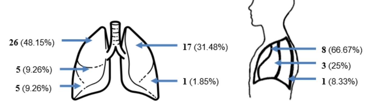
Figure 1. Number of cases corresponding to anatomical location of lung and mediastinal tumors (relative percentage in brackets)
Lung tumors were more frequently located in the superior lobes (79.6%), and most of the mediastinal tumors (66.7%) were in the anterior compartment (see Figure 1).
The transthoracic needle biopsy provided sufficient pathologic material for diagnosing 43 patients (79.6%) with lung tumors. Out of these, 32 cases were identified as lung cancer, the rest were found to be either tuberculosis or pulmonary fibrosis. In the case of mediastinal tumors, the transthoracic biopsy provided a conclusive diagnosis for 10 patients (83.3%) (see Table 3).
Table 3. Morphological diagnosis of lung and mediastinal tumors, respectively
| Number | |
|---|---|
| Lung tumors (N=54) | |
| Adenocarcinoma | 13 |
| Squamous cell carcinoma | 11 |
| Tuberculosis | 7 |
| Large cell carcinoma | 5 |
| Foci of pulmonary fibrosis | 4 |
| Small cell carcinoma | 3 |
| Inconclusive | 11 |
| Mediastinal tumors (N=12) | |
| Non-Hodgkin’s lymphoma | 5 |
| Mediastinal lymphoma | 3 |
| Hodgkin’s lymphoma | 2 |
| Inconclusive | 2 |
Lung cancer was identified in 32 (59.3%) of the 54 patients initially diagnosed with lung tumors. The transthoracic biopsy failed to establish a definitive diagnosis in 13 patients. These cases were subjected to further evaluation by the multidisciplinary board. Despite the associated risks, it was determined that a diagnostic thoracotomy was necessary in 6 of these cases. In 7 instances, the circumstances were deemed inoperable. Complications seen after percutaneous transthoracic needle biopsy in lung tumors were reported in 5 cases (9,3%). Among these, pneumothorax was described in 3 patients (5,6%), who all required subsequent pleural cavity drainage. Hemoptysis occurred in 2 patients (3,7%), which was subsequently stopped by administering hemostatic agents. In patients with mediastinal tumors, pneumothorax was reported in 2 cases (16,7%) following the percutaneous biopsy.
4. Discussion
A significant advantage of this method is the possibility of performing the procedure under only local anesthesia, which is essential for patients suffering from various comorbidities that generally don’t allow undergoing general anesthesia. The good results and low rate of adverse events seen in transthoracic percutaneous puncture in our patients are evident.
In our study, for patients considered unfit for more invasive diagnostic procedures, FGTNB established a conclusive diagnosis in 79.6% of lung tumors and 83.3% of mediastinal tumors, with a rate of manageable adverse effects of 9,3% and 16.7% for lung and mediastinal tumors, respectively. Patients with tumors in the superior lung or the anterior area of the mediastinum were more suitable for the procedure, as the approach was more straightforward, and the risk of adverse events was lower.
A cohort study of 100 patients comparing the accuracy, safety, and effective dose (ED) of FGTNB with CT-guided needle lung biopsy (CTNLB) procedures demonstrated that the adequacy rate of was 95%. The overall sensitivity of FGTNB for detecting malignancy was 87%. Overall accuracy was also 87%. The specificity and positive predictive value were 89% and 99%, respectively, but with a pneumothorax complication rate of 25%. FGTNB is faster, taking only 15 minutes (fluoroscopy-guided CT biopsy of lung lesions ranged from 15 to 41 minutes, with a mean of 23.8 min). FGTNB has proven to be a real-time imaging modality and is associated with low radiation dose to the patient and operator. With the use of a multiplanar fluoroscopic unit, it is usually possible to document the correct placement of the needle tip within the lesion, including small lesions (5).
However, another cohort study of 235 patients using CTNLB demonstrated that the overall diagnostic accuracy was 95.4%, with 95.52% sensitivity, 100% specificity. Complications occurred in 51 patients, and the overall complication rate was 21.7%. The most frequent complication was minor pneumothorax with a rate of 19.1%. (6)
Other studies have demonstrated that the effectiveness of this method is 86% with radiological guidance and 95% when using CT guidance. When using CT as a guiding method, sufficient pathological material was obtained for cytological examination in 89% of patients and for histological diagnosis in 93% of patients. (7,8).
Over the last few years, improvements in imaging, needles, and procedural techniques have all reduced complication rates. Some rare complications include air embolism and tumor seeding, seen in less than 1% of cases. Factors associated with increased rate of complications include the patient’s age, comorbidities, tumor size and location, and the number of punctures performed (9,10).
The limitations of our study were performing the puncture only with the use of radioscopy, which did not allow us to compare the effectiveness of using CT guidance. We also only compared a relatively small number of patients, and these results generally only apply to patients with severe comorbidities who had contraindications for other, more traditional diagnostic interventions.
5. Conclusions
For patients with lung or mediastinal tumors where obtaining a surgical tissue biopsy is not feasible, and the CT-guided biopsy is not available, the fluoroscopic guided transthoracic biopsy may provide the necessary material for pathologic diagnosis with a satisfactory success rate and relatively low risk of manageable adverse events.
Abbreviations:
CT – Computed Tomography
FGTPB – Fluoroscopic Guided Transthoracic Puncture Biopsy
CTNLB – CT Guided Needle Lung Biopsy
Statements:
Author’s contributions: MV, SD, and PC conceived the discussed topic; MV, CP, SD, MA, and MEC drafted the initial paper; MV, MA, MEC, and CC made the final revisions to the final paper.
Consent for publication: As the corresponding author, I confirm that the manuscript has been read by and approved for submission by all authors.
Funding: This study did not receive any specific grant from the funding agencies in the public, commercial, or not-for-profit sector.
Conflicts of Interest: The authors declare no conflict of interest.
References:
- Thapa A, Subedi K, Suwal S, Chataut D, et al. CT guided lung biopsy: Diagnostic yield and complications, using 18G coaxial semi-automatic core needle. Nepal Journal of Medical Sciences 2019;4(1):20-25.
- Kurban LA, Gomersall L, Weir J, Wade P. Fluoroscopy-guided percutaneous lung biopsy: avaluable alternative to computed tomography. Acta Radiol. 2008 Oct;49(8):876-82. doi:10.1080/02841850802225893. PMID: 18618301.
- Modi P, Uppe A. Lung Biopsy Techniques and Clinical Significance. 2022 Jun 28. In:Stat Pearls [Internet]. Treasure Island (FL): StatPearls Publishing; 2023 Jan–. PMID: 33085300.
- Arakawa, H., Y. Nakajima, Y. Kurihara, H. Niimi, T. Ishikawa, CT-guided transthoracic needle biopsy: A comparison between automated biopsy gun and fine needle aspiration, Brain and Language, vol. 51, nr. 7. pp. 503-506, 2006, doi: 10.1016/s0009-9260(96)80191-7.
- Dvorak, Petr, et al. “CT-Guided Biopsy of the Mediastinal Masses. Can Anatomical Relationships Predict Complications?” Biomedical Papers, vol. 163, no. 3, 23 Sept. 2019, pp.220–226, biomed.papers.upol.cz/artkey/ bio-201903-0005_ct-guided-biopsy-of-the-mediastinal-masses-can-anatomical-relationships-predict-complications.php, https://doi.org/10.5507/bp.2018.058. Accessed 24 Sept. 2023.
- Sarajlic, Vesna, et al. “Diagnostic Accuracy and Complication Rates of Percutaneous CT-Guided Coaxial Needle Biopsy of Pulmonary Lesions.” Diagnostic and Interventional Radiology, 22 Mar. 2021, https://doi.org/10.5152/ dir.2021.20844. Accessed 12 May 2021.
- Swischuk, J. L. et al. Percutaneous transthoracic needle biopsy of the lung: review of 612 lesions., In. J. Vasc. Interv. Radiol., vol. 9, nr. 2, pp. 347-52, 2008, doi: 10.1016/s1051- 0443(98)70279-9.
- Sakai, Hitomi, and Masayuki Takeda. “Percutaneous transthoracic needle biopsy of the lung in the era of Precision Medicine.” Journal of Thoracic Disease, vol. 11, no. S9, 2019,https://doi.org/10.21037/jtd.2019.03.20.
- Klein, Jeffrey S., Gregory Salomon, și Ernest A. Stewart, Transthoracic needle biopsy with a coaxially placed 20 gauge automated cutting needle: Results in 122 patients, In. Radiology, vol. 198, nr. 3, pp. 715-720, 1996, doi: 10.1148/radiology.198.3.8628859
- Wiener, Renda Soylemez, Daniel C. Wiener, și Michael K. Gould, Risks of transthoracic needle biopsy: How high? Clinical Pulmonary Medicine, vol. 20, nr. 1. NIH Public Access, pp.29-35, ian. 2013, doi: 10.1097/ CPM.0b013e31827a30c1.


