


Author(s) :
Miruna-Ioana Lazăr1, Ioana-Miruna Stanciu1, Claudiu Mihai Ciuciureanu3, Cristina Mihaela Olaru1, Cornelia Nițipir2
1 Department of Paediatrics, Marie Sklodowska Curie Emergency Hospital for Children, Bucharest, Romania
2 Department of Oncology, “Carol Davila” University of Medicine and Pharmacy, 020021 Bucharest, Romania
3 University of Medicine and Pharmacy “Carol Davila”, Bucharest, Romania
Corresponding author: Ioana-Miruna Stanciu, Email: ioana-miruna.stanciu@drd.umfcd.ro
Publication History: Received - , Revised - , Accepted - , Published Online - 1 October 2023.
Copyright: © The author(s). Published by Casa Cărții de Știință.
User License: Creative Commons Attribution – NonCommercial (CC BY-NC)

 PDF
PDF
 Share
Share
Highlights
-
Aspergillus infection can “mask” an underlying lung adenocarcinoma: Cavitary lesions and an aspergilloma-like picture may delay cancer suspicion, especially when imaging lacks a clear mass or lymphadenopathy.
-
Clinical and microbiology workups can be misleading or late-positive: Initial sputum cultures/bronchoscopy may be negative for malignancy (and even for fungi), while symptoms (fever, cough, hemoptysis) overlap with both infection and cancer.
-
Definitive diagnosis often comes only after surgery and histopathology: In the presented case, lobectomy for presumed chronic aspergillosis revealed Aspergillus colonization plus occult, poorly differentiated primary lung adenocarcinoma, triggering delayed oncologic staging/therapy decisions.
Abstract
Although infrequent, lung cancer can be disguised by the synchronous presence of a fungal infection, most notably by the Aspergillus species. Some cases of incidental adenocarcinoma diagnosed during surgical treatment of pulmonary aspergillosis have been reported, showing just how these fungi can hide more serious, underlying pathology.
This review aims to raise awareness about this occurrence to accelerate the correct etiological diagnosis, establish a timely oncological treatment, and improve the overall morbidity and mortality in lung cancer patients. In addition, we present a rather alluring case of concurrent adenocarcinoma and Aspergillus infection to display how these two entities can appear together.
1. Introduction
Despite significant advances in detection measures and therapeutical management, primary lung cancer remains the most fatal type of malignancy globally, regardless of gender. The high mortality rate of broncho-pulmonary neoplasia could undoubtedly be attributed to delayed diagnosis, taking into consideration the variety of non-specific symptomatology and imaging appearances (1)(2)(3).
Adenocarcinoma represents the most frequent subtype of lung neoplasia, comprising around 40% of the cases worldwide. It tends to develop within the peripheral parenchyma, thus being more challenging to diagnose by bronchoscopy with biopsy. Moreover, several case reports describing an association between the synchronous presence of cavitary bronchogenic carcinoma and chronic fungal infections, particularly aspergillosis, have been published (4)(5).
Aspergillosis entails a spectrum of different clinical presentations of an infection with a prominent, pervasive, opportunistic type of mold. Symptomatology depends mainly on the immunological status of the affected host. The most involved species are represented by A. fumigatus and A. flavus, affecting different organ systems. Symptoms depend mainly on the primary infection site and route, ranging from the lungs, brain, bones, and skin(6).
Immunocompetent individuals hardly suffer from lung aspergillosis, as innate immunity prevents disease establishment. Previously scarred lung tissue or impaired pulmonary function are predisposing factors for an acute or chronic infection following the inhalation of Aspergillus conidia. Hematological cancers are also widely acknowledged risk factors for aspergillosis, as they often manifest through relative neutropenia.
We present a case of lung adenocarcinoma paired with a chronic Aspergillus infection, which initially disguised the neoplasia, thus prolonging the diagnosis and initiation of treatment. We also offer a literature review of similar cases to emphasize this relatively infrequent yet critical association (4, 7).
2. Case report
A 54-year-old woman, never-smoker, without any relevant history of acute or chronic lung disease, presented with persistent fever, spasmodic cough, and breathing difficulties that had occurred two weeks prior and worsened despite levofloxacin treatment prescribed by her primary care physician.
There was no history of weight loss or chest pain. The patient’s medical record revealed primary hypertension, treated appropriately, thyroidectomy for benign goiter, and a hysterectomy for symptomatic uterine fibroma.
On admission, the patient was pale and febrile (38.5 °Celsius), with a BMI of 24. She was hypotensive and in acute respiratory distress, with frequent dry cough, tachypnoea (30 breaths/minute), and hypoxia (peripheral oxygen saturation of 88% in room air), without any palpable lymphadenopathy. She denied any relevant personal medical and occupational history, including tuberculosis, HIV infection, and exposure to silicates. She also denied any family history of lung disease.
Laboratory findings disclosed an elevated number of leukocytes with a predominance of neutrophils (70%), hypochromic microcytic anemia, thrombocytosis, and elevated inflammatory markers (elevated C-reactive protein, high erythrocyte sedimentation rate and hyperfibrinogenaemia). A COVID-19 antigen test was performed on admission with a negative result. Considering the presumptive diagnosis of an acute pulmonary infection, a chest x-ray was ordered, which showed a single cavitary lesion in the right pulmonary area with a diameter of approximately 4 centimeters (see Figure 1).

Figure 1. Chest X-ray on admission showed a hyper-transparent circumscribed lesion with a thick, anfractuous contour adjacent to the right pulmonary hilum.
Following the results of her chest x-ray, a chest computed tomography was ordered, which confirmed the cavitary lesion in the apical segment of the right lower lobe. It further described the presence of ground-glass opacities at this level. There was also a minimal pleural effusion on the posterior side of the right hemithorax (12 mm thick), accompanied by bronchiectasis in the same area (see Figure 2). No masses or mediastinal/ hilar lymphadenopathy were described. At this point, antibiotic therapy was changed to ensure broad-spectrum coverage.
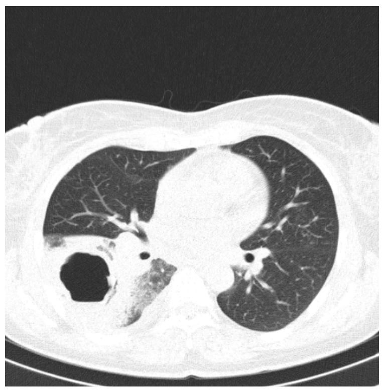
Figure 2. CT scan of the chest showing a cavitary lesion within the lower right lobe surrounded by ground-glass opacities.
Sputum samples were difficult to obtain because of the dry cough, which prompted repeated specimens. Bacterial and fungal cultures, gram stain, and Ziehl-Neelsen stain were all negative. Tuberculosis (TB) infection was ruled out because of the negative result of nucleic acid amplification tests (GeneXpert), negative cultures on the Lowenstein-Jensen medium, and negative interferon-gamma release assays. Concomitant HIV infection was also ruled out because of the absence of HIV-specific antibodies.
Bronchoscopy showed hyperemia of the right lower lobe lining, with associated seromucous secretions, factoring out proliferative lesions. Microscopy examination of the bronchial aspirate showed extracellular Gram-positive diplococci, Gram-negative bacilli, neutrophils, and degraded epithelial cells. Notably, there was no initial bacterial growth on the culture medium. Cytology of the sputum specimen was negative for any atypical cells.
After twenty days of hospitalization and systemic administration of antibiotic therapy, the patient developed an increase in temperature to around 40 degrees Celsius, with general malaise, leukopenia, thrombocytopenia, and a change in cough now with hemoptysis. The cultures from the sputum sample were now positive for colonies of Aspergillus spp. In response to these new findings, voriconazole was added to the intravenous treatment regimen. The sputum microbiological cultures were also positive for Methicillin-resistant Staphylococcus aureus (MRSA), Klebsiella, and Pseudomonas aeruginosa.
After treatment initiation for these pathogens, the patient displayed a favorable clinical evolution, with remission of fever and an expected drop in WBC count and inflammatory markers. Another CT scan of the chest with contrast was performed three months after admission, showing dimensional regression of the cavitary lesion, which measured around 3 cm (see Figure 3).
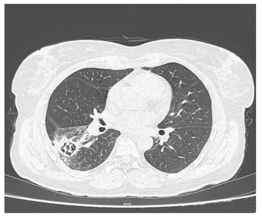
Figure 3. CT scan depicting the evolution of the cavitary lesion after exactly three months of systemic antibiotic therapy.
Following medical treatment, a surgical plan was introduced. A right lower lobectomy was performed, displaying incomplete resolution of the abscess. The histopathological examination of the specimen revealed the presence of bronchiectasis (colonized by dichotomic hyphae identified as Aspergillus spp.) and chronic inflammatory infiltrate surrounding the three-centimeter cavity. Alongside this was described fibrosis, granulation tissue, and necrotic tissue, all suggestive of Aspergillus colonization. The cavity was identified as an Aspergilloma but was unexpectedly surrounded by areas of poorly differentiated malignant cells, constituting an occult adenocarcinoma. Further immunohistochemical testing confirmed the presence of a primary lung adenocarcinoma.
Since malignancy involvement was not suspected before the surgery, hilar and mediastinal lymph nodes were not removed during the procedure. As a result, lymph node involvement could not be excluded, and post-operatory endobronchial ultrasound was not feasible. Following a multidisciplinary approach, systemic adjuvant chemotherapy for non-small cell lung carcinoma was initiated. The patient is now undergoing adjuvant treatment with Cisplatin/Pemetrexed (Cisplatin 75 mg/m2 day 1, Pemetrexed 500 mg/m2 day 1, every 21 days, for four cycles) for a T2aNxM0 (stage IB) poorly differentiated lung adenocarcinoma.
3. Material & methods
We searched “Google Scholar” and “Pub-Med” for similar cases of simultaneous occurrence of primary lung adenocarcinoma and Aspergillus to assess the similarities and difficulties encountered in establishing a diagnosis. While doing so, we acknowledged that lung cancer, in general, not necessarily a particular subtype prone to cavitation, can be associated with concomitant Aspergillus infection. This enforces our objective of advocating for the meticulous elimination of lung cancer, especially when dealing with previously healthy and immunocompetent individuals newly diagnosed with pulmonary aspergillosis.
Several case reports, mini-reviews, and relevant abstracts have been published. Despite this, it would be beneficial for an entire article to display the relationship between concomitant Aspergillosis and lung cancer. Data from these case reports were notably included and analyzed in one extensive review by Nilsson et al., from 2011 (4), which identified over twelve years three more cases of incidental adenocarcinoma coexisting with an Aspergillus infection (see Table 1).
4. Discussion
Upon further review, we have identified eleven other case reports with a positive diagnosis of primary lung adenocarcinoma associated with Aspergillus spp., colonization, or invasion (8–17). There is a significantly increased prevalence in men (10:1), with a median age of 59 and a mean age of 57. Seven patients are chronic tobacco users, as opposed to our case. Two case reports (10, 16) provide no data about the patient’s smoking history. This could hint that adenocarcinoma co-infected with Aspergillus is more prevalent among smoking men.
Lung cancer is more frequently diagnosed among smokers, but this is not the case for the adenocarcinoma subtype, which is notorious for its incidence in non-smokers or never-smokers (2, 18). We believe the link is not necessarily between adenocarcinoma and smoking but between the latter and the co-presence of adenocarcinoma and Aspergillus. The cause might be the persistent destruction of alveolar septa and the generation of dilated bullous spaces, which leads to a favorable colonization site for the saprophytic, opportunistic fungi (11).
Table 1. Primary lung adenocarcinoma associated with Aspergillus infection (* – the period is unspecified, making pack years impossible to calculate; ** – Only tuberculin intradermal reaction test was positive, along with a cavitary lesion on X-ray situated within the upper right pulmonary lobe)
| Author, year |
Gender | Age | Pack year |
Acute/ subacute/ chronic symptoms |
Lesion type | Location | Maximum diameter |
TB tests | Presumptive diagnosis |
Bronchoscopy |
|---|---|---|---|---|---|---|---|---|---|---|
| Smahi et al, 2011 |
M | 60 | 25 | Subacute | Cavitary | RUL | 45 mm | Negative | Aspergilloma | Negative |
| Boyd et al, 2012 |
M | 48 | 32 | Subacute | Cavitary | RUL | 85 mm | Negative | Pulmonary abscess |
Negative |
| McGregor et al, 1989 |
M | 64 | ? | Chronic | Cavitary | LUL | 45 mm | Negative | Fungal infection/ Carcinoma |
Negative |
| Itano et al, 2005 |
M | 77 | 56 | Asymptomatic | Opacity | RUL | 17 mm | Negative | Carcinoma | – |
| Saleh et al, 2008 |
M | 54 | Smoker | Subacute | Cavitary | RUL | 15 mm | Negative | Aspergilloma | Negative |
| Mays et al, 1966 |
M | 45 | 50 | Chronic | Cavitary | RUL | 25 mm | Negative** | Tuberculosis | – |
| El Hage and Fagel, 2020 |
F | 82 | Heavy smoker |
Acute | Opacity | RUL | – | Negative | ABPA | Adenocarcinoma +Aspergillus |
| Marcelis et al, 1981 |
M | 38 | 1 pack a day* |
Unspecified | Cavitary | RUL | 30 mm | Negative | Carcinoma | Negative |
| Fujita et al, 2018 |
M | 82 | ? | Asymptomatic | Cavitary | LLL | – | Negative | Chronic progressive pulmonary aspergillosis |
Adenocarcinoma +Aspergillus |
| Yoshitomi et al, 2000 |
M | 60 | 0 | Acute | Opacity (atelectasis) |
LLL | – | – | Carcinoma | Adenocarcinoma +Aspergillus |
| Yoshitomi et al, 2000 |
M | 53 | 0 | Asymptomatic | Opacity | LUL | 65 mm | – | Carcinoma | Adenocarcinoma +Aspergillus |
| Our case | F | 54 | 0 | Acute | Cavitary | RLL | 40 mm | Negative | Pulmonary abscess |
Negative |
One patient presented with dizziness, which was initially interpreted as acute labyrinthitis. The same patient reported a brownish productive cough for over 20 years and never sought treatment. (13). The rest of the patients presented with various subacute and chronic symptoms, including productive cough (8, 9, 12, 14, 17), hemoptysis (8–10, 12, 15), unintentional weight loss (12, 13), while concomitantly displaying thoracic and right arm pain, alongside generalized urticarial rash (15). Three patients were utterly asymptomatic (11, 16, 17).
Productive cough, hemoptysis, and weight loss are superposable clinical features of a potential neoplasia. In this case, even though paraclinical tests strongly suggest a pulmonary fungal infection, we must emphasize that ruling out concomitant cancer must be prioritized (7, 19). However, our patient presented with acute symptoms, leukocytosis, and systemic inflammatory syndrome. The initially negative bacterial and fungal cultures, undetectable hilar and mediastinal lymphadenopathy, and pulmonary mass led to the presumptive diagnosis of a pulmonary abscess. This was also the case for another patient (9) who presented with fever, hemoptysis, and subacute productive cough for five weeks.
Early diagnosis for some of the patients (8, 12, 13, 16) was “Aspergilloma,” mainly due to the characteristic signs on chest radiography and tomography, showing the distinct air-crescent sign (20). One patient (14) presented with branching opacities, considered specific for allergic broncho-pulmonary aspergillosis (ABPA). These patients showed no paraclinical disease resolution despite administration of systemic corticosteroids, antifungal treatment, and favorable clinical progression. One patient (11) who presented with an opacity rather than a cavity on radiographic imaging had a preliminary diagnosis of cancer, confirmed by histopathological examination of the lung tissue.
All the other patients, including ours that initially presented with a cavitary lesion on chest x-ray or CT had no initial suspicion of malignancy until biopsy and histopathologic exam. Considering the positive outcomes of the Mantoux test, despite negative cultures for acid-fast bacilli, one patient (13) was initially diagnosed with pulmonary tuberculosis and subsequently underwent tuberculostatic treatment.
The patient who presented with thoracic pain referring to his right arm (15) had a strong suspicion of an excavated tumor colonized with Aspergillus from the beginning, which didn’t show common aspects on shoulder x-ray, electromyography, alongside bone, cerebral, and hepatic scintigraphy. In other words, only four out of twelve (33,33%) early diagnoses were correct based solely on clinical presentation and imaging. This shows the importance of keeping concomitant fungal infections and malignancies high on the differential.
Regarding extensive tests for the detection of Aspergillus, four out of twelve patients had relevant bloodwork requested: the antifungal antibodies level was tested in three patients, one of which had positive serology for Aspergillus—the other patient presented with an elevated galactomannan (GM) antigen level, which is specific for aspergillosis.
Eight out of twelve (66%) patients presented with a cavitary lesion initially, possibly suggesting that over half of the adenocarcinoma was excavated, providing a favorable environment for germinating Aspergillus conidia into hyphae. Another theory is that the concurrent fungal infection with a pre-existent adenocarcinoma could have led to tumoral cavitation, as suggested by Nilsson et al. This could be because of the fungi’s secretion of specific lytic enzymes (4), creating further cavitation of the lung parenchyma.
Five of these cavities were first diagnosed as Aspergillomas despite the normal immunological status of the hosts and lack of pre-existing pulmonary disease. The hyper-transparent lesions were predominantly within the right upper pulmonary lobe (7/12). In contrast, two were found in the left lower lobe. There is a well-known predilection of Aspergillus to invade the upper pulmonary lobes, meaning that right and left upper lobe cavities were strongly compatible with a diagnosis of Aspergilloma, tuberculous hole, or both, although the latter being less probable because of the absence of risk factors and negative tests for Mycobacterium Tuberculosis. However, bronchial carcinomas occur more frequently in the upper lobes (13), complicating the differential diagnosis even further in the absence of a mass or lymphadenopathy. Aspergillus was masking an underlying neoplasia in nine out of twelve patients.
The average diameter of the cavitary lesions was approximately 40 mm, with a minimum value of 15 mm and a maximum of 85 mm. Every patient in this study underwent diagnostic bronchoscopy, which identified both Aspergillusand atypical cellularity in one-third of the patients. Moreover, transbronchial and CTguided biopsies were also performed, which were normal. Consequently, this led to a missed initial preoperative lung cancer diagnosis in most patients.
A definitive diagnosis was established by assessing the pathology reports from the surgical biopsy specimen. The histopathological report that belonged to our case described a fungus ball surrounded by necrotic and granular tissue, along with chronic inflammatory infiltrates and islets of atypical cells, constituting poorly differentiated adenocarcinoma. The presence of hyphae was described inside the cavity, confirming the pre-emptive diagnosis of Aspergilloma. Seven other cases (9, 10, 12–14, 17) presented with necrotic tissue on their biopsy examination. One of them disclosed (10) foci of bacterial proliferation and exudate, in addition to the dichotomic hyphae.
In contrast, another patient’s cavity (11) was lined by bronchiolar epithelium, which suggested bronchiolar dilation near the atypical glandular and epithelial cells with a high mitotic index. One patient’s (12) pathological exam revealed central necrosis of the adenocarcinoma, populated by fragmented fungal filaments. Regarding prognosis and tumoral grading, almost all adenocarcinomas were poorly differentiated, which generally implies a poor prognosis compared to a well-differentiated cancer. (21).
5. Conclusions
The case we described of an occult adenocarcinoma, initially hidden by a nidus of chronic fungal infection, was fascinating. Her initial lack of response to treatment showcased the importance of always suspecting concomitant pathologies, especially in patients unresponsive to suspected disease treatment guidelines.
Three months of systemic antibiotic and antifungal medication administration without clinical improvement led to the surgical excision of the affected pulmonary lobe and subsequent histopathological analysis. As an adenocarcinoma was not initially suspected, it only further complicated oncologic management after the fact, as the surgical treatment was thought to be only for the unresponsive Aspergillus infection.
This case displays one of many examples that could have been managed better if clinical suspicion of concomitant fungal infection and lung cancer remained high. Even in immunocompetent patients, it remains vital to emphasize the importance of a possible underlying proliferative process in a preliminary diagnosis of pulmonary Aspergillosis.
List of abbreviations:
ABPA – Allergic broncho-pulmonary aspergillosis BMI – Body mass index COVID-19 – Coronavirus disease 2019 CT – Computed Tomography HIV – Human immunodeficiency virus MRSA – Methicillin-resistant Staphylococcus aureus TB – Tuberculosis WBC – White blood cell
Statements:
Authors’ contributions: MIL and IMS conceived and planned the analysis. MIL, CMO and CN contributed to the interpretation of the results. MIL took the lead in writing the manuscript. CN and CMO made the final approval. All authors provided critical feedback and helped shape the research, analysis and manuscript
Consent for publication: As the corresponding author, I confirm that the manuscript has been read and approved for submission by all named authors.
Conflict of interests: The authors declare no conflict of interest.
Funding sources: None.
Statement of Ethics: The article was approved by the Ethics Committee of Elias Universitary Emergency Hospital Bucharest
References:
- Oliver AL. Lung Cancer: Epidemiology and Screening. Surg Clin North Am. 2022;102:335–344.
- Rivera GA, Wakelee H. Lung Cancer in Never Smokers. Adv Exp Med Biol. 2016;893:43–57.
- Feather A, Randall D, Waterhouse M, et al. Kumar & Clark’s clinical medicine.
- Nilsson JR, Restrepo CS, Jagirdar J. Two cases of endobronchial carcinoid masked by superimposed aspergillosis: a review of the literature of primary lung cancers associated with Aspergillus. Ann Diagn Pathol. 2011;17:131–136.
- Hutchinson BD, Shroff GS, Truong MT, et al. Spectrum of Lung Adenocarcinoma. Semin Ultrasound CT MR. 2019;40:255–264.
- Vuong MF, Hollingshead CM, Waymack JR. Aspergillosis. Pediatric Transplant and Oncology Infectious Diseases. 2023;170-180.e3.
- Chabi ML, Goracci A, Roche N, et al. Pulmonary aspergillosis. Diagn Interv Imaging. 2015;96:435–442.
- Smahi M, Serraj M, Ouadnouni Y, et al. Aspergilloma in combination with adenocarcinoma of the lung. World J Surg Oncol. Epub ahead of print 27 February 2011. DOI: 10.1186/1477-7819-9-27.
- Boyd M, Ojha S, Goyos J, et al. Invasive pulmonary aspergillosis and lung adenocarcinoma: Case report. Thorac Cancer. 2013;4:212–214.
- McGregor DH, Papasian CJ, Pierce PD. Aspergilloma Within Cavitating Pulmonary Adenocarcinoma. Am J Clin Pathol. 1989;91:100–103.
- Itano H, Andou A, Date H, et al. Non-small cell lung cancer coexisting with pulmonary aspergilloma. Jpn J Thorac Cardiovasc Surg. 2005;53:513–516.
- Saleh W, Ostry A, Henteleff H. Aspergilloma in combination with adenocarcinoma of the lung. Can J Surg. 2008;51:E3.
- Mays E. Edward, Hawkins A. Josepth. Cavitary Bronchiolar Carcinoma with an Intracavitary Aspergilloma. Am Rev Respir Dis. 1966;95:1056–1060.
- El Hage H, Fagel L. A Case of Endobronchial Aspergilloma Coexisting With Lung Adenocarcinoma. Cureus. 2020;12:e11736–e11736.
- Marcelis L, Ardichvili D, Vanhaeverbeek M. Aspergillome Greffe Dans Une Neoplasie Pulmonaire Excavee. http://dx.doi.org/101080/22953337198111718786. 2016;36:72–76.
- Fujita K, Uchida N, Horimoto K, et al. Two cases of cavitary lung cancer with concomitant chronic infectious disease. Respir Med Case Rep. 2018;24:122–124.
- Yoshitomi A, Terada S, Fujita H, et al. Aspergillosis with non-cavitary lung cancer. Kansenshogaku Zasshi. 2000;74:536–540.
- Oliver AL. Lung Cancer: Epidemiology and Screening. Surg Clin North Am. 2022;102:335–344.
- Kousha M, Tadi R, Soubani AO. Pulmonary aspergillosis: a clinical review. Eur Respir Rev. 2011;20:156–174.
- Collins J. CT signs and patterns of lung disease. Radiol Clin North Am. 2001;39:1115–1135.
- Kuhn E, Morbini P, Cancellieri A, et al. Adenocarcinoma classification: patterns and prognosis. Pathologica. 2018;110:5–11.

