


Author(s) :
Hajar Benabdesselam1, Samia Khalfi1, Kaoutar Soussy1, Wissal Hassani1, Fatima Zahra Farhane1, Zineb Alami1, Touria Bouhafa1
1 Department of Radiotherapy and Brachytherapy, Oncology Hospital, CHU Hassan II, Fez
Corresponding author: Hajar Benabdesselam, Email: hajar.benabdesselam44@gmail.com
Publication History: Received - 1 October 2025, Revised - 10 December 2025, Accepted - 31 December 2025, Published Online - 31 December 2025.
Copyright: © 2025 The author(s). Published by Casa Cărții de Știință.
User License: Creative Commons Attribution – NonCommercial (CC BY-NC)

 PDF
PDF
 Share
Share
Abstract
Wilms tumor, also known as nephroblastoma, is the most common renal cancer in infants and young children. Typical treatment combines surgery, chemotherapy, and sometimes radiotherapy. Prognosis is generally favorable when the disease is detected and treated early.
We report a typical case of Wilms tumor in a 12-month-old infant and discuss the specific aspects of management in this case. Our patient underwent abdominal ultrasound (instead of MRI due to limited resources) and CT scan, which revealed a large lower-pole left renal mass suggestive of nephroblastoma. She subsequently received neoadjuvant chemotherapy according to the GFA/nephro 2005 protocol, followed by nephrectomy with removal of lombo-aortic and mesenteric lymph nodes. Histopathology confirmed a blastemal-type Wilms tumor, high-risk, stage II (SIOP 2001 classification), with intra-abdominal rupture. Radiotherapy was indicated due to high-risk stage and rupture, with a total dose of 12.5 Gy in 10 fractions.
1. Introduction
Wilms tumor (nephroblastoma) is a malignant renal tumor arising from embryonal-like tissue. It is the most common pediatric renal tumor (>90%) and accounts for 5–14% of all childhood cancers. It is specific to early childhood, most frequently between 1 and 5 years. The tumor grows rapidly and locally, with potential lung metastases.
Its treatment is specific in this age group due to renal immaturity, which requires adjusted doses and carries an increased risk of long-term effects. Specialized care improved treatment outcomes, with cure rates around 90%.
Management typically involves initial chemotherapy, nephrectomy, and, depending on risk factors, postoperative chemotherapy and/or radiotherapy.
We report the case of a 12-month-old infant treated in our department, highlighting the particularities of external radiotherapy in such a young patient.
2. Case Presentation
The 12-month-old infant was born at term, without major complications or other associated diseases. She had no relevant family history of oncologic disease.
The initial symptoms were abdominal distension and postprandial vomiting. Physical examination revealed a firm abdominal-pelvic mass with contralateral venous circulation.
An abdominal ultrasound and a thoraco-abdominopelvic CT scan were performed instead of an MRI due to the unavailability of a timely appointment. The CT scan revealed a large left renal mass in the lower pole, causing ureteropelvic dilatation, suggestive of a nephroblastoma (Figure 1).
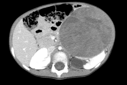
Figure 1. Axial CT image revealed a massive tumor of the left kidney
The patient received neoadjuvant chemotherapy per GFA/nephro 2005 protocol with carboplatin and etoposide (6 cycles) because it was considered a stage II with risk of intra-abdominal rupture. This protocol was used because it aligned with institutional practice, and it was adapted to local resources. More recent recommendations were issued by UMBRELLA SIOP-RTSG 2017. Treatment side effects included leukopenia and thrombocytopenia, which were managed with growth factor support. She subsequently underwent nephrectomy with lombo-aortic and mesenteric lymphadenectomy (10 regional nodes were excised). Postoperative recovery was uneventful.
Histology and immunohistochemistry confirmed a blastemal-type Wilms tumor, high-risk, stage II (SIOP 2001 classification), with intra-abdominal rupture.
Two months post-surgery, the infant was in good health. Radiotherapy was performed to the whole abdomen using 3D-conformal radiotherapy (6 MeV photons). The total dose was 12.5 Gy in 10 fractions (1.25 Gy/fraction) over 16 days (April 14–May 5, 2023).
The CTV encompassed the entire abdominal cavity, and the PTV was defined by adding a 1cm margin to the CTV. Dosimetric planning involved two anteroposterior fields and one right lateral field to spare the right kidney.
Dose constraints were respected: right kidney (the contralateral kidney): Dmean = 11.4 Gy, V12=0, V15 = 0. Liver Dmean = 0 Gy. Dmax spinal cord: 12 Gy and homogeneous dose distribution across bone marrow. (Figure 2)
The treatment was interrupted at the fifth radiotherapy session due to febrile neutropenia and grade 2 anemia, for which the patient was hospitalized and received a platelet transfusion and antibiotics. Another side effect was localized erythema. Following radiotherapy, the patient received adjuvant chemotherapy and remained without evidence of relapse.
Figure 2. Dosimetry image of our patient showing the coverage of the different volumes
3. Discussion
Wilms tumor usually occurs in children < 5 years of age, but it may sometimes be diagnosed in older children and exceptionally in adults. It accounts for nearly 4% of cancers in children < 15 years. Synchronous and bilateral tumors are observed in nearly 5% of patients (1,2).
It is most often revealed by a random mass syndrome. More rarely, hematuria (20%) or a cracking or tumoral rupture with acute abdomen occurs (2).
The workup includes clinical abdominal examination, followed by ultrasound and MRI. The definitive diagnosis is based on the anatomo-pathological examination.
In Europe, according to the guidelines of the International Society of Pediatric Oncology (SIOP), needle biopsy is not systematically done unless the child is over 10 years old. Between 6 months and 9 years, the child is treated with a standard preoperative chemotherapy regimen (3).
Radiological imaging is of fundamental importance in the diagnosis of renal tumors, particularly to identify children who may benefit from initial surgical treatment or to indicate a biopsy for preoperative histopathological confirmation before starting cytotoxic treatment. However, there are no pathognomonic imaging results that clearly differentiate Wilms tumors from other renal tumors, nor among the heterogeneous group of Non-Wilms Renal Tumors (4,5). MRI or high-quality computed tomography can reveal important features to help predict histological diagnosis, in combination with clinical features, and to inform the choice of initial therapeutic approaches (e.g., preoperative empirical chemotherapy, biopsy, etc.) (5).
In our case, the UMBRELLA protocol was followed: chemotherapy first, followed by surgery, radiation, and adjuvant chemotherapy.
The UMBRELLA protocol continues to recommend actinomycin D and vincristine for preoperative use in newly diagnosed patients with Wilms tumor aged 6 months, based on the results of the SIOP trials, which showed a reduction in tumor size with these agents (6,7). This benefit was also independently observed in the UKW3 randomized controlled trial conducted by the UK Children’s Cancer and Leukemia Group (UKCCLG) (8).
After preoperative chemotherapy, radical nephrectomy is the standard of care for children with Wilms tumor according to the UMBRELLA protocol, which specifies surgical guidelines and emphasizes the importance of collecting a minimum of 7 local lymph nodes for precise staging (9,10).
The reason for nephrectomy in infants at first is that a greater proportion of renal tumors in this age group are mesogenic congenital tumors, congenital mesoblastic nephroma, or malignant rhabdoid tumors that require surgery alone. (11,12)
Complete abdominal radiation therapy is indicated in the following cases: intermediate risk or high risk tumors with a major tumor rupture (visible on imaging or during surgery), preoperative or peroperatory tumor rupture, or macroscopic peritoneal deposits, according to the scientific societies: SIOP–RTSG, National Comprehensive Cancer Network 2024 (NCCN), and French Radiation Oncology Society (SFRO) (13-15).
Postoperative chemotherapy for Wilms tumor is similar in infants to that of older children who have had a nephrectomy, with drug doses adjusted according to age and body weight, based on the experience of previous SIOP studies (12).
It should be noted that in children with a single kidney or who have received nephrotoxic radiation or chemotherapy, long-term monitoring of liver function is essential, particularly when the liver has been exposed to significant doses of radiation therapy or hepatotoxic agents. Monitoring height, weight, and pubertal development is an essential part of follow-up, as multimodal treatments can disrupt bone, muscle, or hormonal growth.
4. Conclusion
Treatment strategies for very young children with Wilms’ tumor are based on a carefully tailored combination of chemotherapy, surgery, and, less commonly, radiation therapy. Chemotherapy is generally reduced to limit renal, hepatic, and bone marrow toxicity, while surgery aims to remove the tumor while preserving as much of the renal parenchyma as possible. Radiotherapy is only used when strictly necessary due to its potential impact on growth and developing organs. The overall goal is to maintain optimal oncological efficacy while minimizing long-term effects.
The prognosis is generally favorable thanks to modern therapies, especially when the disease is detected and treated at an early stage. Regular follow-up is essential to monitor for recurrence or complications.
Abbreviations
3D-conformal – Three-Dimensional Conformal (Radiotherapy)
CT – Computed Tomography
CTV – Clinical Target Volume
Dmax – Maximum Dose
Dmean – Mean Dose
GFA – Groupement Français d’Action (French Pediatric Oncology Group protocol)
Gy – Gray (unit of absorbed radiation dose)
MeV – Megaelectron-volts (unit of photon energy)
MRI – Magnetic Resonance Imaging
NCCN – National Comprehensive Cancer Network
PTV – Planning Target Volume
SFRO – Société Française de Radiothérapie Oncologique (French Radiation Oncology Society)
SIOP – International Society of Paediatric Oncology
SIOP–RTSG – International Society of Paediatric Oncology – Renal Tumor Study Group
UKCCLG – UK Children’s Cancer and Leukaemia Group
UMBRELLA – International protocol for childhood renal tumor treatment
V12 / V15 – Volume of the organ receiving at least 12 Gy / 15 Gy
Statements
Authors’ Contributions: HB contributed to the interpretation of the results and drafted the manuscript. SK provided final approval. All authors provided essential comments and contributed to the development of the research, analysis, and manuscript.
Consent for publication: As a corresponding author, I confirm that the manuscript has been read and approved for submission by all the authors listed.
Conflicts of Interest: The authors declare no conflicts of interest.
Funding sources: None.
Ethical approval: The manuscript does not contain studies conducted on humans or animals., therefore ethical approval was not asked.
Written informed consent for publication Informed consent was obtained from the patient for the publication of this case report and accompanying images.
References
-
Louis DN, Perry A, Wesseling P, et al. The 2021 WHO classification of tumors of the central nervous system: a summary. Neuro Oncol. 2021;23(8):1231-51. doi:10.1093/neuonc/noab106.
-
d’Andon A, Vassal G, Hartmann O, Couanet D, Pein F. Wilms tumor (nephroblastoma). Villejuif: Institut Gustave-Roussy; [date unknown].
-
Watson T, Oostveen M, Rogers H, Pritchard-Jones K, Olsen Ø. The role of imaging in the initial investigation of paediatric renal tumours. Lancet Child Adolesc Health. 2020;4(3):232-41. doi:10.1016/S2352-4642(19)30340-2.
-
Ayaz E, Ozcan HN, Oguz B, et al. Beyond Wilms tumor: imaging findings and outcomes of rare renal tumors in children. Pediatr Radiol. 2022;52(13):2557-67. doi:10.1007/s00247-022-05422-7.
-
Watson T, Oostveen M, Rogers H, Pritchard-Jones K, Olsen Ø. The role of imaging in the initial investigation of paediatric renal tumours. Lancet Child Adolesc Health. 2020;4(3):232-41. doi:10.1016/S2352-4642(19)30340-2. (Note: This is a duplicate of Ref 3; check if a different paper was intended).
-
Weirich A, et al. Survival in nephroblastoma treated according to the trial and study SIOP-9/GPOH with respect to relapse and morbidity. Ann Oncol. 2004;15(5):808-20.
-
de Kraker J, et al. Reduction of postoperative chemotherapy in children with stage I intermediate-risk and anaplastic Wilms tumour (SIOP 93–01 trial): a randomised controlled trial. Lancet. 2004;364(9441):1229-35.
-
Mitchell C, et al. Immediate nephrectomy versus preoperative chemotherapy in the management of non-metastatic Wilms’ tumour: results of a randomised trial (UKW3) by the UK Children’s Cancer Study Group. Eur J Cancer. 2006;42(15):2554-62.
-
Shamberger RC, et al. Surgery-related factors and local recurrence of Wilms tumor in National Wilms Tumor Study 4. Ann Surg. 1999;229(2):292-7.
-
Kieran K, et al. Lymph node involvement in Wilms tumor: results from National Wilms Tumor Studies 4 and 5. J Pediatr Surg. 2012;47(4):700-6.
-
van den Heuvel-Eibrink MM, et al. Characteristics and survival of 750 children diagnosed with a renal tumor in the first seven months of life: a collaborative study by the SIOP/GPOH/SFOP, NWTSG, and UKCCSG Wilms tumor study groups. Pediatr Blood Cancer. 2008;50(6):1130-4.
-
Brok J, Treger TD, Gooskens SL, van den Heuvel-Eibrink MM, Pritchard-Jones K. Biology and treatment of renal tumours in childhood. Eur J Cancer. 2016;68:179-95.
-
van den Heuvel-Eibrink EM, Hol JA, Pritchard-Jones K, van Tinteren H, Furtwängler R. Justification of Wilms tumor treatment in the UMBRELLA SIOP–RTSG 2016 protocol. International Society of Pediatric Oncology — Renal Tumor Study Group (SIOP–RTSG); 2016.
-
National Comprehensive Cancer Network. Wilms Tumor (Nephroblastoma) (Version 1.2024). NCCN Clinical Practice Guidelines in Oncology. Plymouth Meeting: NCCN; 2024.
-
Société Française de Radiothérapie Oncologique (SFRO). Pediatric radiotherapy procedures guide: neuroblastomas and nephroblastomas. Paris: SFRO; [accessed on September 30, 2025].