


Author(s) :
Sanyamita Jain1, Piyush Kumar1, Navitha Silambarasan1
1 Shri Ram Murti Smarak Institute of Medical Sciences, Bareilly, Up, India
Corresponding author: Piyush Kumar, Email: piykumagr@gmail.com
Publication History: Received - May 1, 2025, Revised - August 14, 2025, Accepted - August 31, 2025, Published Online - August 31, 2025.
Copyright: © 2025 The author(s). Published by Casa Cărții de Știință.
User License: Creative Commons Attribution – NonCommercial (CC BY-NC)

 PDF
PDF
 Share
Share
Highlights
- Replanning with dose constraints for the temporal lobes and hippocampi significantly reduced radiation exposure to these structures in nasopharyngeal carcinoma patients treated with radiotherapy.
- The study showed that it is feasible to spare these brain regions without compromising the radiation dose to the tumor or other organs at risk.
- The authors recommend the routine contouring of the temporal lobes and hippocampi in treatment planning to potentially reduce neurocognitive side effects in patients undergoing radiotherapy for nasopharyngeal carcinoma.
Abstract
Background: The temporal lobe and the hippocampus can receive high radiation doses due to their proximity to the target volumes in nasopharyngeal cancers radiation therapy. However, they are infrequently delineated during the planning process. The aim of this study was to retrospectively analyze doses received by these structures and compared with replanning.
Material and methods: Twelve NPC patients treated between 2017–2023, were included. Temporal lobes and hippocampi were retrospectively contoured and replanned. Dose constraints were applied: temporal lobe—Dmax< 69 Gy, D1cc< 58 Gy, D2cc< 60.3 Gy, V45< 15.1 cc, V70< 1.13 cc; hippocampus—Dmax< 16 Gy, D100%< 9 Gy, D40%< 7.3 Gy.
Results: The study included 12 patients. The analysis of temporal lobe doses revealed that (pre- vs. post-replanning) Dmax <69Gy was achieved in 2 vs. 11 patients (62.9–73.3 vs. 62.5–70.5 Gy, p=0.09), D1cc <58 Gy achieved in 3 vs. 9 patients (51-68.3 Gy vs 50.2 -59.1.3 Gy ,p=0.01), D 2cc <60.3 Gy achieved in 6 vs. 11 (51.6-70.4Gy vs 49.6-64.09Gy)p=0.02), V45 achieved in 9 vs. 12 (5.8-25.3 vs 4.3-11.6, p=0.01), V70 achieved in 11 vs. 12 (0-1.4Gy vs 0-0.0001Gy p=0.009). Amongst hippocampal dosimetric parameters (right/left) D100%< 9 Gy could be achieved in 1 (8.9–17.2Gy/ 5.5–18.7Gy ,both sides) vs. 5 patients (5.7–10.2 /4.7–12.0.7Gy; p=0.02, p=0.04); Dmax < 16 Gy: 0 (26 –58.2Gy /26- 72.9Gy) vs. 2 (right, 14.9 –30.13 Gy; p<0.001) and 1 (left, 15.3 –33.2Gy, p<0.001) after replanning ; D40%< 7.3 Gy: 0 (17.4-34.8/ 14.1-39.2 Gy, p=0.04) vs. 1 (7-14.9/6.4-22.5, p<0.001 ) after replanning. PTV and OAR parameters remained clinically acceptable after replanning.
Conclusion: Replanning significantly reduced radiation exposure to the temporal lobes and hippocampi without compromising target coverage. Routine contouring of these structures is recommended to minimize neurocognitive toxicity in NPC patients undergoing chemoradiation.
1. Introduction
Radiotherapy, along with concurrent chemotherapy, represents the mainstay of management in nasopharyngeal carcinoma (NPC). Intensity modulated radiotherapy (IMRT) shows obvious advantages over conventional radiotherapy by producing highly conformal dose distributions. However, due to the anatomical proximity between the nasopharynx and the cerebrum, dosimetric “hot spots” can occur in temporal lobes (TLs), even when using conformal techniques such as IMRT. Temporal lobe necrosis (TLN) a relatively common in NPC, being reported in various percentages (1–56%) in long-term survivors. The risk is particularly higher for those with skull-base or intracranial invasion (1).
Hippocampal sparing is usually done in cases of metastatic brain tumors treated with whole-brain radiation therapy (WBRT), as radiation-induced injury to the hippocampus leads to deficits in learning, memory, and spatial processing (2). In one study, deterioration of memory function occurred in 30%–60% patients with primary brain tumors 8–18 months after cranial irradiation (3). Irradiation of the brain parenchyma results in hypoxic injury, especially affecting the CA-1 sub-region of the hippocampus, which is crucial for memory formation and recall. The neural stem cell component of the hippocampus may explain its susceptibility to ionizing radiation damage and subsequent neurocognitive decline (4,5).
The RTOG 0933 study demonstrated that WBRT using IMRT with hippocampal sparing was associated with improvement of memory loss (6). Hippocampal sparing has been primarily studied in primary brain tumors and brain metastasis (6), as well as in nasopharyngeal (7,8), maxillary sinus (9), pituitary (10), oropharyngeal (11), and base-of-skull tumors (13). In our department, a previous study (14) compared the feasibility of hippocampal sparing in treating brain tumors with three-dimensional conformal radiotherapy (3D-CRT) and IMRT, and their effects on NCF, found that the anatomical location of the tumor significantly influences the decision to implement hippocampal sparing.
Hippocampal sparing is not routinely practiced in cases of nasopharyngeal cancer. Lowering the radiation dose to the hippocampus could help preserve the neurocognitive capacity and contribute to an improved quality of life.
Our study aimed to evaluate the feasibility of temporal lobe and hippocampal sparing IMRT in nasopharyngeal carcinoma patients treated with chemoradiation.
2. Materials and methods
2.1. Study setting and description
This retrospective observational study was conducted at the Department of Radiation Oncology, Shri Ram Murti Institute of Medical Sciences. We included in the study patients with carcinoma nasopharynx, treated from 2017 to 2023 using IMRT. The prescribed radiation dose was 70 Gy in 35 fractions, 2 Gy/fraction, five days a week, along with concurrent chemotherapy (cisplatin 35 mg/m2).
2.2. Target volume delineation and organs at risk
A CT scan was done for radiotherapy planning, with an axial CT slice thickness of 3 mm. Target volume delineation was performed according to guidelines by Lee et al (15). All patients were treated according to the same protocols as recommended by the International Commission on Radiation Units and Measurements Reports (ICRU) Report 83 (16).
2.3. Dose prescription and Organ at Risk evaluation
The planning objective was a minimum dose of 95% and a maximum dose of 107% relative to the prescribed dose. The doses that the PTV and OAR received were determined from cumulative dose–volume histograms.
The following OAR dosimetric constraints were prescribed at the time of treatment planning: PRV Brainstem: Dmax <54 Gy; PRV spinal cord: Dmax<50 GY; mandible: Dmax<70 Gy; optic nerve: Dmax<54 Gy; parotid gland: Dmean<26 Gy; PRV cochlea: Dmean<50 Gy as per QUANTEC guidelines (17).
2.4. Temporal Lobe and Hippocampal delineation and prescription for IMRT planning
We contoured the temporal lobe bilaterally, in a single volume according to guidelines by Sun et al (18) and set the following dose constraints: Dmax < 69Gy (19), D 1cc< 58Gy (20) , D 2cc <60.3cc (21), V70<1.13cc (22)
For the hippocampus we used the guidelines by Scoccianti et al. (22) and contoured in selected patients. We then determined whether the following dose constraints were achieved: Dmax<16 Gy, D100%<9 Gy, dose to 40% of the hippocampal volume<7.3 Gy (23).
2.5. Radiotherapy planning
Dose evaluation to the hippocampus and temporal lobe was made in previously planned and delivered IMRT patients (Plan A). All patients were then replanned by incorporating temporal lobe and hippocampal dose constraints in the planning process (Plan B) The dosimetric parameters were then compared between the two groups. For planning and contouring, we used Varian Eclipse version 13.6 Treatment Planning System. Dosimetric calculations were done using the anisotropic analytical algorithm.
2.6. PTV Dosimetric parameters
The PTV dosimetric parameters evaluated to assess plan quality included V57, Dmax, Dmean, D2%, D50%, D98%, pConformity index (pCI) (24), and homogeneity index (HI).
Homogeneity index
HI was calculated using the following formula:
\( HI = \frac{{D_{2\%} – D_{98\%}}}{{D_{50\%}}} \)
where D2%, D50%, and D98% are the absolute dose delivered to 2%, 50%, and 98% of the PTV, respectively. An HI value of zero indicates a homogeneous distribution.
Conformity index
Conformity index (CI) is a measure of the degree of conformity of the absorbed dose distribution to the PTV. In this work, Paddick’s CI was used for evaluation:
\text{PCI} = \frac{(TV_{PIV})^2}{TV \times PIV}
where TVPIV, TV, and PIV are the prescribed isodose volume over the target volume, the target volume, and the prescription isodose volume, respectively.
2.7. Statistical analysis
Statistical significance was determined using the unpaired t-test, where a p-value of <0.05 was considered statistically significant.
3. RESULTS
The study included a total of 12 patients, mostly male, staged T2-3 N+ (Table 1). At the time of initial treatment planning (Plan A), dose coverage of the PTV was optimal and dose constraints for OARs were met for all patients.
Table 1: Patient characteristics of the study population
| Patient Characteristics | Value
(N=12) |
% |
| Sex | ||
| Male | 11 | 91.7 |
| Female | 1 | 8.3 |
| T stage | ||
| T1 | 2 | 16.7 |
| T2 | 4 | 33.3 |
| T3 | 6 | 50 |
| T4 | 0 | 0 |
| N stage | ||
| N0 | 4 | 33.3 |
| N1 | 2 | 16.7 |
| N2 | 3 | 25 |
| N3 | 3 | 25 |
3.1. Replanning impact on doses received by the temporal lobe
After contouring the temporal lobe, it was possible to achieve lower doses while preserving target coverage and without overpassing dose constraints for the other organs at risk for more patients. A comparison of the values of the two plans for each patient is presented in Table 2.
Table 2: Temporal lobe dosimetric parameters before (Plan A) and after replanning (Plan B) for each patient.

Dmax=maximum dose, D1cc= dose received by 1cc volume, D2cc= dose received by 2cc volume, V70=volume receiving 70 Gy dose; Dose constraints: Dmax<69Gy, D1cc<58 Gy, <60.3 Gy, V70<1.13cc; light blue = constraints met, dark blue = not achieved constraints.
When comparing dosimetric parameters’ mean values in the whole group, Dmax and V70 were lower after replanning, but the statistical significance was not met. However, D1cc and D2cc were significantly decreased in the new irradiation plans (Table 3).
Table 3. Mean temporal lobe dosimetric parameters patients before (Plan A) and after replanning (Plan B).
| Temporal Lobe Dosimetric Parameter | Plan A | Plan B | p-value | ||
| Mean | SD | Mean | SD | ||
| DMax | 68.5 | 5.7 | 64.8 | 4.4 | 0.09 |
| D1cc | 61.8 | 8.48 | 53.8 | 5.3 | 0.01 |
| D2cc | 59.3 | 9.39 | 51.5 | 6.5 | 0.02 |
| V70 | 0.29 | 0.49 | 0.01 | 0.05 | 0.09 |
Dmax=maximum dose, D1cc= dose received by 1cc volume, D2cc= dose received by 2cc volume, V70=volume receiving 70 Gy dose, and SD=standard deviation
3.2. Replanning impact on doses received by the hippocampus
After contouring the right and left hippocampus, it was possible to achieve lower doses while preserving target coverage and without overpassing dose constraints for the other organs at risk for more patients. A comparison of the values of the two plans for each patient is presented in Table 4.
Table 4: Hippocampus dosimetric parameters before replanning (Plan A) and after replanning (Plan B) for each patient

D100%= dose received by 100% volume, Dmax= dose received by 1cc volume, D40%= dose received by 40% volume,
Dose constraints: D100%<9Gy, Dmax<16 Gy, D40%<7.3 Gy
light blue = constraints met, dark blue = not achieved constraints.
When comparing dosimetric parameters’ mean values in the whole group, D100% was lower after replanning, however it was more difficult to achieve Dmax and D40%. It was also noted that all the values upon replanning were much lower and highly statistically significant.(Table 5).
Table 5. Mean Hippocampus dosimetric parameters patients before and after replanning.
| Organ at Risk | Dosimetric Parameter | Mean +/-SD (Before Replanning) | MEAN +/-
SD (After Replanning) |
P value | ||
| Right
Hippocampus |
D100% | 12.5 | 5.3 | 8.64 | 3.8 | 0.02 |
| DMAX | 47.5 | 12.3 | 27.9 | 11.1 | 0.0005 | |
| D40% | 25.9 | 6.9 | 12.8 | 2.3 | 0.04 | |
| Left
Hippocampus |
D100% | 13.3 | 4.5 | 9.4 | 3.75 | 0.04 |
| DMAX | 45.05 | 13.5 | 26 | 9.3 | 0.0006 | |
| D40% | 27.2 | 8.4 | 14.5 | 4.8 | 0.0002 | |
| Combined Hippocampus | D max | 49.3 | 11.7 | 28.04 | 10.09 | 0.0001 |
Dmax=maximum dose, D100%=dose received by 100% volume, within the target volume, D40%=dose received by 40% volume, and SD=standard deviation
3.3. General impact of replanning after contouring the temporal lobe and hippocampus
No significant difference between PTV parameters and the initially considered OAR was observed after replanning, as shown in Supplementary Tables 1 and 2, respectively.
However, safe doses could be obtained in the temporal lobes after contouring and including them as OAR during replanning in more patients compared with the original plan, which was retrospectively analyzed (Figure 1).
Hippocampal sparing parameters were almost never within constraints in the initial plans, except for D40% of less than 7.3 Gy in half of the patients. Hippocampal D100% and Dmax were respected with replanning in almost half of the cases when only one side was spared (Figure 2). However, no constraints could be met when considering the whole hippocampus.

Figure 1. Number of patients for which temporal lobe dosimetric constraints were respected before (Plan A) and after contouring and replanning, respectively (Plan B)
Dmax=maximum dose (<69Gy), D1cc= dose received by 1cc volume (<58 Gy), D2cc= dose received by 2cc volume (<60.3 Gy), V70=volume receiving 70 Gy (<1.13cc);
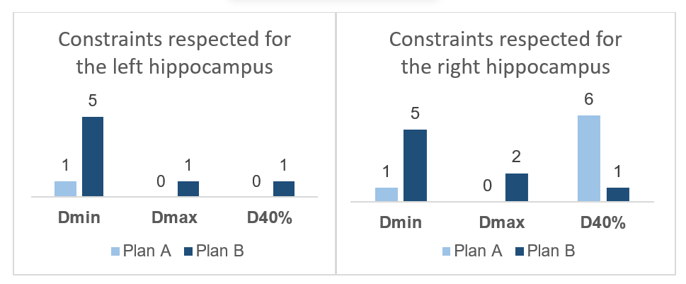
Figure 2. Number of patients for which hippocampus dosimetric constraints were respected before (Plan A) and after contouring and replanning, respectively (Plan B)
D100%=Dose received by 100% volume (<9 Gy) Dmax=maximum dose (<16 Gy), D40%=dose received by 40% volume (<7.3 Gy)
4. Discussion
Temporal lobe and hippocampus are both crucial structures for memory and recall. In our study when we compared dosimetric parameters before and after replanning we found that Dmax and V70 were lower after replanning, but the statistical significance was not met, while D1cc and D2cc were significantly decreased in the new irradiation plans. D1cc and D2cc are volumetric and robust to small calculation noise, ie optimization shrinks these volumes effectively, while Dmax is a single point (or near single -voxel) metric a tiny residual spike from leaf transmission, contouring differences or calculation grid can keep Dmax high even when hotspot volume is trimmed, moreover, Dmax and V70 vary more between patients, the effect size maybe smaller relative to variance which more difficult to reach statistical significance in a small sample size.
It was also found that despite prescribing constraints, it was more difficult to achieve hippocampal dosimetry because it is a smaller volume, so even a tiny hotspot produces a more significant dosimetric change, it also has stricter clinical constraints achieving which may require more complex planning which may be limited by OAR tradeoffs. There were differences between achieved constraints in the left and right hippocampus which could be attributed to differences in the PTV volumes which would be asymmetrical, also depending on beam entry and fluence modulation where the disease is well lateralized, more beam fluence would pass towards that temporal region and the hippocampus would receive a higher scatter dose. The decrease of doses was statistically significant for hippocampus as mean values, but still, dosimetric constraints were achieved in very few patients because hippocampus is a small volume structure, a very small hotspot can increase the Dmax above limit even when the mean dose improves. More so, the variability was statistically significant since the overall dose in all patients decreased upon replanning.
Zhou et al (29) compared the radiation-induced temporal lobe injury in patients with nasopharyngeal carcinoma (NPC) treated with intensity-modulated radiotherapy (IMRT) or two-dimensional conventional radiotherapy (2D-CRT). The actuarial 5-year incidence was 16% and 34.9% (P<0.001) for the IMRT and 2D-CRT groups, respectively. Where both T stage (P<0.001) and radiation technique (P<0.001) were independent predictors, T1, T2, and T3 disease had a significantly higher risk when treated with 2D-CRT (P = 0.005, 0.016, <0.001, respectively). Similarly, Kam et al. (30) compared IMRT with 2D-RT and three-dimensional conformal radiotherapy (3D-CRT) treatment plans, demonstrating the feasibility of temporal lobe sparing and a more conformal dose distribution in IMRT. In our group of 12 patients, temporal Lobe Dmax was less than 69 Gy in 4 patients before contouring the volume and including it in the optimization process and in 9 patients after replanning.
Kong (19) et al. in a cohort, retrospective study including 132 patients, 42 necrotic and 222 normal (temporal lobe) TLs, advised (Tolerance dose 5/5) TD5/5 for temporal lobe Dmax = 69.0 ± 1.6, TD5/5 for D1cc = 62.8 ± 2.2.
Feng et al (22) showed that D2cc of the temporal lobe, fraction size of prescription, T stage, and chemotherapy were the independent predictive factors for TLI. The logistic dose-response model has indicated the TD5/5 and TD50/5 of D2cc are 60.3 Gy and 76.9 Gy, respectively. D2cc of the temporal lobe, fraction size of prescription, T stage, and chemotherapy were the possible independent predictive factors for TLI after IMRT of NPC. Similarly, Lu et al (21). In a Cohort study, retrospective analysis including 188 patients, 94 with necrotic and 282 normal TLs demonstrated AUC for V70 in predicting TLN, prescribed V70<1.13 cc, which were evaluated in this study.
The hippocampus is critical for declarative memory, radiation results in hypoxic injury, which is crucial for memory formation and recall (10). Neurocognitive decline in head-neck cancer due to incidental irradiation is sparingly discussed. The studies by Gondi et al (6,23) demonstrate the neurocognitive decline with hippocampal D40%EQD2 of more than 7.3 Gy and with the hippocampal Dmax exceeding 16 Gy and the D100 exceeding 9 Gy in brain irradiation. Han et al (31) evaluated the dosimetric feasibility of using hippocampus (HPC) sparing intensity-modulated radiotherapy (IMRT) in patients with locally advanced nasopharyngeal carcinoma (NPC). They demonstrated that to achieve the sparing of NPC, PTV Parameters such as HI and CI do not need to be compromised. The dose (Dmax, D2%, D40%, Dmean, Dmedian, D98% and Dmin) and volume (V5, V10, V15, V20, V30, V40 and V50) parameters for the HPC were significantly lower in the HPC sparing plans (p<0.05), except for Dmin (P = 0.06) and V5 (P = 0.12). In our study, we evaluated Dmax, D40%, and D100% in nasopharyngeal cancers during plan evaluation. We had similar results to this previously mentioned study, where, even though in most plans the dose constraints of the Hippocampi were not met, the values were significantly lower in HS plans compared to plans where constraints were given.
Dunlop et al (32) demonstrated the feasibility of generating clinically acceptable and deliverable radiotherapy plans for HNC that either specifically spare the bilateral hippocampi ie Hippocampal Sparing RT (HSRT) or spare the whole brain in addition to the hippocampi, he demonstrated significant reductions in hippocampal doses relative to standard plans were achievable in HSRT which was also seen in our study 5 patients could achieve right hippocampus D100%<9 GY (7.6–19.5 Gy) (p value = 0.02), two patients could achieve Dmax<16 Gy (14.9 –30.13 Gy) (p value = 0.0005), and one patient could achieve D40%<7.3 Gy (7-14.9) (p value = 0.04), five patients could achieve left hippocampus D100%<9 GY (7.0–18.5 Gy) (p value = 0.04), one patient could achieve Dmax<16 Gy (15.9 –33.2 Gy) (p value = 0.0006), and one patient could achieve D40%<7.3 Gy (6.4-22.5) (p value = 0.0002) all which demonstrated highly statistically significant dose reduction when compared the plans where HS was not practiced.
Peternal et al. (33) conducted a systematic literature review comparing hippocampal D40% in conventional and HS RT plans. In four previous studies (n = 79), researchers reduced D40% hippocampal radiation doses in HS plans compared to conventional RT on average from 24.9 Gy to 12.6 Gy. Among 12 NPC patients included in this in silico study, statistically significant differences between HS and conventional VMAT plans were observed in hippocampal EQD2 Dmax (23.8 vs. 46.4 Gy) D100%, (3.8 vs. 4.6 Gy), Dmean (8.1 vs. 15.1 Gy), and D40% (8.3 vs. 15.8 Gy). PTV coverage and OAR doses were similar, with less homogeneous PTV coverage in HS plans (p = 0.038). This translated to a lower probability of memory decline in HS plans (interquartile range 15.8–29.6 %) compared to conventional plans (33.8–81.1 %) based on the NTCP model (p = 0.002).
In our study, a drastic reduction in EQD2 Dmax (47.5 vs. 27.9 Gy), D100% 12.5 vs. 8.6 Gy), and D40% (25.9 vs. 12.8 Gy) was noted for right hippocampus, EQD2 Dmax (45.05 vs. 26Gy), D100% (13.3 vs. 9.4 Gy), and D40% (27.2 vs. 14.5.8 Gy) were noted for left hippocampus, all of which were statistically significant.
The strength of the study is that it highlights the hippocampus and Temporal Lobe as OARs in patients with Nasopharyngeal cancer, where it is not routinely delineated. It emphasizes that when delineated and dose constraints are included in the planning process, hippocampal and temporal lobe sparing is feasible.. This can potentially decrease the risk of neurocognitive decline and thereby improve the quality of life.
The limitations of this study include a small sample size (12 patients) and a lack of clinical or radiological follow-up, which would help establish a clinical correlation. The small sample size could be due to the rarity of the condition being studied or resource constraints. The lack of follow-up could be attributed to logistical challenges. The findings from a small cohort may not be generalizable to the broader population of patients with nasopharyngeal carcinoma, and the results are to be interpreted with caution. Larger studies are needed to confirm these results and ensure they apply to diverse patient populations.
A prospective study with a larger sample size and comprehensive follow-up protocols, along with baseline neurocognitive function testing, will further help strengthen the evidence base and will help in generalization of the results and ensure they apply to diverse patient populations.
5. Conclusion
Considering temporal lobe and hippocampus as OARs in nasopharygeal cancer patients significantly decrease the received doses to these volumes without compromising target coverage or increasing the dose to other OARs. Temporal lobe dose limits seem easier to achieve compared to hippocampus. Therefore, we conclude that both the hippocampus and the temporal lobe should be routinely delineated, and dose constraints should be given to both these neural structures in Nasopharyngeal carcinoma patients undergoing radiotherapy.
ABBREVIATIONS
2 – DCRT two – dimensional conventional radiotherapy
3 – DCRT – three-dimensional conformal radiotherapy
AAA – anisotropic analytical algorithm
CI – conformity index
CTV – clinical target volume
Dmin – Minimum Dose
D100% – dose received by 100% volume
Dmax – maximum dose
D40% – Dose received by 40% of the volume
D1cc – dose received by 1cc volume
D2cc – dose received by 2cc volume
ENE – Extra Nodal Extension
HI – homogeneity index
HSRT – Hippocampal Sparing Radiotherapy
HS – Hippocampal Sparing
ICRU – International Commission on Radiation Units and Measurements Reports
IMRT – intensity – modulated radiation therapy
NCF – neurocognitive function
OAR – organ at risk
TV – planning target volume
PRV – Planning Risk Volume
QUANTEC – Quantitative Analyses of Normal Tissue Effects in the Clinic
SD – standard deviation
TPS – Treatment Planning System
NPC – nasopharyngeal carcinoma
TL – temporal lobe
TLN Temporal lobe necrosis
TD5/5 – Tolerance dose 5/5
VMAT – Volumetric Modulated Arc Therapy
V70 – volume receiving 70 Gy dose
WBRT – whole-brain radiation therapy
STATEMENTS
Authors contributions: Concepualization: PK, SJ; Methodology: PK, SJ; Data curation: SJ, NS; Formal Analysis: PK, SJ; Writing and editing: SJ
Funding: None.
Conflicts of Interest: The authors have no conflicts of interest to declare.
REFERENCES
- Zhou X, Liu P, Wang X. Temporal lobe necrosis following radiotherapy in nasopharyngeal carcinoma: new insight into the management. Front Oncol. 2021;10:593487. doi:10.3389/fonc.2020.593487.
- Abayomi OK. Pathogenesis of irradiation-induced cognitive dysfunction. Acta Oncol. 1996;35:659-63. doi:10.1080/028418602760169389.
- Meyers CA, Brown PD. Role and relevance of neurocognitive assessment in clinical trials of patients with CNS tumors. J Clin Oncol. 2006;24:1305-9. doi:10.1200/JCO.2005.04.6086.
- Monje ML, Mizumatsu S, Fike JR, et al. Irradiation induces neural precursor-cell dysfunction. Nat Med. 2002;8:955-62. doi:10.1038/nm749.
- Mizumatsu S, Monje ML, Morhardt DR, et al. Extreme sensitivity of adult neurogenesis to low doses of X-irradiation. Cancer Res. 2003;63:4021-7.
- Gondi V, Mehta MP, Pugh S, et al. Memory preservation with conformal avoidance of the hippocampus during whole-brain radiation therapy for patients with brain metastases: primary endpoint results of RTOG 0933. Int J Radiat Oncol Biol Phys. 2013;87:1186. doi:10.1200/JCO.2014.57.2909.
- Lee PW, Hung BK, Woo EK, et al. Effects of radiation therapy on neuropsychological functioning in patients with nasopharyngeal carcinoma. J Neurol Neurosurg Psychiatry. 1989;52:488-92. doi:10.1136/jnnp.52.4.488.
- Leung SF, Kreel L, Tsao SY. Asymptomatic temporal lobe injury after radiotherapy for nasopharyngeal carcinoma: incidence and determinants. Br J Radiol. 1992;65:710-4. doi:10.1259/0007-1285-65-776-710.
- Sakata K, Aoki Y, Karasawa K, et al. Analysis of the results of combined therapy for maxillary carcinoma. Cancer. 1993;71:2715-22. doi:10.1002/1097-0142(19930501)71:9<2715::aid-cncr2820710905>3.0.co;2-0.
- Grattan-Smith PJ, Morris JG, Shores EA, et al. Neuropsychological abnormalities in patients with pituitary tumours. Acta Neurol Scand. 1992;86:626-31. doi:10.1111/j.1600-0404.1992.tb05500.x.
- Seol SW, Lee BH, Sita TL, et al. Hippocampus sparing volumetric modulated arc therapy in patients with loco-regionally advanced oropharyngeal cancer. Phys Imaging Radiat Oncol. 2022;24:71-5. doi:10.1016/j.phro.2022.09.008.
- Meyers CA, Geara F, Wong PF, et al. Neurocognitive effects of therapeutic irradiation for base of skull tumors. Int J Radiat Oncol Biol Phys. 2000;46:51-5. doi:10.1016/s0360-3016(99)00376-4.
- Chaturvedi D, Kumar P, Chauhan AK, Kumar P, Nigam J, Sivaji SN, Silambarasan N. Comparison of hippocampal sparing intensity modulated radiotherapy plans in patients of brain tumors treated by three-dimensional conformal radiotherapy. SRMS J Med Sci. 2022;7:22-30. doi:10.21761/jms.v7i01.6.
- Lee AW, Ng WT, Pan JJ, Poh SS, Ahn YC, AlHussain H, et al. International guideline for the delineation of the clinical target volumes (CTV) for nasopharyngeal carcinoma. Radiother Oncol. 2018;126(1):25-36. doi:10.1016/j.radonc.2017.10.032.
- Hodapp N. The ICRU Report 83: prescribing, recording and reporting photon-beam intensity-modulated radiation therapy (IMRT). Strahlenther Onkol. 2012;188:97-9.
- Marks LB, Ten Haken RK, Martel MK. Guest editors introduction QUANTEC: a users guide. Int J Radiat Oncol Biol Phys. 2010;76:S1-2.
- Sun Y, Yu XL, Luo W, Lee AW, Wee JT, Lee N, et al. Recommendation for a contouring method and atlas of organs at risk in nasopharyngeal carcinoma patients receiving intensity-modulated radiotherapy. Radiother Oncol. 2014;110(3):390-7. doi:10.1016/j.radonc.2013.10.035.
- Kong C, Zhu XZ, Lee TF, Feng PB, Xu JH, Qian PD, et al. LASSO-based NTCP model for radiation-induced temporal lobe injury developing after intensity-modulated radiotherapy of nasopharyngeal carcinoma. Sci Rep. 2016;6:26378. doi:10.1038/srep26378.
- Zeng L, Huang SM, Tian YM, Sun XM, Han F, Lu TX, et al. Normal tissue complication probability model for radiation-induced temporal lobe injury after intensity-modulated radiation therapy for nasopharyngeal carcinoma. Radiology. 2015;276:243-9. doi:10.1148/radiol.14141721.
- Lee AW, Ng WT, Pan JJ, et al. International guideline on dose prioritization and acceptance criteria in radiation therapy planning for nasopharyngeal carcinoma. Int J Radiat Oncol Biol Phys. 2019;105(3):567-80.
- Feng M, Huang Y, Fan X, Xu P, Lang J, Wang D. Prognostic variables for temporal lobe injury after intensity modulated-radiotherapy of nasopharyngeal carcinoma. Cancer Med. 2018;7:557-64. doi:10.1002/cam4.1291.
- Lu L, Sheng Y, Zhang G, Li Y, OuYang PY, Ge Y, et al. Temporal lobe injury patterns following intensity modulated radiotherapy in a large cohort of nasopharyngeal carcinoma patients. Oral Oncol. 2018;85:8-14. doi:10.1016/j.oraloncology.2018.07.020.
- Scoccianti S, Detti B, Gadda D, et al. Organs at risk in the brain and their dose constraints in adults and in children: a radiation oncologist’s guide for delineation in everyday practice. Radiother Oncol. 2015;114:230-8. doi:10.1016/j.radonc.2015.01.016.
- Gondi V, Hermann BP, Mehta MP, Tome WA. Hippocampal dosimetry predicts neurocognitive function impairment after fractionated stereotactic radiotherapy for benign or low-grade adult brain tumors. Int J Radiat Oncol Biol Phys. 2013;85:348-54. doi:10.1016/j.ijrobp.2011.10.021.
- Paddick I. A simple scoring ratio to index the conformity of radiosurgical treatment plans. J Neurosurg. 2000;93 Suppl 3:219-22. doi:10.3171/jns.2000.93.supplement. PMID:11143252.
- Lee AW, Ng SH, Ho JH, Tse VK, Poon YF, Tse CC, et al. Clinical diagnosis of late temporal lobe necrosis following radiation therapy for nasopharyngeal carcinoma. Cancer. 1988;61:1535-42. doi:10.1002/1097-0142(19880415)61:8<1535::AID-CNCR2820610809>3.0.CO;2-E.
- Hsu YC, Wang LF, Lee KW, Ho KY, Huang CJ, Kuo WR. Cerebral radionecrosis in patients with nasopharyngeal carcinoma. Kaohsiung J Med Sci. 2005;21:452-9. doi:10.1016/S1607-551X(09)70150-0.
- Belka C, Budach W, Kortmann RD, Bamberg M. Radiation induced CNS toxicity: molecular and cellular mechanisms. Br J Cancer. 2001;85:1233-9.
- Zhou GQ, Yu XL, Chen M, Guo R, Lei Y, et al. Radiation-induced temporal lobe injury for nasopharyngeal carcinoma: a comparison of intensity-modulated radiotherapy and conventional two-dimensional radiotherapy. PLoS One. 2013;8(7):e67488. doi:10.1371/journal.pone.0067488.
- Kam MK, Chau RM, Suen J, Choi PH, Teo PM. Intensity-modulated radiotherapy in nasopharyngeal carcinoma: dosimetric advantage over conventional plans and feasibility of dose escalation. Int J Radiat Oncol Biol Phys. 2003;56(1):145-57. doi:10.1016/s0360-3016(03)00075-0.
- Han G, Liu D, Gan H, Denniston KA, Li S, et al. Evaluation of the dosimetric feasibility of hippocampal sparing intensity-modulated radiotherapy in patients with locally advanced nasopharyngeal carcinoma. PLoS One. 2014;9(2):e90007. doi:10.1371/journal.pone.0090007.
- Dunlop A, Welsh L, McQuaid D, Dean J, Gulliford S, Hansen V, et al. Brain-sparing methods for IMRT of head and neck cancer. PLoS One. 2015;10(3):e0120141. doi:10.1371/journal.pone.0120141.
- Peternel M, Jenko A, Peterlin P, Petrovič L, Strojan P, Plavc G. Comparison of conventional and hippocampus-sparing radiotherapy in nasopharyngeal carcinoma: in silico study and systematic review. Clin Transl Radiat Oncol. 2024;46:100751. doi:10.1016/j.ctro.2024.100751.