


Author(s) :
Simona Natarîș1, Alexandru Iacob2, Bogdan Fetica3
1 Oncology Institute “Prof. Dr. Ion Chiricuta”, Cluj-Napoca, Romania
2 Department of Radiation Oncology, RTC Amethyst, Cluj-Napoca, Romania
3 Pathology Department, Oncology Institute “Prof. Dr. Ion Chiricuta” Cluj-Napoca, Romania
Corresponding author: Simona Natarîș, Email: simona_stir@yahoo.com
Publication History: Received - May 17, 2025, Revised - August 10, 2025, Accepted - August 23, 2025, Published Online - August 23, 2025.
Copyright: © 2025 The author(s). Published by Casa Cărții de Știință.
User License: Creative Commons Attribution – NonCommercial (CC BY-NC)

 PDF
 Share
Highlights
- Neuroendocrine endometrial cancer is a rare and aggressive form of uterine cancer that presents particular diagnostic and treatment challenges.
- The definitive diagnosis of mixed endometrioid adenocarcinoma and small cell neuroendocrine carcinoma (SCNEC) was only made after a second-opinion review, which proved the absence of the specific EWSR1 gene rearrangement characteristic of PNET.
- A specialized pathological review and advanced diagnostic techniques like FISH, increase the probability of an accurate diagnosis, which is mandatory for guiding appropriate treatment and predicting patient outcomes.
Abstract
Small cell neuroendocrine carcinoma (SCNEC) of the endometrium is a rare, high-grade neuroendocrine neoplasm with aggressive biological behavior. It commonly coexists with endometrioid carcinoma and is often misdiagnosed as undifferentiated carcinoma, carcinosarcoma, primitive neuroectodermal tumor (PNET), or carcinoid tumor due to similar morphological features.
We report the case of a 52-year-old female initially diagnosed with a primitive neuroectodermal tumor not otherwise specified (PNET-NOS) associated with endometrioid carcinoma. The patient underwent surgical resection followed by adjuvant chemotherapy; however, the disease progressed during treatment. A second pathological review was requested, leading to the diagnosis of mixed endometrial adenocarcinoma with a small cell neuroendocrine carcinoma component, FIGO stage IIIC2. This final diagnosis was based on the distinct pathological and immunohistochemical findings, along with the absence of EWSR1 gene rearrangement on fluorescence in situ hybridization (FISH) analysis, effectively excluding PNET.
The patient is currently receiving second-line treatment for progressive disease.
1. Introduction
Endometrioid cancer is one of the most common cancers in females worldwide and accounts for 7% of new cancer diagnoses and 4% of cancer deaths (2). The most common subtypes of endometrial cancer are endometrioid and serous carcinomas.
Primitive neuroectodermal tumors (PNETs) belong to a spectrum of diseases known as the Ewing family of tumors, characterized by small round cell tumors of neuroectodermal origin. PNETs of the genital tract are rare, with only around 70 cases published in the literature (1).
Small cell neuroendocrine carcinoma (SCNEC) is a particular histological subtype that accounts for 0.8% of endometrial carcinomas, and coexists with endometrioid carcinoma and atypical endometrial hyperplasia (2, 3).
In retrospective studies of neuroendocrine endometrial carcinoma (NEC) cases, 89% were reclassified, indicating substantial diagnostic disagreement with the original assessments. It is often misinterpreted as undifferentiated carcinoma, a sarcomatoid component of carcinosarcoma, PNET, and carcinoid tumor (5).
This case report illustrates such diagnostic challenges: the initial histopathological interpretation suggested a mixed uterine tumor (PNET associated with endometrial carcinoma), but the final diagnosis was revised to mixed endometrial adenocarcinoma with a component of SCNEC.
2. Case presentation
In October 2024, a 52-year-old Romanian female patient presented to her general practitioner with post-menopausal vaginal bleeding. Her obstetric history included one pregnancy and one birth, with menopause occurring nine years prior. She had no personal or family history of gynecological or oncological diseases.
Physical exam revealed a mobile uterus measuring approximately 8 cm, free adnexal areas, without cervical lesions or other lesions detectable by digital rectal examination. An endometrial curettage was performed, showing a grade III endometrioid endometrial carcinoma (EEC). The magnetic resonance imaging (MRI) report revealed a uterine mass of 6/9/7 mm CC/AP/LL and two enlarged internal iliac lymph nodes (LNs) measuring 2,8 /2,6/2 cm and 3,3/1,4/2,3 cm, respectively. There was no evidence of extra-uterine spread. A thoracoabdominal CT scan showed no suspicious secondary lesions.
The patient underwent total abdominal hysterectomy with bilateral adnexectomy, and bilateral pelvic, obturator and para-aortic lymphadenectomy. The intervention was performed in an oncology institute by a surgeon specialized in oncological-gynecological pathology. The postoperative course was uneventful, and the patient was discharged shortly after the surgery with recommendations for outpatient follow-up.
Ten days after surgery, the patient presents to the emergency department with right flank abdominal pain. An abdomino-pelvic CT scan is performed, revealing grade I/II ureterohydronephrosis associated with lithiasis. A CT urogram confirmed right ureteral lithiasis and left renal lithiasis. Symptoms were relieved after ureteral stenting in the Urology Department. Biological markers were tested, revealing high CA125 levels 72.5 U/mL (nv 0-35u/mL). No baseline CA125 levels were available for comparison.
Complete histopathological and immunohistochemical report (Table 1) concluded that the tumor was a primitive neuroectodermal tumor NOS (PNET-NOS) associated with a high-grade endometrioid endometrial carcinoma. The tumor was invading less than half of the myometrium, displaying lymphatic invasion, but no vascular or perineural invasion. It was staged as pT1a N1 L1 V0 Pn0 R0. The parametrium and adnexal structures were free of malignancy. A total of 31 LNs were excised, of which 8 presented PNET metastases: four of 24 bilateral iliac nodes and four of the seven para-aortic nodes.
Two months after surgery, the patient was referred for adjuvant external beam radiotherapy. Postoperative imaging assessment included a brain MRI and a CT scan of the chest, abdomen, and pelvis. No brain metastases were found. However, two lesions measuring 17 mm and 28 mm in the right external and common iliac regions, respectively, were identified. Both had minimal contrast enhancement, raising differential considerations between adenopathy and lymphocele. In parallel, tumor marker CA125 showed an increasing trend (110 U/mL).
PET/CT was performed to assess disease extension accurately, confirming progressive disease with FDG-avid LNs, including: adenopathy of 24 mm interaortocaval and 22mm retrocaval (SUVmax 8.78), left lateral aortic adenopathy of maximum 21.4mm, left common and internal periiliac adenopathy of up to 16mm (SUV 3.12), periobturator and right external periiliac adenopathy of up to 30mm (SUVmax 4.60).
Based on these findings, the multidisciplinary team recommended adjuvant chemotherapy (Carboplatin + Etoposide) and external beam radiation therapy (EBRT) to the pelvis and para-aortic nodes (45 Gy/ 25 fractions) with an additional simultaneous integrated boost (SIB) up to 55-57.5Gy in 25 fractions. The EBRT should have been followed by intravaginal brachytherapy (15 Gy/ 3 fractions).
Additionally, considering the rare histological subtype and the aggressive behavior, a second pathological opinion was requested to confirm the diagnosis. The second pathological result was received during adjuvant treatment.
The patient received six cycles of adjuvant chemotherapy with Carboplatin AUC5 (total dose: 600mg) + Etoposide 100mg/ mp (total dose=190 mg), which were well tolerated, without acute toxicity. Upon completion of the first line systemic treatment, a follow-up PET-CT showed progressive disease. An inter-aorto-caval, retrocaval, and left latero-aortic adenopathic conglomerate measuring 58/33/39mm, highly FDG-avid, was identified. Peri-obturator and right external iliac adenopathies were noted, measuring up to 26/30mm, and were also metabolically active (SUV 6.14). External radiotherapy was not performed due to the lack of response to systemic treatment, with an increased risk of distant micrometastases outside the irradiation field. Therefore, the multidisciplinary team recommended 2nd line systemic treatment with Topotecan.
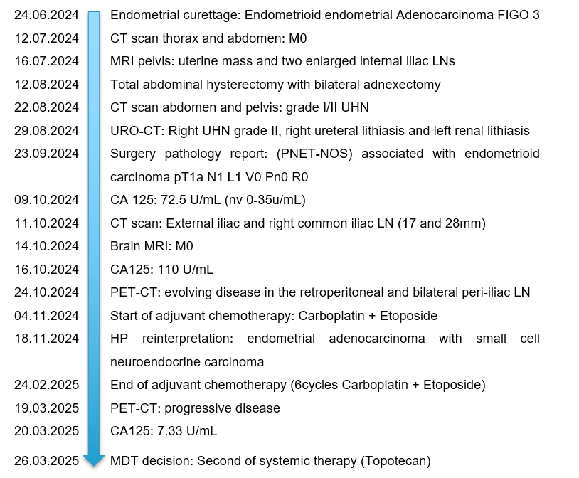
Figure 1. Timeline of the disease and the received therapy
PNET-NOS – Primitive neuroectodermal tumor; UHN – ureterohydronephrosis, LN – lymph nodes
During the first-line systemic treatment, the reinterpretation of the resection specimen performed by an oncology-expert pathologist described malignant tumor proliferation composed of epithelioid-like cells arranged in insular patterns along with small cells with basophilic nuclei with numerous mitoses and apoptotic features without apparent differentiation (Figures 2 and 3). The tumor was infiltrating the inner half of the myometrium (5 mm out of 15 mm).

Figure 2. Small cell neuroendocrine carcinoma component (a- x4, b- x10, c-x20).
Figure 3. HE staining (a), and immunostaining with synaptophysin (b), chromogranin (c), and CD 56 (d) as markers for neuroendocrine tumors, all positive in the SCNEC component.
The immunohistochemical profile is summarized in Table 1, compared to the IHC profile of the first histopathological result.
Table 1. Comparative IHC profile of the two tumor types
| 1st IHC profile-PNET | 2nd IHC profile-SCNEC | |
| Synaptophysin; Chromogranin;
FLI1; CD99. |
Diffuse positive on the small cell component |
Focally positive
Focally positive
– Positive on the endometrioid adenocarcinoma component and focally on the small cell component |
| CD56;
Calretinin. |
Isolated positive on the small cell component |
Focally positive |
| p16 | Diffuse positive | Diffusely positive |
| GFAP | Positive isolated cells | – |
| PAX8 | Diffuse positive, low intensity on the carcinoma component | Negative on the small cell component |
| CK18 | Diffuse positive on the carcinoma component | focally positive |
| CK AE1/AE3 | Negative on the tumoral cells | focally positive |
| p53;
ER; CK8; EMA |
Positive isolated on the carcinoma component |
– |
| S100 | ||
| Desmin | ||
| CD10 | ||
| LCA | negative | negative |
| Melan A | ||
| TTF1 | ||
| Cyclin D1 | ||
| Vimentin | – | focally positive |
| Reticulin | – | positive in the peritumoral stroma with delimitation of carcinomatous islands |
FISH testing was performed. Using the ZytoLight SPEC EWSR 1 Dual Color Break Apart Probe to assess the presence of EWSR 1 (22q12) gene rearrangements from a formalin-fixed paraffin-embedded tissue block. A total of 100 tumor cell nuclei from representative areas showed signals for both ends of the gene were evaluated. A cell was considered positive if the two signals for the ends of the genes were separated by a space greater than twice the diameter of the individually measured signals. A threshold of 5% positive nuclei was used to determine EWSR 1 rearrangements. The findings revealed no evidence of EWSR1 rearrangement. Therefore, the morphological appearance correlated with the immunohistochemical profile, and the lack of EWSR 1 rearrangement in FISH analysis corresponds to a mixed endometrial adenocarcinoma with an endometrioid component of 10% and a small cell neuroendocrine carcinoma component on 90% of the surface, Ki 67 80%, pT1N2aL1V0Pn0R0 FIGO III C2.
We are in the setting of a mixed endometrial adenocarcinoma, in which the neuroendocrine component was initially interpreted as PNET-NOS. The patient underwent surgical resection followed by six cycles of adjuvant chemotherapy with Etoposide and Carboplatin. Despite adequate treatment, the disease progressed at the level of the LNs. Based on the multidisciplinary evaluation, second-line systemic therapy with Topotecan was initiated. It is notable that, despite progressive disease on CT scans, the CA125 tumor marker decreased to normal values (7.33 U/mL), likely influenced by the low proportion (10%) of the endometrioid component within the mixed tumor.
3. Discussion
The rarity of neuroendocrine tumors presents a diagnostic challenge, as accurate diagnosis is crucial for an appropriate treatment plan and prognosis (Table 2). Metastases are commonly found at diagnosis, with 56-68% of patients presenting with FIGO stage III or IV disease (6).
Table 2. Differences between the clinical management- Endometrial SmCC and PNET
| SNEC | PNET | |
| TUMOR TYPE | · High-grade neuroendocrine carcinoma
· Often mixed with other histologies (e.g., endometrioid or serous). |
· A rare tumor of neural crest origin
· Part of the Ewing sarcoma family Central or peripheral type. |
| Treatment Approach | ||
| Surgery | · TH/BSO and lymphadenectomy. | · TH/BSO with staging |
| Chemotherapy | · Platinum-based regimens
· Similar to small cell lung cancer |
· VAC/IE
· similar to Ewing sarcoma |
| Radiation | · Often postoperatively,
· Especially for N+ |
· May be used for local control, Especially for R1 or R2 |
| PROGNOSIS | · Poor
· Early metastasis and aggressive behavior. |
· Poor
· Variable outcomes depending on response to chemotherapy |
SCNEC – Small Cell Neuroendocrine Carcinoma; PNET – Endometrial Primitive Neuroectodermal Tumor; TH/BSO – bilateral salpingo-oophorectomy; VAC/IE – vincristine, doxorubicin, cyclophosphamide alternating with ifosfamide and etoposide; N+ – Positive lymph nodes; R1 tangential resection margin; R2 – residual macroscopic disease.
Half of the SCNECs in the uterine body coexist with endometrioid carcinoma, which can result in misdiagnosis. These cases may be interpreted as initial cytology indicated grade III endometrial carcinoma, or as poorly differentiated carcinoma because of the strong cellular and structural atypia and poor differentiation. SCNECs are rarely diagnosed preoperatively, as typical findings of SCNECs, such as nuclear molding, are not commonly observed (2). In our case, initial cytology indicated grade III endometrial carcinoma, but the histopahological report of the resection specimen confirmed the neuroendocrine component (Figure 2)
In many such cases, a high-grade component may be detected histologically but misinterpreted as the solid component of endometrioid carcinoma, undifferentiated carcinoma, the sarcomatoid component of carcinosarcoma, or as a PNET (5). In our case, the SCNEC was initially diagnosed as PNET.
The name PNET was first used by Hart and Earle in 1973 to describe a group of small round cell tumors with variable ependymal, glial, and neural differentiation that developed from fetal neuroectodermal cells (7). Primitive neuroectodermal tumors are included in the family of Ewing’s sarcomas, characterized by the chromosomal translocation (11.22) (q24; 12) that originates in the Ewing sarcoma breakpoint region 1- friend leukemia integration 1 transcription factor (EWS-FLI) gene fusion (4). Ewing’s sarcoma and PNET belong to a heterogeneous group of bone and soft tissue tumors in which PNET exhibits neural differentiation and Ewing’s sarcoma remains undifferentiated (8,7)
Typically, immunohistochemistry for three markers- CD56, synaptophysin, and chromogranin- plays a central role in the diagnostic algorithm of Small Cell Neuroendocrine carcinoma (Figure 3). Synaptophysin is considered the most specific and sensitive marker of neuroendocrine differentiation, whereas PNETs typically express CD99, friend leukemia integration 1 transcription factor (FLI1), and vimentin. PNETs, in contrast to NECa, frequently show evidence of neuroectodermal differentiation such as fibrillary background, rosettes, ganglion cells, astrocyte-like cells and are cytokeratin- negative. Although PNETs may be synaptophysin positive, they are typically chromogranin negative (4, 9). In our case, most of the neuroendocrine-related immunohistochemical markers were positive. However, FISH analysis revealed the lack of EWSR1 rearrangement, excluding the diagnosis of PNET.
There is no evidence that routine gynecological examinations improve the detection or prognosis of these rare tumors.
Prospective data collection and multicenter collaboration are needed to understand such rare entities better and to develop more effective treatment strategies.
From a therapeutic point of view, the question arises whether earlier identification of SCNEC histology might have led to a different choice of chemotherapy and altered the disease course. There are currently no guidelines for adjuvant therapy for endometrial SCNEC, and conventional pharmacotherapy is exploited from small-cell lung cancer, including etoposide, irinotecan, and amrubicin. NCCN guidelines specify that the first-line therapy for small-cell lung cancer is a combination of etoposide and a platinum-based agent (cisplatin or carboplatin), and the same regimens are generally used for endometrial SCNEC (1). In uterine PNET, there is no consensus on optimal chemotherapeutic regimen, but case series have demonstrated that adjuvant chemotherapy consisting of platinum and etoposide may be effective in advanced stage (1).
4. Conclusion
Neuroendocrine endometrial cancer is a rare and aggressive form of uterine cancer that presents particular diagnostic and treatment challenges (Table 2). Neuroendocrine and neuroectodermal histology share overlapping microscopic features such as protein S-100 and vimentin, associated with neural differentiation, which is often misinterpreted, leading to misdiagnosis and suboptimal management. However, neuroendocrine tumors typically show positive staining for neuroendocrine markers chromogranin A, synaptophysin, and CD56, which are not commonly present in neuroectodermal tumors. Conversely, neuroectodermal tumors, including Ewing’s sarcoma and PNETs, may express CD99 and FLI-1 and often present pathognomonic chromosomal translocations, such as EWSR1 gene fusions. In certain situations, detection of these translocations using advanced techniques like FISH or PCR is the only marker that can accurately differentiate these tumors.
In rare tumor types, a second review of the pathologic specimen by a specialized pathologist is highly recommended, as it may change the therapeutic approach and significantly impact patients’ outcomes.
ABBREVIATIONS
CC – Craniocaudal
CT – Computed Tomography
EBRT – External Beam Radiation Therapy
EEC – Endometrioid Endometrial Carcinoma
FIGO – International Federation of Gynecology and Obstetrics
FISH – Fluorescence In Situ Hybridization
FLI1 – Friend Leukemia Integration 1
Gy – Gray (unit of absorbed radiation dose)
HE – Hematoxylin and Eosin
IHC – Immunohistochemical
LN – Lymph Node
MDT – Multidisciplinary Team
MRI – Magnetic Resonance Imaging
NEC – Neuroendocrine Endometrial Carcinoma
NOS – Not Otherwise Specified
PCR – Polymerase Chain Reaction
PET/CT – Positron Emission Tomography/Computed Tomography
PNET – Primitive Neuroectodermal Tumor
SCNEC – Small Cell Neuroendocrine Carcinoma
SIB – Simultaneous Integrated Boost
SUV – Standardized Uptake Value
TH/BSO – Total Hysterectomy with Bilateral Salpingo-Oophorectomy
UHN – Ureterohydronephrosis
VAC/IE – Vincristine, Doxorubicin, Cyclophosphamide alternating with Ifosfamide and Etoposide
STATEMENTS
Authors’ contributions: SN and AI conceived and planned the analysis. BF contributed to the interpretation of the results. SN took the lead in writing the manuscript. All authors provided critical feedback and helped shape the research, analysis, and manuscript.
Consent for publication: As the corresponding author, I confirm that the manuscript has been read and approved for submission by all named authors.
Conflict of interest: The authors declare no conflict of interest.
Founding source: None
Patient consent: Written informed consent was obtained from the patient for publication of this case report and any accompanying images.
REFERENCES
1. Iida T, Muramatsu T, Kajiwara H, et al. Small Cell Neuroendocrine Carcinoma of the Endometrium with Difficulty Identifying the Original Site in the Uterus. Tokai J Exp Clin Med. 2020;45(4):156-161
2. Liu X, Tian Y, Yan S, et al. Neuroendocrine carcinoma of the endometrium: a retrospective analysis of data from a single center. BMC Cancer. 2024;24(1):636. Published 2024 May 24. doi:10.1186/s12885-024-12393-5
3. Benitez Delgado T, Laseca-Modrego M, Gonzalez Garcia-Cano D, Rave Ramirez A, Arencibia-Sánchez O. Uterine Primitive Neuroectodermal Tumor. Cureus. 2021;13(7):e16437. doi:10.7759/cureus.16437
4. Pocrnich CE, Ramalingam P, Euscher ED, Malpica A. Neuroendocrine Carcinoma of the Endometrium: A Clinicopathologic Study of 25 Cases. Am J Surg Pathol. 2016;40(5):577-586. doi:10.1097/PAS.0000000000000633
5. Winarto H, Calvin D, Kusuma F, et al. Diagnostic and therapeutic challenge of neuroendocrine endometrial carcinoma: a case report. Pan Afr Med J. 2024;48:92. doi:10.11604/pamj.2024.48.92.36130
6. Venizelos ID, Zafrakas M, Dragoumis K, Tzevelekis P, Kellartzis D, Bontis J. Primitive neuroectodermal tumor (PNET) of the uterine isthmus. Eur J Gynaecol Oncol. 2004;25(3):384-386.
7. Aminimoghaddam S, Seifirad S, Abbasi Dezfouli G, et al. Uterine primitive neuroectodermal tumor. Arch Iran Med. 2015;18(4):260-262.
8. Zhou, H., Tao, L., Chen, D., Gao, R., Zhao, L., Li, G., Chen, H., Ren, Y., Li, X., Wang, Y., & Pang, L. (2017). Immunohistochemical findings in small cell neuroendocrine carcinoma of the cervix: a report of two cases. Translational Cancer Research, 6(4), 860-868. doi:10.21037/tcr.2017.07.15
9. Ko ML, Jeng CJ, Huang SH, Shen J, Chen SC, Tzeng CR. Large cell neuroendocrine carcinoma of the uterine cervix associated with adenocarcinoma. Taiwan J Obstet Gynecol. 2007;46(1):68-70. doi:10.1016/S1028-4559(08)60111-4
10. Odunsi K, Olatinwo M, Collins Y, Withiam-Leitch M, Lele S, Spiegel GW. Primary primitive neuroectodermal tumor of the uterus: a report of two cases and review of the literature. Gynecol Oncol. 2004;92(2):689-696. doi:10.1016/j.ygyno.2003.09.029