


Author(s) :
Edvina E. Pîrvu1, Cristina E. Cocioabă1
1Medical Oncology Department, Colțea Clinical Hospital, Bucharest, Romania
Corresponding author: Edvina E. Pîrvu , Email: edvinapirvu@gmail.com
Publication History: Received - , Revised - , Accepted - , Published Online - December 2022.
Copyright: © The author(s). Published by Casa Cărții de Știință.
User License: Creative Commons Attribution – NonCommercial (CC BY-NC)

 PDF
PDF
 Share
Share
Highlights
-
Reactivation of tuberculosis may occur during PD-1 blockade, representing a rare but clinically significant complication of nivolumab therapy.
-
Prompt multidisciplinary evaluation and initiation of antituberculous therapy allowed safe reintroduction of nivolumab with durable oncologic response.
-
In high tuberculosis-incidence countries, clinicians should maintain a high index of suspicion for TB reactivation during immune checkpoint inhibitor treatment.
Abstract
Nivolumab, a fully human immunoglobulin G4 (IgG4) monoclonal antibody PD-1 immune checkpoint inhibitor and other immune checkpoint inhibitors are used to promote activation of anti-tumor immuno response in the fight against cancer. Recently published case reports raised awareness on a particular adverse effect of immunotherapy: reactivation of latent Mycobacterium tuberculosis infection. This case report describes a 67-year old Caucasian male who presented with concomitant tuberculosis infection of the pleura and pericardium with nivolumab therapy for non-small cell lung cancer. He received antituberculous treatment, with favorable evolution. With no available guidelines for the management of tuberculosis during PD-1/PD-L1 blockade, a high index of suspicion should exist when the evolution of the patient takes an unexpected turn. This approach should be applied especially in countries with a high incidence of tuberculosis.
1. Introduction
Immune checkpoint inhibitors are transforming lung cancer treatment, providing long-lasting responses and improved survival, but with a new spectrum of toxicities. The modulation of the immune response due to immune checkpoint inhibitors and the immune depression induced by cancer can reactivate latent tuberculosis (TB) via excessive tumor necrosis factor-α (TNF-α) secretion (1,2). The diagnosis of tuberculosis could be challenging, with many non-specific symptoms such as weight loss, anorexia, cachexia, new interstitial infiltrates possibly owing to both cancer progression and infection. Case reports of tuberculosis reactivation under immunotherapy are increasing in the medical literature with two theoretical hypotheses that could explain this phenomenon: a hypersensitive response similar to immune reconstitution inflammatory syndrome or immune checkpoint–related lymphopenia (3).
2. Case presentation
We present the case of a Caucasian male undergoing treatment for advanced-stage lung cancer, and who, following three months of nivolumab treatment developed tuberculous pericarditis and pleurisy. Romania is a country with a particularly high prevalence of tuberculosis. Thus, this case adds to the current medical literature on the association of nivolumab therapy for advanced cancer with the concomitant development of acute tuberculosis.
Main characteristics & concerns: A 67 year-old former smoker male presented in July 2016 with weight loss (5 kilograms in 2 months), coughing and progressive dyspnea. The symptoms developed insidiously in the last 8 weeks.
Comorbidities & interventions: The patient had moderate obstructive airway disease as a result of a 35-pack-year history and a history of duodenal ulcer, no personal familial history of cancer and no exposure to occupational carcinogens. He was an occasional drinker. He had no known drug allergies and was on home medication with pantoprazole 40 mg/day.
Examination & investigations: At presentation, the patient was evaluated using the Eastern Cooperative Oncology Group (ECOG) scale and had a performance status (PS) of 1. Clinical examination revealed reduced air entry and dullness to percussion in the right upper lung. He was afebrile, with a good nutritional status. The chest radiography showed a 3 centimeters spiculated mass in the apex of the right lung. Haemato-biochemistry screening was within normal range. Due to the high suspicion of cancer, a diagnostic mediastinoscopy with biopsy of the nodal station 4 R was performed. Computed tomography (CT) scans of the head, thorax and abdomen performed for staging established the stage as T2N3M0 (Figure.1).
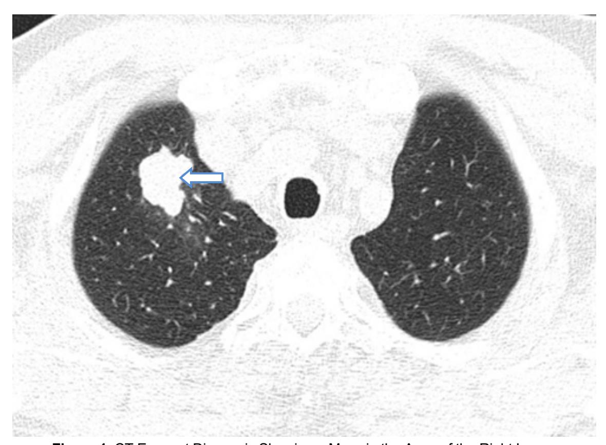
Figure 1. CT Exam at Diagnosis Showing a Mass in the Apex of the Right Lung.
Diagnosis: The histology showed a moderately differentiated adenocarcinoma with an immunohistochemical profile, which was consistent with lung cancer (CK7-positive, CK20-negative, and TTF1-positive).
Treatment and Outcomes: The case was discussed with the multidisciplinary cancer care team and according to the standard of care at the time, the patient received definitive chemoradiation with a regimen consisting of taxane and platinum salt, that it is known from previous studies to provide a better survival compared with sequential chemoradiation (4).
From October 2016 he started chemotherapy with paclitaxel 175 mg/m2 and cisplatin 75 mg/m2 in day 1, every 3 weeks, for 4 cycles, concurrent with radiotherapy to a total dose of 66 Gray in 33 fractions (Figure 2). Towards the end of the treatment, the patient presented severe hematologic toxicities (grade III anemia, grade III thrombocytopenia, grade II neutropenia), which required admission to the hospital and blood transfusion, with full recovery.
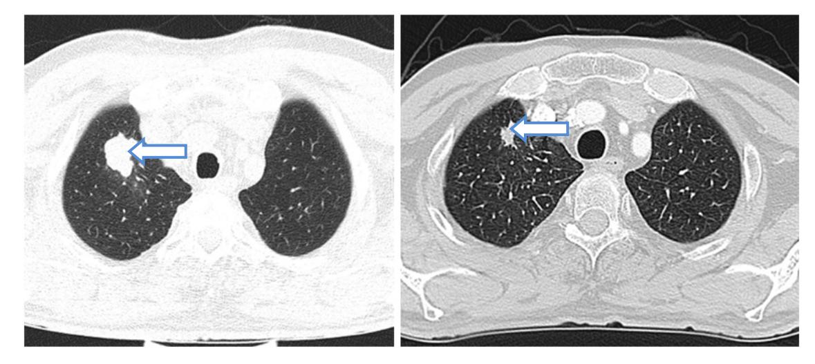
Figure 2. CT Exams Comparing the Pulmonary Mass at Diagnosis (right) and the Pulmonary Mass after Chemoradiation (left).
In February 2017, a follow-up CT of the thorax described new pseudo-nodular lesions in the lower lobes. The patient was asymptomatic. He was referred to pulmonology to exclude an infection. Cultures were negative for Grampositive and Gram-negative bacterium and Koch’s bacillus. At the time of progression to metastatic disease, the tumor was tested and was negative for ALK fusions and EGFR mutations. No PDL-1 testing was available in Romania at that time and immunotherapy was not approved in the first line metastatic setting. Due to previous toxicities, pemetrexed monotherapy 500 mg/m2 every 3 weeks was started in April 2017, with clinical benefits (no dyspnea and 3 kilograms weight gain). The sequential CT scans indicated stable disease, and the patient had a good tolerance to this treatment for 18 months, when the primary tumor and mediastinal lymph nodes showed progression on imaging.
In November 2018 nivolumab monotherapy 240 mg every 2 weeks flat dose was initiated. After three months, a CT scan was performed to evaluate the response to treatment. It described regression of the primary tumor, mediastinal nodes and pulmonary metastasis, with bilateral pleurisy and pericardial effusion (Figure 3). Diagnostic and therapeutic pericardiocentesis and thoracentesis were performed, and the fluids were diagnosed as exudates, with frequent lymphocytes, frequent reactive mesothelial cells and rare erythrocytes. Because both pericardial and pleural fluid collections were rapidly growing, with persistent dyspnea, the patient was referred to thoracic surgery. In May 2019 the patient underwent left thoracoscopy, with pericardial window and pleural biopsy. The pathology report from the pericardium and pleura described fibro-adipose tissue with epithelioid granulomas and chronic inflammatory infiltrate. Subsequently, a quantiFERON TB test was performed, which detected the presence of Mycobacterium tuberculosis.

Figure 3. CT exam Showing Pleurisy and Pericardial Effusion After the Initiation of Treatment with Nivolumab.
Based on the positive result and knowing that the most common cause of granulomatous inflammation worldwide is tuberculosis (5), with Romania being highly endemic, the pulmonologist started the patient on antituberculous treatment. The treatment was initiated in June 2019 and consisted of rifampin 600 mg/day, isoniazid 300 mg/day, ethambutol 1200 mg/day and pyrazinamide 1500 mg/day for 6 months concomitant with prednisolone 32 mg/day for the first 30 days. Nivolumab was stopped during the first month of antituberculous treatment and reinitiated in July 2019 at the same dose, with complete resolution of the pleural and pericardial effusions on serial CT scans and echocardiograms and with significant clinical benefit; the patient was asymptomatic.
Follow-up: The patient sustained partial radiological response on the primary tumor, mediastinal nodes and pulmonary metastasis, with no evidence of progression, and with excellent treatment tolerance until January 2021, when he progressed on the primary tumor and pulmonary metastasis after 26 months of stable disease on nivolumab. Docetaxel 75 mg/m2 monotherapy was started for a total of 4 cycles. Unfortunately, the patient progressed soon after the treatment ended, with severe deterioration of the performance status and succumbed to his death in July 2021.
3. Discussion
Strengths & limitations: The current case report, is the first one in Romania which to our knowledge describes reactivation of tuberculosis during nivolumab therapy for non-small cell lung cancer. However, a diagnosis bias for TB may be raised by the lack of a positive TB culture from the patient’s pleral and pericardial fluids. On the other hand, the association between biopsy proved granulomatous inflammation involving mediastinal lymph nodes and a positive quantiFERON TB test combined with a favorable response to anti TB therapy provides important arguments in favor of an ongoing TB infection. In this patient, interpreting the pleural and pericardial fluid collections as progressive disease would have been an error with potential fatal consequences. In countries with limited resources, where the possibilities to rebiopsy in the metastatic setting are scarce, a paradoxical response should always be interpreted with caution and, if possible, a new biopsy should be performed. Discussing the case in the multidisciplinary team allowed for a correct diagnosis and prompted timely initiation of antituberculous treatment, with favorable outcomes.
Medical Literature: The rate of tuberculosis among cancer patients receiving anti-PD1/PD-L1 agents was estimated to 1 in 1000 patients according to the French prospective registry managed at the Gustave Roussy Cancer Center (6). Giving the significantly higher incidence of tuberculosis in Romania (74/100 000 in 2016) (7), a higher rate of tuberculosis among patients treated with immunotherapy is expected. Unfortunately, in Romania currently there is no functional cancer registry; hence no data on the rate of tuberculosis among cancer patients receiving immunotherapy is available. Additionally, there are no regional guidelines to address this challenging association.
According to 2018 World Health Organization (WHO) guidelines (8), cancer patients are not included in the screening of latent tuberculosis due to the lack of evidence. However, increasing evidence suggests a significantly high correlation between the PD-1/PD-L1 blockade therapy in cancer patients and the incidence of active TB. (9,10,11). A recent systematic review showed that the TB rate in patients receiving PD-1/PD-L1 blockade therapies was 2,000 cases per 100,000, a rate that was 35 times higher than in the general population. Moreover, the mortality rate of the infected patients was considerably higher than currently expected (in the 18 patients for whom the clinical outcome was recorded, 77.8% were cured or achieved remission, and 22.2% died of TB).
Overall, the systematic review suggests that the administration of PD-1/PD-L1 blockade therapies to patients interfered with the immunologic control of latent TB infections, that led to increased activity of tuberculosis bacilli and active TB disease (12). The mechanism behind this association remains elusive. In the majority of exposed individuals, a stable relationship forms between host immune cells and Mycobacterium Tuberculosis (MTB) preventing disease progression. However, an emerging concept is that an excessive immune response to MTB may be equally harmful. Hence the anti PD-1/PD-L1 therapy may contribute to TB reactivation. For instance, a recent study revealed that hypoxia within TB lesions led to the up-regulated expression of PD-1/PD-L1 axis proteins. Meanwhile, results of another study focusing on PD-1/PD-L1 blockade immunotherapy indicated that inhibition of this pathway could stimulate multiple types of immune cells within granulomas (i.e. T cells, macrophages, dendritic cells), thereby resulting in granuloma collapse and excessive immunopathology. These results are consistent with the results of studies of MTB-infected patients and of human 3D cell culture-based TB models that suggest that PD-1 inhibition accelerates MTB growth via excessive TNF-α secretion. Taken together, these results highlight the important role of PD-1 in finetuning the balance between pro- and antiinflammatory responses against tubercle bacilli to avoid tissue damage (2,13). Intragranulomatous hypoxia has been described as a possible intimate correlative factor. PD-1 and its ligand PD-L1 are expressed in human granulomas suggesting a regulatory role at the site of disease (14), TB granulomas are hypoxic and PDL1 is up-regulated by hypoxia further suggesting a mechanistic link between hypoxia and the PD-1/PD-L1 axis within TB lesions (15)(16). Preclinical data showed that hypoxia increases the expression of PD-1 and its ligands, and that PD-1 inhibition increases MTB growth (2). TNF- α seems primarily responsible for this effect whereas TNF- α neutralization reverses the anti-PD-1 induced phenotype (17). Taken together all this new incoming information may sustain the recommendation for routine monitoring of latent TB in patients receiving ICIs-based immunotherapies (12).
Generally, in the case of active tuberculosis, immunotherapy is interrupted for a limited period of time, as well as any further immunosuppression while the anti-tuberculosis treatment is promptly started. Since there is no guideline, the exact moment of (re)starting the PD-1/PD-L1 inhibitors after the adequate anti-TB treatment is not known; nevertheless, the literature suggests resuming or initiating immunotherapy 2–4weeks after anti-tuberculosis treatment. Additionally, the hepatic toxicity of antituberculostatic drugs should be considered and monitored carefully (18).
4. Conclusions
To the best of our knowledge, this is the first case report in Romania of an acute TB infection during immunotherapy in a patient with lung cancer. The anti-TB specific treatment was effective and allowed for the continuation of nivolumab with a good and durable clinical response. Emerging data confirm that tuberculosis reactivation can occur during immunotherapy and it must be actively monitored allowing for a timely diagnosis and initiation of specific treatment, particularly in countries with a high TB incidence. Involvement of the multidisciplinary team early in the course of the disease is essential for the correct diagnosis and management of such cases.
Abbreviations:
ALK – anaplastic lymphoma kinase
CK 7 – cytokeratin 7
CK 20 – cytokeratin 20
CT – computed tomography
ECOG – eastern cooperative oncology group
EGFR – epidermal growth factor receptor
ICIs – immune checkpoint inhibitors
MTB – mycobacterium tuberculosis
PD-1 – programmed cell death protein 1
PD-L1 – programmed death ligand 1
TB – tuberculosis
TNF- α – tumor necrosis factor alfa
TTF1 – thyroid transcription factor 1
WHO – world health organization
Statements:
Authors’ contributions: EP wrote the manuscript, CC revised the text.
Consent for publication: As the corresponding author, I confirm that the manuscript has been read and approved for submission by all co-authors.
Conflict of interest: All authors declare having no competing interests associated with this publication.
Funding Sources: This research did not receive any specific grant from funding agencies in the public, commercial, or not-for-profit sector.
Informed Consent: The informed consent was obtained from the patient for publication and any accompanying images.
Ethical Approval: The treatment strategy was approved by the institutional/local tumor board (name).
References:
- Picchi H, Mateus C, Chouaid C, Besse B, Marabelle A, Michot JM, et al. Infectious complications associated with the use of immune checkpoint inhibitors in oncology: reactivation of tuberculosis after anti PD-1 treatment. Clin Microbiol Infect. (2018) 24:216–8. doi: 10.1016/j.cmi.2017.12.003
- Tezera LB, Bielecka ML, Ogongo P et al. Anti-PD-1 immunotherapy leads to tuberculosis reactivation via dysregulation of TNF-α; eLife. 2020; 9: e52668.Published online 2020 Feb 24. doi: 10.7554/eLife.52668
- T. Reungwetwattana, A.A. Adjei Anti-PD-1 antibody treatment and the development of acute pulmonary tuberculosis J Thorac Oncol, 11 (2016), pp. 2048-2050
- Walraven I, Damhuis RA, Ten Berge MG, et al. Treatment Variation of Sequential versus Concurrent Chemoradiotherapy in Stage III Non-Small Cell Lung Cancer Patients in the Netherlands and Belgium. Clin Oncol (R Coll Radiol) 2017;29:e177-85. 10.1016/j.clon.2017.07.012
- James DG. A clinicopathological classification of granulomatous disorders Postgraduate Medical Journal 2000;76:457-465.
- https://www.gustaveroussy.fr/fr/reisamic, accessed on the 15.04.2020
- Int J Tuberc Lung Dis. 2019 Feb 1;23(2):226-231. doi: 10.5588/ijtld.18.0270. Tuberculosis remains a public health problem in Romania. Golli AL, Niţu MF, Turcu F, Popescu M, Ciobanu-Mitrache L, Olteanu M
- Latent tuberculosis infection: updated and consolidated guidelines for programmatic management. Geneva: World Health Organization; 2018. Licence: CC BY-NC-SA 3.0 IGO.
- Im Y, Lee J, Kim SJ, Koh WJ, Jhun BW, Lee SH. Development of Tuberculosis in Cancer Patients Receiving Immune Checkpoint Inhibitors. Respir Med (2020) 161:105853. doi: 10.1016/j.rmed.2019.105853
- Fujita K, Kim YH, Kanai O, Yoshida H, Mio T, Hirai T. Emerging Concerns of Infectious Diseases in Lung Cancer Patients Receiving Immune Checkpoint Inhibitor Therapy. Respir Med (2019) 146:66–70. doi: 10.1016/j.rmed.2018.11.021
- Chan GH, Gwee YX, Low JL, Huang Y, Chan ZY, Choo JR, et al. Immune Checkpoint Inhibition for Non-Small Cell Lung Cancer in Patients With Pulmonary Tuberculosis or Hepatitis B: Experience From a Single Asian Centre. Lung Cancer (2020) 146:145–53. doi: 10.1016/j.lungcan.2020.05.020
- Kewei Liu, Dongpo Wang, Cong Yao, Min Qiao, Qing Li, Weicong Ren, Shanshan Li, Mengqiu Gao and Yu Pang.Increased Tuberculosis Incidence Due to Immunotherapy Based on PD-1 and PD-L1 Blockade: A Systematic Review and Meta-Analysis. Front. Immunol. (19 May 2022) Sec. Systems Immunology https://doi.org/10.3389/fimmu.2022.727220
- Lu D, Ni Z, Liu X, Feng S, Dong X, Shi X, et al. Beyond T Cells: Understanding the Role of PD-1/PD-L1 in Tumor-Associated Macrophages. J Immunol Res (2019) 2019:1919082. doi: 10.1155/2019/1919082
- Elkington PT, Bateman AC, Thomas GJ, Ottensmeier CH. Implications of tuberculosis reactivation after immune checkpoint inhibition. American Journal of Respiratory and Critical Care Medicine. 2018;198:1451–1453. doi: 10.1164/rccm.201807-1250LE
- Belton M, Brilha S, Manavaki R, Mauri F, Nijran K, Hong YT, Patel NH, Dembek M, Tezera L, Green J, Moores R, Aigbirhio F, Al-Nahhas A, Fryer TD, Elkington PT, Friedland JS. Hypoxia and tissue destruction in pulmonary TB. Thorax. 2016;71:1145–1153. doi: 10.1136/thoraxjnl-2015-207402
- Noman MZ, Desantis G, Janji B, Hasmim M, Karray S, Dessen P, Bronte V, Chouaib S. PD-L1 is a novel direct target of HIF-1α, and its blockade under hypoxia enhanced MDSC-mediated T cell activation. The Journal of Experimental Medicine. 2014;211:781–790. doi: 10.1084/jem.20131916.
- Tezera LB, Bielecka MK, Chancellor A, Reichmann MT, Shammari BA, Brace P, Batty A, Tocheva A, Jogai S, Marshall BG, Tebruegge M, Jayasinghe SN, Mansour S, Elkington PT. Dissection of the host-pathogen interaction in human tuberculosis using a bioengineered 3-dimensional model. eLife. 2017a;6:e21283. doi: 10.7554/eLife.21283
- Ramappa V, Aithal GP. Hepatotoxicity Related to Anti-tuberculosis Drugs: Mechanisms and Management. J Clin Exp Hepatol. 2013 Mar;3(1):37-49. doi: 10.1016/j.jceh.2012.12.001. Epub 2012 Dec 20. PMID: 25755470; PMCID: PMC3940184


