


Author(s) :
Konstantin Gordon1,2, Igor Gulidov1, Danil Gogolin1, Olga Lepilina2, Olga Golovanova3, Alexey Semenov1, Sergey Dujenko4, Kira Medvedeva1,
Sergey Koryakin4, Sergey Ivanov5, Andrey Kaprin6
1 Department of Proton Therapy, A. Tsyb Medical Radiological Research Center, Obninsk, Russia
2 Department of Histology, Cytology, and Embryology, Medical Institution, People’s Friendship University of Russia, Moscow, Russia
3 Department of Dosimetry, A. Tsyb Medical Radiological Research Center, Obninsk, Russia
4 Department of Radiophysics, A. Tsyb Medical Radiological Research Center, Obninsk, Russia
5 A. Tsyb Medical Radiological Research Center, Obninsk, Russia
6 National Medical Research Radiological Center, Moscow, Russia
Corresponding author: Konstantin Gordon, Email: dr_gordon@yahoo.com
Publication History: Received - , Revised - , Accepted - , Published Online - 2021.
Copyright: © The author(s). Published by Casa Cărții de Știință.
User License: Creative Commons Attribution – NonCommercial (CC BY-NC)

 PDF
PDF
 Share
Share
Highlights
-
Sinonasal esthesioneuroblastoma (ENB) is an uncommon intranasal malignancy (often locally advanced at diagnosis) with a strong tendency for loco-regional recurrence; relapses near the skull base create major constraints because critical organs-at-risk (optic nerves/chiasm, brainstem, temporal lobes) limit re-irradiation dosing.
-
A 49-year-old woman previously treated in 2001 with partial resection + 60 Gy/30 fractions (Co-60) presents in 2016 with radiographic recurrence extending to the skull base and involving visual structures; surgery is not feasible and biopsy is not obtainable due to tumor location, so the relapse is managed based on imaging and multidisciplinary decision-making.
-
The center delivers spot-scanning proton therapy (IMPT) 50 Gy(RBE) in 25 fractions, with image guidance and immobilization; treatment planning reconstructs the prior 2D plan and estimates cumulative BED to OARs while maintaining target coverage (reported V95 ~92%) and limiting maximum doses to optic pathways/chiasm and brainstem.
Abstract
Head and neck (H&N) cancers are in the 7-8th place among all types of cancer. Despite novel approaches in cancer treatment, most of the patients have a high risk of loco-regional recurrence.
Esthesioneuroblastoma (ENB) is a very rare H&N neoplasm, accounting for only 3-6% of all intranasal tumors. These tumors usually are presented with locally advanced stages and required radiotherapy as a part of the comprehensive treatment. Loco-regional failures represent a challenge, and re-irradiation can be effective for some groups of patients. We present a case of re-irradiation with protons of sinonasal tumor in a 49-years old patient with recurrent ENB, involving visual structures. We achieved a 5-year positive local control after the proton therapy (PT), without significant toxicity. The case shows an example of re-irradiation as an essential area for PT application, given the need to maximize the treatment’s efficacy and minimize the risk of severe toxicity.
1. Introduction
Esthesioneuroblastoma (ENB) was first described in 1924 by Berger et al. [1]. ENB is a rare tumor originating from the olfactory neuroepithelium of a sinonasal cavity, accounting for 3% of all intranasal cancers [2]. ENBs have various biological behaviors, from benign to highly aggressive. Even if benign, most cases are diagnosed with locally advanced tumors, infiltrating skull base, orbits, intracranial foramens, etc. These features usually make impossible the radical surgery. Thus, radiotherapy (RT) is recommended as adjuvant or primary treatment in advanced tumors. The dose usually ranges from 50 to 66 Gy, while higher levels can damage the organs at risk (OAR), located in a skull base [3]. Locoregional recurrence after prior therapy continues to be the most frequent pattern of failure in EBNs. Stereotactic RT-techniques can be used in case of small recurrence [4]. With its known dosimetric and radiobiological advantages, proton therapy can be effectively used to irradiate large tumor volumes.
2. Case report
In January 2016, a 49-year-old woman with a relapse of EBN, raised from the right side of the nasal cavity and extended to the skull base, was presented to the institutional tumor board. She had her first treatment for EBN (Hyams grade 2, Kadish-Morita stage D) in 2001, with partial resection, followed by radiotherapy 60Gy in 30 fractions (Fx), provided via 60Co-unit. The patient had stable disease until 2016, when tumor relapsed and started to grow, so the patient was transferred from a regional hospital to A.Tsyb MRRC Proton Center. As biopsy from the recurrent tumor (rT4bN0M0) was not available due to localization, so we accepted radiographic signs as sufficient to confirm the relapse.
The recurrent tumor (Fig. 1A) involved optical nerves and chiasm, even though the patient had no vision problems. The surgery was not feasible, and without biopsy, the chemotherapy could not be properly selected. The case was discussed within the tumor board, and since there were no MRIsigns of brain necrosis, re-irradiation with protons was recommended.
The 2nd RT-course was delivered in January-February 2016 via a fixed horizontal spot-scanning synchrotron-based proton beam in a seated position (5). The thermoplastic mask and daily image guidance were used to reduce positioning errors. A dose of 50 Gy in 25 Fx was chosen as our compromise between effective tumor control rate and a vision loss risk, even we considered the dose from prior course as not very relevant after 15 years interim.
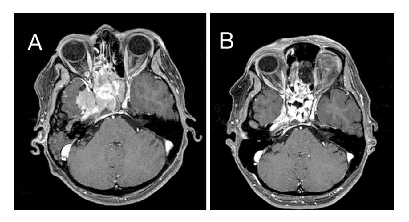
Fig. 1. Diagnostic MR-imaging before PT in 2016 (A), VGTV=110.8 cm3, and after 5 years, in 2021 (B), VGTV=93.9 cm3.
Table 1. Approximate cumulative BED to the OARs (α/β = 3)
| 2001 (60Co,60Gy) | 2016 (PT,50GyRBE) | Total BED | |
|---|---|---|---|
| Optic nerve R (Dmax) | 120 | 78.6 | 198.6 |
| Optic nerve L (Dmax) | 120 | 85.5 | 205.5 |
| Chiasma (Dmax) | 120 | 72 | 192 |
| Temporal lobe R (Dmean) | 68 | 6 | 72 |
| Temporal lobe L (Dmean) | 26 | 2.6 | 28.6 |
| Whole-brain (Dmean) | 20 | 3 | 23 |
| Brain stem (Dmax) | 120 | 92.6 | 212.6 |
| Cochlea R (Dmean) | 56 | 32.6 | 88.6 |
| Cochlea L (Dmean) | 56 | 23.6 | 79.6 |
General tumor volume (GTV) was delineated based on MRI and CT coregistration, and a 3 mm margin was added to make PTV (planning tumor volume). The GTV volume was 110.8 cm3, and the final PTV was 214.3 cm3. Prior 2D RT-plan from 60Co-machine was reconstructed and roughly estimated. The biological effective doses (BED) to the critical structures were calculated to approximate the cumulative limits (Table 1), taking in account also the long interval from the first RT-course.
The PT-dose distribution is shown in Figure 2. The V95 (volume receiving 95% of the prescribed dose) was 92.4%. The maximum dose (Dmax) to the brain stem was 47.9 GyRBE (relative biological efficacy, equal to 1.1). The right visual nerve received 43.6 GyRBE (Dmax), and the left nerve had 46.9 GyRBE. The dose to the chiasma was 40.7 GyRBE. Mean doses to the right and left cochleas were 21.8 GyRBE and 17.0 GyRBE, respectively. Mean dose to the right temporal lobe was 4.5 GyRBE, to the left – 0.7 GyRBE.
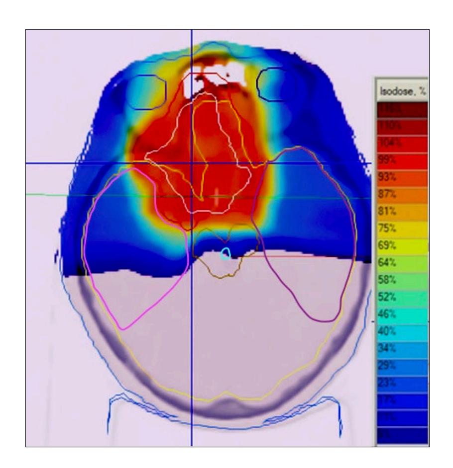
Fig. 2 Dose distribution of the proton therapy plan (Intensity-modulation PT, JSC Protomtm TPS).
The patient tolerated PT very well. The acute toxicity was presented by local nasal mucositis (grade 1-2). At 6 months after reirradiation, the follow-up MRI described an 18% decrease in volume (stable disease). The last check-up was performed in February 2021 showing that the recurrent tumor is still controlled (Fig. 1B). No late toxicity has occurred during follow-up (e.g., vision problems or neurocognitive dysfunction), so patient’s quality of life was preserved.
3. Discussion
Local recurrence is the main problem in the management of ENB, reported in 1 /3 of the patients [3]. The choice of treatment depends upon the type of relapse and prior treatment received. Interest in re-irradiation is growing along with the development of new and precise RT techniques, e.g., intensity-modulated RT or proton therapy. Even though re-irradiation is still challenging: professionals have to find a frail balance between RT-dosing and OAR sparing. For small tumor volumes, far enough from the critical structures, stereotactic RT can usually be an effective and safe option [4,6]. Takiar et al. assumed the irradiation volume > 50 cm3 as an indication of poor outcomes, associated with severe toxicity. The PT was shown as an effective treatment with reasonable toxicity in limited series, even for large tumor extension [7]. In 2016, Phan et al. published the results of proton re-irradiation in H&N cancers in 60 patients, demonstrating 1year overall survival (OS) 83.8% and grade 3 late toxicity rate of 16.7% [8]. The study by McDonald et al. included 61 patients, retreated with PT for H&N recurrence or second primary tumor, and 2-year OS was 32.7%. Grade ≥3 toxicities were recorded in 14.7% [9]. Romesser et al. described a 25.1% risk of failure in 12 months and a favorable toxicity profile after PT re-treating of 91 patients [10].
Regarding our case, PT re-irradiation helped achieve 5-year tumor local control without remarkable brain toxicity or vision loss, obviously with cumulative doses to the OARs potentially maxed out.
4. Conclusion
In selected patients, re-RT can be an effective treatment, with acceptable risks of adverse events. From our point of view, PT is the optimal choice for re-irradiation of large tumor volumes, as illustrated in this case.
Abbreviations
CT – computer tomography
ENB – esthesioneuroblastoma
Fx – fraction
GTV – gross tumor volume
Gy – Gray
H&N – head and neck
MRI – magnetic resonance imaging
OAR – organ at risk
PT – proton therapy
PTV – planning tumor volume
RBE – relative biological efficacy
RT – radiotherapy
Statements
Authors’ contributions: KG, IG conceived and planned the case report. KG, AS, OG, DG,
OL, KM, SK, SD carried out the treatment and planning. KG, IG contributed to the interpretation
of the results. KG, IG took the lead in writing the manuscript. SI, AK made the final approval. All
authors provided critical feedback and helped shape the research, analysis and manuscript.
Consent for publication: As corresponding author, I confirm that the manuscript has been
read and approved for submission by all named authors.
Conflict of interests: The authors have no conflicts of interest to declare.
Written informed Consent for Publication of case. The informed consent was obtained
from the patient for publication of this case report and any accompanying images.
Funding Sources: none.
Ethical Approval: The treatment strategy in this case was approved by a local tumor board.
Statement of Ethics: The accompanying manuscript does not contain any studies carried
out by the authors on humans or animals.
Availability of data: The data used to support the findings of this study is restricted by the
Ethical Committee of A. Tsyb MRRC in order to protect patients privacy. Data is available upon
request from the corresponding author for researchers, who meet the criteria for access to
confidential data
References
- Berger L, Luc R, Richard D. L’esthesioneuroepitheliome olfactif. Bull Assoc Fr Etude Cancer. 1924;13:410–421
- Spirin D, Kobyakov G, Cherekaev G, Nazarov N, Kadasheva A, Vetlova R, et al. Epidemiology, diagnosis,
clinical symptoms, and classification of malignant primary skull base tumors. Zhurnal voprosy neirokhirurgii imeni N.
N. Burdenko. 2016;80(3):106–113. doi: 10.17116/neiro2016803106-113 [Russian]. - Kumar R. Esthesioneuroblastoma: Multimodal management and review of literature. World J Clin Cases.
2015;3(9):774-778. doi:10.12998/wjcc.v3.i9.774 - Van Gompel JJ, Carlson ML, Pollock BE, Moore EJ, Foote RL, Link MJ. Stereotactic radiosurgical salvage
treatment for locally recurrent esthesioneuroblastoma. Neurosurgery. 2013;72(3):332-340.
doi:10.1227/NEU.0b013e31827fcdc2
80 - Gulidov I, Gordon K, Gogolin D, Mardynsky Yu, Lepilina O, Neledov D, et al. Reirradiation of intracranial tumors with active beam scanning protons. Siberian Journal of Oncology. 2017;16(5):63–70. doi:10.21294/1814- 4861-2017-16-5-63-70 [Russian]
- Gogineni E, Zhang I, Rana Z, et al. Quality of Life Outcomes Following Organ-Sparing SBRT in Previously Irradiated Recurrent Head and Neck Cancer. Front Oncol. 2019;9:836. Published 2019 Sep 10. doi:10.3389/fonc.2019.00836
- Takiar V, Garden AS, Ma D, Morrison WH, Edson M, Zafereo ME, et al. Reirradiation of Head and Neck Cancers With Intensity Modulated Radiation Therapy: Outcomes and Analyses. Int J Radiat Oncol Biol Phys. 2016;95(4):1117-1131. doi:10.1016/j.ijrobp.2016.03.015
- Phan J, Sio TT, Nguyen TP, Takiar V, Gunn GB, Garden AS, et al. Reirradiation of Head and Neck Cancers With Proton Therapy: Outcomes and Analyses. Int J Radiat Oncol Biol Phys. 2016;96(1):30-41. doi:10.1016/j.ijrobp.2016.03.053
- McDonald MW, Zolali-Meybodi O, Lehnert SJ, Estabrook NC, Liu Y, Cohen-Gadol AA, et al. Reirradiation of Recurrent and Second Primary Head and Neck Cancer With Proton Therapy. Int J Radiat Oncol Biol Phys. 2016;96(4):808-819. doi:10.1016/j.ijrobp.2016.07.037
- Romesser PB, Cahlon O, Scher ED, Hug EB, Sine K, DeSelm C, et al: Proton beam reirradiation for recurrent head and neck cancer: Multi-institutional Report on feasibility and early outcomes. Int J Radiat Oncol Biol Phys 95:386-395, 2016. http: //dx.doi.org/10.1016/j.ijrobp.2016.02.036


Table 1. Approximate cumulative BED to the OARs (a/b = 3)
| Optic nerve R (Dmax) | Optic nerve L (Dmax) | Chiasma
(Dmax) |
Temporal lobe R
(Dmean) |
Temporal lobe L
(Dmean) |
Whole-brain
(Dmean) |
Brain stem (Dmax) | Cochlea R (Dmean) | Cochlea L (Dmean) |
|
| 2001 (60Co,60Gy) |
120 | 120 | 120 | 68 | 26 | 20 | 120 | 56 | 56 |
| 2016 (PT,50GyRBE) |
78.6 | 85.5 | 72 | 6 | 2.6 | 3 | 92.6 | 32.6 | 23.6 |
| Total BED | 198.6 | 205.5 | 192 | 72 | 28.6 | 23 | 212.6 | 88.6 | 79.6 |