


Author(s) :
Ouaddane Alami Salem1, Fatima-Ezzahra Alouan1, Bauer Nicholas1
1 Radiotherapy Service, Oncology Hospital, Emile Muller Hospital Of The South Alsace Regional Hospital Group, Mulhouse, France
Corresponding author: Salem Ouaddane Alami, Email: salem.ouaddanealami@usmba.ac.ma
Publication History: Received - 16 July 2025, Revised - 7 October 2025, Accepted - 20 October 2025, Published Online - 29 December 2025.
Copyright: © 2025 The author(s). Published by Casa Cărții de Știință.
User License: Creative Commons Attribution – NonCommercial (CC BY-NC)

 PDF
PDF
 Share
Share
Highlights
- Exclusive radiotherapy can be a highly effective, non-invasive curative treatment for unresected mandibular solitary bone plasmacytoma in elderly patients.
- Using Helical Tomotherapy and a dose of 46 Gy normalized to the 50% isodose line, target volume coverage (98% of PTV) was excellent, while sparing critical organs at risk.
- The patient achieved complete resolution of radiodermatitis and a 62% reduction in tumor volume at the six-month follow-up, underscoring the high radiosensitivity and local control rates (over 90%) of this rare plasma cell neoplasm.
Abstract
Background: Solitary plasmacytoma (SP) is a rare plasma cell tumor causing localized monoclonal plasma cell proliferation without systemic disease. Mandibular cases present diagnostic and management difficulties due to both anatomical complexity and the risk of progression to multiple myeloma (MM). This report evaluates the clinical outcome of exclusive radiotherapy in an unresected solitary bone plasmacytoma (SBP) of the mandible in an adult patient.
Case Presentation: We report the case of an 80-year-old man with a solitary bone plasmacytoma (SBP) of the mandible. He first presented with dental mobility and mandibular swelling. Imaging revealed a left parasymphyseal mandibular lesion initially suspected to be osteomyelitis. Histopathological analysis confirmed a mature plasma cell neoplasm. Extensive staging, including bone marrow biopsy and FDG-PET, confirmed the lesion’s solitary nature. The patient refused surgery. Exclusive conformational radiotherapy was delivered to a total dose of 46 Gy in 23 fractions with good tolerance. Post-treatment evaluation showed significant tumor regression and a stable clinical condition.
Conclusion: This case demonstrates that exclusive radiotherapy can achieve significant tumor regression and clinical stability in a solitary mandibular plasmacytoma. Comprehensive diagnostic evaluation and multidisciplinary management are crucial for such rare clinical situations. Further research is needed to refine strategies for these complex cases.
1. Introduction
Solitary plasmacytoma (SP) is histologically characterized by a localized accumulation of monoclonal neoplastic plasma cells, without signs of systemic plasma cell proliferation, absence of bone marrow dissemination, and a unique lesion. It is a rare form of plasma cell neoplasm, accounting for 5 to 10% of all plasma cell neoplasms according to the literature (1-2). It can be classified into two groups based on its location: solitary bone plasmacytoma (SBP) or intramedullary plasmacytoma, and extramedullary plasmacytoma (EMP) (1-3). The bone forms are the most common and primarily affect the vertebrae, as well as the ribs, clavicles, sternum, and long bones (4). Mandibular involvement has been rarely described in the literature.
The frequent progression of solitary bone plasmacytoma to multiple myeloma, reported in 55% of cases in the literature, continues to raise questions about its nosological authenticity (5).
Based on a case of solitary bone plasmacytoma of the mandible, complemented by a literature review, we aim to revisit the diagnostic, therapeutic, and evolutionary aspects of this condition.
The aim of this case report is to examine the role of exclusive radiotherapy in the curative treatment of an unresected mandibular solitary plasmacytoma in an elderly patient.
2. Case presentation
Our patient is an 80-year-old man with a history of hypertension and diabetes, both of which are well controlled with medical treatment. The disease started approximately three months prior to admission in the Radiation Oncology department, with the onset of localized swelling and dental mobility, prompting him to consult his dentist. A panoramic dental X-ray and a CT scan of the facial bones were performed, revealing a semi-recent left parasymphyseal mandibular fracture with significant mandibular osteocondensation and an associated anterior periosteal reaction. These findings were highly suggestive of chronic osteomyelitis complicating the aforementioned fracture. There was also evidence of a phlegmon involving the soft tissues surrounding the left mandibular body in the fracture area, without a clearly organized collection.
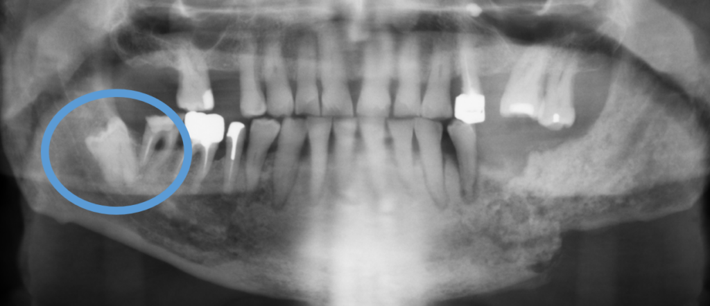
Figure 1. Left mandibular osteolytic lesion on orthopantomogram in a patient with plasmacytoma.
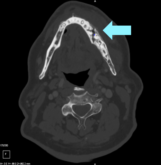
Figure 2. CT scan slice showing osteolysis of the left mandibular ramus caused by the solitary plasmacytoma.
The patient was then referred to an Ear-Nose-Throat (ENT) Doctor who completed the assessment with a Magnetic Resonance Imaging (MRI) of the facial bones. The imaging revealed a fracture of the horizontal branch of the left mandible, with significant surrounding soft-tissue infiltration and moderate enhancement, findings compatible with osteomyelitis. No true mass effect or cervical lymphadenopathy was noted.
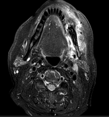
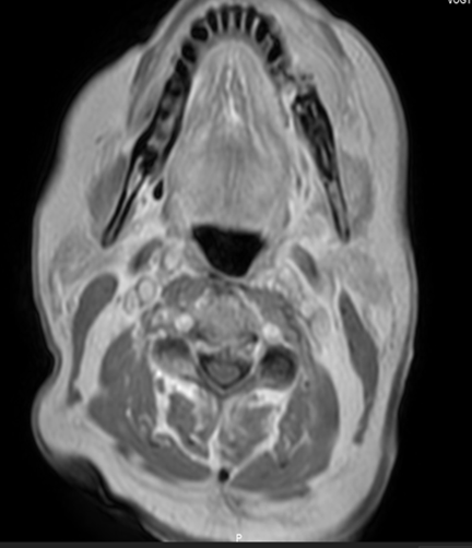
Figure 3. Axial MRI images: T2-weighted (A) and T1-weighted (B) sequences showing a left mandibular lesion with high signal intensity, suggestive of a solitary plasmacytoma.
The ENT Doctor subsequently performed a surgical biopsy of the left mandibular stump, which revealed a mature plasma cell neoplasm with lambda and IgM monotypy. The cells were CD138-positive and CD20-negative, with a Ki-67 index of 1–2%. Immunofixation showed the presence of two monoclonal immunoglobulins (IgM kappa and IgM lambda). The biological workup revealed no hypercalcemia or renal dysfunction. Bone marrow analysis showed a polymorphic marrow without cytological evidence of plasma cell tumor infiltration.
As part of the staging workup, an FDG-PET scan confirmed that the mandibular plasmacytic lesion was solitary, with a few benign-appearing lymph nodes in the cervico-thoracic region. The case was discussed in a multidisciplinary hematology meeting, and given the patient’s refusal of surgery, it was decided to proceed with exclusive radiotherapy.
Figure 4: PET-CT scan slice showing hypermetabolism corresponding to the left mandibular fracture involvement, with a SUVmax of 8.6 compared to a hepatic reference of 3.6.
Upon admission to our department, the clinical examination found a patient in good general condition (Performance Status 1), without local pain (Visual Analog Scale, VAS score 0), weighing 85 kg, and standing 1.80 m tall.
The locoregional examination revealed hypoesthesia in the territory of the left mandibular branch (V3), along with a slight leftward deviation of the mouth.
There was also a swelling of the lower left mandibular region, in the context of prior dental avulsions (teeth 27 and 34), with ongoing healing progressing well.
A planning CT scan was then performed using a five-point thermoplastic mask and knee supports to ensure immobilization. Alignment was performed using wall lasers set to the level of the tragus. The images were transferred from the treatment planning system (TPS) to the contouring console.
Helical tomotherapy was selected for treatment delivery, utilizing 6 MV photon beams generated by a linear accelerator equipped with a multileaf collimator (MLC) to conform the dose precisely to the target volumes. Target volume delineation included the GTV-T (Gross Tumor Volume – Tumor), corresponding to the visible tumor mass; the CTV-T (Clinical Target Volume – Tumor), defined by adding a 5 mm margin around the GTV-T to account for potential microscopic spread; and the PTV-T (Planning Target Volume – Tumor), obtained by expanding the GTV-T with a 10 mm margin to compensate for setup uncertainties and patient motion. A total dose of 46 Gy was prescribed to the PTV, normalized to the 50% isodose line, in accordance with ICRU Report 83. The maximum dose (Dmax) remained below 105% of the prescribed dose. Target volume coverage was excellent, with more than 98% of the PTV receiving the prescribed dose. Organs at risk (OARs) were delineated using rigid image fusion with MRI to enhance anatomical accuracy. These included the brain, eyes, crystalline lenses, optic nerves, brainstem, cochleae, oral cavity, limbs (if within the field), larynx, parotid glands, submandibular glands, pharyngeal constrictor muscles, skin, pituitary gland, and spinal cord. All dose constraints to the OARs were respected. Daily patient positioning was verified using on-board cone-beam computed tomography (CBCT) fused with the planning CT to ensure precise alignment and treatment accuracy. The overall treatment duration was 32 days, completed without interruption. During the course of radiotherapy, the patient developed grade 2 radiomucositis (according to CTCAE), which was managed with mouthwashes containing sodium bicarbonate, xylocaine, and corticosteroids. Grade 2 radiodermatitis also developed and was treated with emollients and adhesive dressings; both conditions improved with appropriate symptomatic management. At the six-month follow-up visit, clinical evaluation confirmed significant tumor regression, as observed at the three-month post-treatment assessment. However, hypoesthesia in the V3 (mandibular) territory persisted. A follow-up MRI revealed a 62% reduction in tumor volume compared to the initial imaging. To date, no late toxicities have been observed, and the radiodermatitis that developed during treatment has completely resolved.
3. Discussion
Plasma cell neoplasms account for 1 to 2% of all human malignancies. Several clinical forms exist, among which solitary plasmacytoma represents less than 10% of these proliferations. It was first described by Schridde in 1905 (6-7). Solitary plasmacytoma is classified into two groups. The most common is solitary bone plasmacytoma (SBP), which frequently evolves into a multiple form. It commonly affects the axial skeleton and rarely involves the jaws. Only about 4.4% of SBPs occur in the mandible, most often in areas rich in bone marrow such as the body, angle, and ramus of the mandible (8). The second group is solitary extramedullary plasmacytoma (SEP) (9).
The etiology of solitary plasmacytoma remains uncertain, but several hypotheses have been proposed, including radiation exposure, chemical agents, viral infections, and genetic factors. Cytogenetic studies have identified losses of chromosomes 13, 1p, and 14q, and gains of chromosomes 19p, 9q, and 1q. Interleukin-6 is considered a key growth factor in the pathogenesis of this condition (10).
SBP most often occurs in patients aged 50 to 80 years, with a mean age of onset around 60. It is rare before the age of 40 (11). The condition is more frequent in men, with a reported male-to-female ratio of 2:1 (12).
The clinical presentation of solitary plasmacytoma of the mandible (SPM) is not specific; facial swelling, dental mobility, and sensory disturbances are the most frequent clinical signs. In rare cases, a solitary plasmacytoma of the mandible may be discovered during a pathological fracture, post-extraction bleeding, or a dental joint disorder in the case of condylar localization (13).
Malignant plasma cells produce cytokines and release an osteoclast-activating factor that stimulates osteoclasts to resorb bone; consequently, on radiographic examination, they appear as well-defined osteolytic lesions with either unilocular or multilocular radiolucency (10-14). According to Lae et al., three radiographic patterns have been described in SBP: multilocular ‘soap bubble’ lesions, unilocular radiolucency (as seen in our patient) with a cystic appearance, and poorly defined destructive bone resorption (15). CT helps to refine the radiological image and assess the extension toward cortical and soft tissues. MRI remains a valuable imaging modality for detecting bone-involved plasmacytomas, assessing soft tissue involvement and bone marrow infiltration, and evaluating disease throughout the body. Solitary plasmacytomas show low signal intensity on T1-weighted images and high signal intensity on T2-weighted images, with homogeneous enhancement after gadolinium contrast injection (16).
In our case, there were signs of periosteal reaction with cortical destruction, potentially extending widely into the surrounding tissues.
Under the microscope, plasma cells exhibit varying degrees of differentiation within a sparsely cellular stroma. Sukpanichnant et al. classified plasmacytomas into mild, moderate, and severe dysplasia based on the degree of plasma cell differentiation (17). Nuclei may be binucleated. The spherical nuclei are eccentrically located and display regular or irregular chromatin margination, often in a cartwheel pattern (18). Occasionally, the chromatin appears coarsely clumped, showing a clock-face configuration. The cells present a pale, globular perinuclear cytoplasmic area known as a hof. Pseudoangiomatous areas, multinucleated giant cells, amyloid deposition, and myxoid changes may also be observed in some cases. Plasma cells may contain intracytoplasmic inclusions (Russell bodies) and intranuclear inclusions known as Dutcher bodies. Immature plasma cells have larger or more irregular nuclei, less condensed chromatin, and may display prominent nucleoli. These immature cells are larger and more pleomorphic, with abundant, mildly basophilic cytoplasm (10).
The neoplastic process is secretory in approximately 99% of cases, producing monoclonal immunoglobulins of either light or heavy chains, which can be detected in the serum or urine (15). In accordance with the literature, the present case revealed elevated serum lambda light chains, exceeding the normal range. To diagnose a case of solitary bone plasmacytoma (SBP) and to exclude multiple myeloma (MM), thorough clinical investigations are required, including skeletal radiological survey, bone marrow aspiration, complete blood count, serum calcium levels, and renal function tests (15). Specific diagnostic criteria for SBP were proposed by Bataille and Sany (Table 1)(19).
The present case meets all of the aforementioned criteria. In addition to laboratory investigations, determining monoclonal restriction to either kappa or lambda light chains is a crucial approach in the evaluation of suspected plasmacytoma.
Table 1. Specific Diagnostic Criteria for Solitary Bone Plasmacytoma (SBP) (19)
| Solitary Myeloma: Criteria for Diagnosis |
| 1) Presence of solitary bone tumor*.
2) A biopsy showing plasma cell histology, 3) Absence of myeloma cells** in bone-marrow examinations. 4) Absence of anemia, hypercalcemia, or renal involvement. 5) Absence of monoclonal component or low monoclonal component (only detected by immunoelectrophoresis) with disappearance after surgery and/or radiation therapy. 6) Normal levels of immunoglobulins or low levels with increase to normal levels after surgery and/or radiation therapy. |
* Radiologic examination, bone scanning, and computed tomography will be useful for such an accurate assessment. ** In sternum and pelvis
From a therapeutic perspective, surgical treatment may be considered for diagnostic purposes, in the presence of neurological complications such as spinal cord compression, or to treat or prevent a pathological fracture in bone weakened by tumor-induced osteolysis. In the specific case of mandibular involvement, surgical intervention is feasible in the vast majority of situations.
Complete resection may be indicated for peripheral, easily accessible lesions; however, mutilating surgery should be avoided, especially since radiotherapy offers comparable efficacy while allowing better preservation of function (5-6-20-21)
Moreover, the radiosensitivity and radiocurability of solitary bone plasmacytoma have been documented for over half a century (19,22). Radiotherapy is considered the standard treatment for solitary bone plasmacytoma. When administered at optimal doses of 40 to 50 Gy, it provides a local control rate exceeding 90%, with excellent tolerance and rapid, durable pain relief.
A dose–response effect was reported by Mendenhall et al. (23), who observed a 94% local control rate at doses above 40 Gy, compared to 69% at lower doses. Similarly, Frassica et al. (24) reported a 15.6% local failure rate in a series of 46 patients when the radiotherapy dose was below 45 Gy, whereas the local control rate was 100% with higher doses.
The addition of chemotherapy is not recommended, as it has not been shown to improve outcomes in terms of recurrence or progression to multiple myeloma (4-5).
Predictive factors for local recurrence appear to include, in addition to insufficient radiotherapy doses, the anatomical location of the solitary bone plasmacytoma — with spinal or pelvic lesions being more difficult to manage than peripheral ones — the persistent presence of monoclonal protein after treatment, as well as tumor size (5).
The median survival rate for solitary plasmacytoma is longer than that for multiple myeloma, due to the localized nature of the disease. However, clinical remission remains rare (25). SBPs carry a high risk of progression to multiple myeloma (65% to 84% at 10 years and nearly 100% at 15 years), whereas SEPs have a lower risk of progression (10% to 30% at 10 years) but a higher risk of local recurrence(26). Scientific data suggest that patients with an SBP larger than 4 to 5 cm are at increased risk of developing multiple myeloma (27).
Due to the progressive nature of SBP and the extended period (6 to 8 months) required to achieve maximal radiotherapy response, periodic monitoring is essential. A complete blood count, serum biochemistry (calcium and creatinine), and systematic evaluation of potential biomarkers such as persistent M-protein, clonal expansion of free light chains (FLC), and the presence of clonal plasma cells are standard practices(10). PET/CT, when available, can be particularly useful for follow-up in skeletal studies
4. Conclusion
Solitary plasmacytoma of the mandible is a rare entity, often difficult to diagnose and manage due to its potential for local aggressiveness and progression to multiple myeloma. Through this case, we highlight the efficacy of exclusive radiotherapy in achieving local control of the disease, even in anatomically complex locations such as the mandible. A thorough diagnostic evaluation, precise treatment planning, and close post-treatment monitoring remain essential. Further studies and larger case series are required to refine therapeutic strategies, particularly for aggressive or atypical forms. This work contributes to the literature supporting the use of radiotherapy as an effective conservative treatment in selected cases.
Abbreviations
CBCT – Cone Beam Computed Tomography
CD138 / CD20 – Cluster of Differentiation (Immunohistochemical markers)
CRAB – Calcium elevation, Renal insufficiency, Anemia, and Bone lesions (Multiple Myeloma diagnostic criteria)
CT – Computed Tomography
CTCAE – Common Terminology Criteria for Adverse Events
CTV-T – Clinical Target Volume – Tumor
Dmax – Maximum Dose
EMP – Extramedullary Plasmacytoma
ENT – Ear, Nose, and Throat
FDG-PET – Fluorodeoxyglucose Positron Emission Tomography
GTV-T – Gross Tumor Volume – Tumor
ICRU – International Commission on Radiation Units and Measurements
IgM – Immunoglobulin M
Ki-67 – Cellular proliferation biomarker
MLC – Multileaf Collimator
MM – Multiple Myeloma
MRI – Magnetic Resonance Imaging
OAR – Organs At Risk
PTV-T – Planning Target Volume – Tumor
SBP – Solitary Bone Plasmacytoma
SEP – Solitary Extramedullary Plasmacytoma
SP – Solitary Plasmacytoma
SPM – Solitary Plasmacytoma of the Mandible
SUVmax – Maximum Standardized Uptake Value
TPS – Treatment Planning System
V3 – Mandibular branch of the Trigeminal Nerve
VAS – Visual Analog Scale (for pain assessment)
VIDE – Vincristine, Ifosfamide, Doxorubicin, and Etoposide (Chemotherapy protocol)
Statements
Authors’ contributions: OAS: Writing – original draft, Writing – review & editing. FEZA Writing – review & editing. NB Conceptualization, supervision, and guidance.
Consent for publication: As the corresponding author, I confirm that the manuscript has been read and approved for submission by all named authors
Funding: This case report has received no financial support.
Conflict of interest: The authors declare that they have no known competing financial interests or personal relationships that could have appeared to influence the work reported in this paper.
Informed consent: The informed consent was obtained from the patient for publication of this case report and any accompanying images.
Statement of Ethics: The case management was approved by the tumor board of the Emile Muller Hospital Of The South Alsace Regional Hospital Group, Mulhouse, France.
References
-
Dimopoulos MA, Moulopoulos LA, Maniatis A, Alexanian R. Solitary plasmacytoma of bone and asymptomatic multiple myeloma. Blood. 2000 Sep 15;96(6):2037-44.
-
Wiltshaw E. The natural history of extramedullary plasmacytoma and its relation to solitary myeloma of bone and myelomatosis. Medicine (Baltimore). 1976 May;55(3):217-38.
-
Frizzera G. Castleman’s disease and related disorders. Semin Diagn Pathol. 1988 Nov;5(4):346-64.
-
Facon T, Yacoub-Agha I, Leleu X. Myélome multiple. Encycl Med Chir. 2003;13-014-E-10:15 p.
-
Kochbati L, Ben Romdhane NK, Mrad K, Nasr C, Ben Salah DE, Ben Romdhane K, et al. Solitary bone plasmocytoma: treatment and outcome features. Cancer Radiother. 2004 Apr;8(2):70-4.
-
Mendenhall WM, Mendenhall C, Mendenhall NP. Solitary plasmacytoma of bone and soft tissues. Am J Otolaryngol. 2003 Nov-Dec;24(6):395-9.
-
Chittemsetti S, Guttikonda VR, Sravya T, Manchikatla PK. Solitary plasmacytoma of the mandible: a rare entity. J Oral Maxillofac Pathol. 2019;23(Suppl 1):136-9.
-
Seoane J, Aguirre-Urizar JM, Esparza-Gómez G, Suárez-Cunqueiro M, Campos-Trapero J, Pomareda M. The spectrum of plasma cell neoplasia in oral pathology. Med Oral. 2003;8(4):269-80.
-
Tsang RW, Campbell BA, Goda GS, Kelsey CR, Kirova YM, Parikh RR, et al. Radiation Therapy for Solitary Plasmacytoma and Multiple Myeloma: Guidelines From the International Lymphoma Radiation Oncology Group. Int J Radiat Oncol Biol Phys. 2018;101(4):794-808.
-
Rao K, Priya NS, Umadevi HS, Smitha T, Reshma V, Grace Shebha A. Solitary bone plasmacytoma of the maxilla – A rare case report. Int J Clin Dent Sci. 2011;2(2):37-40.
-
Piatelli A, Di Alberti L, Scarano A, Piattelli M. Solitary plasmacytoma of the jawbones. Report of a case and review of the literature. Oral Surg Oral Med Oral Pathol Oral Radiol Endod. 1997 Feb;83(2):265-71.
-
Canger EM, Çelenk P, Alkan A, Günhan Ö. Mandibular involvement of solitary plasmocytoma: a case report. Med Oral Patol Oral Cir Bucal. 2007;12(1):E7-9.
-
Florencio M, Jose L, Gil-Diez, Campano FJ, Jose R, Del Hoyo A. Mandibular lesion as the first evidence of Multiple Myeloma. J Craniomaxillofac Surg. 1989;17(7):315-7.
-
Singh A, Singh V, Sharma N. Solitary plasmacytoma of the mandible: a rare case report. Int J Med Dent Sci. 2012;1(1):28-32.
-
Lae ME, Vencio EF, Inwards CY, Unni KK, Nascimento AG. Myeloma of the jaw bones: a clinicopathologic study of 33 cases. Head Neck. 2003 May;25(5):373-81.
-
Pham A, Mahindra A. Solitary Plasmacytoma: a Review of Diagnosis and Management. Curr Hematol Malig Rep. 2019 Apr;14(2):63-9.
-
Sukpanichnant S, Cousar JB, Leelasiri A, Graber SE, Greer JP, Collins RD. Diagnostic criteria and histologic grading in multiple myeloma: histologic and immunohistologic analysis of 176 cases with clinical correlation. Hum Pathol. 1994 Mar;25(3):308-18.
-
Valetti C, Grossi CE, Milstein C, Sitia R. Russell bodies: a general response of secretory cells to synthesis of a mutant immunoglobulin which can neither exit from, nor be degraded in, the endoplasmic reticulum. J Cell Biol. 1991 Nov;115(4):983-94.
-
Bataille R, Sany J. Solitary myeloma: clinical and prognostic features of a review of 114 cases. Cancer. 1981 Aug 1;48(3):845-51.
-
Bencheikh R, Benhammou A, Rabeh G, Benbouzid MA, Boulaich M, Essakali L, et al. Solitary bony plasmocytoma of the mandible. Rev Stomatol Chir Maxillofac. 2007 Apr;108(2):135-8.
-
Moukhlissi M, Bouamama I, Bouchbika Z, Benchekroun N, Benider A. Plasmocytome solitaire osseux. Cancer Radiother. 2011;15(6-7):633.
-
Maalej M, Moalla M. Plasmocytomes solitaires osseux : problèmes nosologiques et thérapeutiques. Tunis Med. 1989;67(11):661-4.
-
Mendenhall CM, Thar TL, Million RR. Solitary plasmacytoma of bone and soft tissue. Int J Radiat Oncol Biol Phys. 1980 Nov;6(11):1497-501.
-
Frassica DA, Frassica FJ, Schray MF, Sim FH, Kyle RA. Solitary plasmacytoma of bone: Mayo Clinic experience. Int J Radiat Oncol Biol Phys. 1989 Jan;16(1):43-8.
-
Suryavanshi H, Patankar S, Dhumal S, Choudhari S. Solitary plasmacytoma of jaw bone: A case report and systematic review of fifty cases from literature. J Oral Maxillofac Pathol. 2021 Jan-Apr;25(1):205.
-
Reed V, Shah J, Mazloom A, Varma DG, Rodriguez MA, Delasalle K, et al. Solitary plasmacytomas: outcome and prognostic factors after definitive radiation therapy. Cancer. 2011 Oct 1;117(19):4468-74.
-
Tsang RW, Gospodarowicz MK, Pintilie M, Bezjak A, Wells W, Hodgson DC, et al. Solitary plasmacytoma treated with radiotherapy: impact of tumor size on outcome. Int J Radiat Oncol Biol Phys. 2001 May 1;50(1):113-20.