


Author(s) :
Yassine Dabir1, Mohamed Ait Erraisse1, Kaoutar Soussy1, Samia Khalfi1, Touria Bouhafa1
1 Department of Radiotherapy, University Hospital Hassan II, Fez, Morocco
Corresponding author: Yassine Dabir, Email: yassinedabir96@gmail.com
Publication History: Received - 14 July 2025, Revised - 26 October 2025, Accepted - 14 December 2025, Published Online - 29 December 2025.
Copyright: © 2025 The author(s). Published by Casa Cărții de Știință.
User License: Creative Commons Attribution – NonCommercial (CC BY-NC)
 Share
Share
Highlights
- Rosai Dorfman Disease can be considered as differential diagnosis for persistent or atypical bone lesions.
- Specific pathology profile (S100+, CD68+, CD163+, and CD1a-) helps distinguishing the condition from similar histiocytic disorders like Langerhans cell histiocytosis.
- Palliative radiotherapy (20Gy} in 10 fractions achieved complete pain resolution and durable local control at a 15-month follow-up.
Abstract
1. Introduction
Rosai–Dorfman disease (RDD) is a rare histiocytic disorder, with approximately 100 new cases reported annually in the United States. It predominantly affects children and young adults, often presenting with painless cervical lymphadenopathy. While the etiology of RDD remains unclear, associations with viral infections such as herpes viruses, Epstein–Barr virus, CMV, and HIV have been suggested, though a definitive link has yet to be established.
Nodal RDD is characterized by the sinus expansion of large histiocytes, whereas extranodal lesions exhibit more fibrosis and fewer histiocytes. Bone involvement, although uncommon, occurs in 5–10% of cases and is typically associated with nodal disease. Diagnosis of RDD involves a comprehensive approach, including medical history, physical examination, CT imaging, FDG-PET-CT scans, and laboratory evaluations.
Recent literature has highlighted a potential relationship between RDD and IgG4-positive plasma cells. Although this association remains debated, acknowledging it helps situate RDD within the spectrum of immune-mediated fibroinflammatory disorders and may guide pathologic review in challenging cases (e.g., increased IgG4-positive plasma cells and IgG4/IgG ratios).
Because RDD can mimic other histiocytic or lymphoproliferative processes, the differential diagnosis should include Erdheim–Chester disease and Langerhans cell histiocytosis, among others. Distinguishing features rely on clinical–radiologic context and immunohistochemical profile (e.g., RDD typically S100+, CD68+, CD163+, and CD1a−), while ECD often shows foamy histiocytes with MAPK pathway mutations and LCH characteristically shows CD1a+ and Langerin+ (CD207) staining.
There is no standardized treatment for RDD; observation may suffice for uncomplicated nodal/cutaneous disease, surgery is often limited to biopsy or local control in unifocal sites, and systemic therapies are used selectively. Radiotherapy has shown benefit in refractory soft tissue and osseous disease, particularly at moderate doses.
2. Case Presentation
A 36-year-old North African female presented with a post-traumatic pertrochanteric fracture of the left femur, managed with internal fixation using a dynamic hip screw. Initial pathology from the surgery was inconclusive. Approximately eight weeks postoperatively, the patient continued to report persistent left thigh pain and lameness. The timeline of these events is shown in Figure 1. A subsequent bone biopsy at the fracture site revealed a polymorphic infiltrate of histiocytes, neutrophils, and plasma cells (Figure 2). The immunophenotype was consistent with extranodal RDD (S100+, CD68+, CD163+, CD1a−), in line with typical RDD profiles.
Following the histologic diagnosis, no systemic therapy was initiated at that time. The patient was placed under clinical surveillance with symptomatic management, resulting in partial improvement in pain and mobility over the next few weeks.
However, approximately five months after diagnosis, the patient developed recurrent left thigh pain with worsening function and reliance on bilateral canes. PET imaging (Figure 3) demonstrated intense hypermetabolic activity surrounding the osteosynthesis material and the adjacent femoral bone, consistent with disease recurrence, prompting referral to the hematology-oncology clinic.
Given symptomatic recurrence with PET-avid disease, palliative external beam radiotherapy was delivered to achieve pain relief and local control in this benign, inflammatory histiocytic proliferation known to be radiosensitive. The patient was treated on a linear accelerator with 6 MV photons. Computed tomography simulation was performed with appropriate lower-limb immobilization. Target volumes encompassed the hypermetabolic lesion and adjacent involved bone with a planning margin. Image-guided verification was used to ensure accurate setup.
The prescribed dose was 20 Gy in 10 daily fractions over two weeks. MRI of the left femur prior to RT showed abnormal signal and contrast enhancement along the mid to distal femoral diaphysis.
Clinical outcome was favorable; the patient reported complete pain resolution by 6 weeks post-RT, with recovered mobility and no need for walking aids. A follow-up PET-CT at 3 months showed persistent but reduced hypermetabolic activity. No acute or late RT-related toxicities were observed during follow-up. At ~15 months after RT, the patient is clinically asymptomatic with no radiographic progression.
Because increased FDG uptake may be influenced by postoperative inflammation or infection, we considered these confounders. The clinical course, absence of systemic signs of infection, and imaging stability favored disease-related activity at baseline with improvement post-RT.

Figure 1. Clinical Timeline of Patient Presentation, Diagnosis, and Treatment
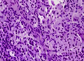
Figure 2. Histopathology of the lesion demonstrates large histiocytes with foamy cytoplasm surrounded by small inflammatory cells (hematoxylin and eosin, magnification 400×).

Figure 3. Pre-radiotherapy PET scan showing SUVmax 10.8 hypermetabolic activity from the coxofemoral region into the femoral shaft.
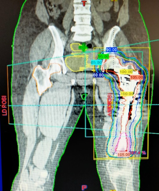
Figure 4. Beam ’s-eye view of the treatment plan (6 MV photons, AP/PA configuration).
3. Discussion
RDD displays diverse clinical manifestations and variable outcomes. Osseous involvement, while uncommon, can be a source of persistent pain and functional limitation. This case underscores key diagnostic and therapeutic points relevant to skeletal RDD.
Rosai-Dorfman Disease (RDD) primarily affects children and young adults, with a higher prevalence observed in males and individuals of African descent. The cutaneous form of RDD is more frequently seen in female Asians (1).
Distinguishing RDD from Erdheim–Chester disease and Langerhans cell histiocytosis requires integration of clinical, radiologic, and immunohistochemical data. ECD typically presents with long-bone diaphyseal osteosclerosis and can harbor MAPK pathway mutations, while LCH shows CD1a and Langerin positivity. In contrast, RDD classically shows positivity for S100 and CD68/CD163, with negativity for CD1a (2)
Selected series have reported increased IgG4-positive plasma cells in a subset of RDD, although the significance of this finding remains uncertain and likely context-dependent. Incorporating IgG4-related disease into the discussion may avoid misclassification in cases with abundant plasma cells and fibrosis (3).
Most patients with classic (nodal) RDD present with bilateral, massive, painless cervical lymphadenopathy (4). Bone involvement in RDD, although uncommon, is documented in 5-10% of cases and is typically associated with nodal disease. Patients frequently present with bone pain, while pathological fractures remain uncommon. The lesions are typically located in the metaphyseal or diaphyseal regions of long bones and appear as osteolytic or mixed lytic-sclerotic on imaging, usually with a narrow zone of transition. The radiologic differential diagnosis is broad and includes chronic osteomyelitis, fibrous dysplasia, lymphoma, and Ewing sarcoma (5). The recurrence of symptoms in this patient, despite initial surgical intervention, underscores the challenge of managing skeletal RDD.
A significant milestone in understanding Rosai-Dorfman disease was the publication of the consensus guidelines by Abla et al. in 2018, which offer comprehensive, structured recommendations for its diagnosis and clinical management. These guidelines emphasize a systematic approach, including a detailed medical history, thorough physical and neurological examinations, and appropriate imaging modalities, particularly CT and PET-CT scans. These tools are essential not only for accurate staging but also for detecting disease recurrence and for informing individualized treatment strategies (5)
The management of RDD lacks a standardized approach, with treatment strategies often tailored to the individual patient’s clinical presentation. Observation is often appropriate, as spontaneous remissions occur in 20-50% of patients with nodal or cutaneous disease (6). Surgical intervention is usually limited to biopsy, but resection can be curative for unifocal disease (7). Chemotherapy has shown mixed results, with low-dose methotrexate and 6-mercaptopurine demonstrating efficacy in some cases (8)
Radiotherapy, although modestly effective, can be beneficial for refractory soft tissue and bone disease (9). As a benign inflammatory histiocytic proliferation, RDD often responds to moderate-dose irradiation through reduction in histiocytic infiltration and inflammatory activity. Published experience—largely case reports/series—describes doses of 20–50 Gy (1.8–2 Gy/fraction) achieving symptom relief and local control in many cases (10). Our choice of 20 Gy/10 fractions balanced efficacy and toxicity for a focal, symptomatic site.
Pain relief within weeks and decreased PET avidity at 3 months align with reports of early symptomatic benefit. To improve interpretability in RDD, response documentation can include pain scores, analgesic use, performance status, and, where feasible, standardized PET criteria (e.g., PERCIST).
Radiotherapy can be combined with or sequenced to surgery (for debulking or stabilization), corticosteroids, and selected immunomodulatory or cytotoxic agents in multifocal or refractory disease, tailored to the extent and symptoms.
Overall, in the absence of standardized protocols, individualized multidisciplinary care remains central, guided by consensus recommendations and case-based evidence.
4. Conclusion
This case illustrates durable symptom control with palliative radiotherapy for recurrent skeletal RDD, with the patient remaining clinically asymptomatic and without radiographic progression at 15 months. The report emphasizes careful differential diagnosis and supports moderate-dose radiotherapy as a safe, effective option within a multidisciplinary strategy.
Statements
Conflicts of interest: There are no conflicts of interest.
Ethical Approval and Consent to Participate: This case report was conducted in accordance with ethical standards. Ethical approval was obtained from the appropriate ethics committee. Written informed consent was obtained from the patient.
Consent for Publication: Written informed consent was obtained from the patient for publication of this case report and images.
Availability of Supporting Data: Available from the corresponding author upon reasonable request.
Competing Interests: The authors declare no competing interests.
Funding: No specific grant was received.
Authors’ Contributions: YDD conception, drafting, and final approval: MAE: technical support, review; KS: data collection, review, and critical revisions; SK: data analysis, review; TB: supervision, critical feedback.
Acknowledgements: We thank the patient for cooperation and consent.
References
-
Kutlubay Z, Bairamov O, Sevim A, Demirkesen C, Mat MC. Rosai-Dorfman disease: a case report with nodal and cutaneous involvement and review of the literature. Am J Dermatopathol. 2014;36(4):353-7.
-
Emile JF, Abla O, Fraitag S, et al. Revised classification of histiocytoses and neoplasms of the macrophage-dendritic cell lineages. Blood. 2016;127(22):2672–2681.
-
Bruce-Brand C, Schneider JW, Schubert P. Rosai-Dorfman-Destombes disease: an overview of histology and pathogenesis with emphasis on IgG4-positive plasma cells. J Clin Pathol. 2020;73(11):697-705.
-
Al-Khateeb TH. Cutaneous Rosai-Dorfman disease of the face: a comprehensive literature review and case report. J Oral Maxillofac Surg. 2016;74(3):528-40.
-
Abla O, Jacobsen E, Picarsic J, Krenova Z, Jaffe R, Emile JF, et al. Consensus recommendations for the diagnosis and clinical management of Rosai-Dorfman-Destombes disease. Blood. 2018;131(26):2877-90.
-
Lima FB, Barcelos PS, Constancio AP, Nogueira CD, Melo-Filho AA. Rosai-Dorfman disease with spontaneous resolution: case report of a child. Rev Bras Hematol Hemoter. 2011;33(4):312-4.
-
Horneff G, Jurgens H, Hort W, Karitzky D, Gobel U. Sinus histiocytosis with massive lymphadenopathy (Rosai-Dorfman disease): response to methotrexate and mercaptopurine. Med Pediatr Oncol. 1996;27(3):187-92.
-
Maklad AM, Bayoumi Y, Tunio M, Alshakweer W, Dahar MA, Akbar SA. Steroid-resistant extranodal Rosai-Dorfman disease of cheek mass and ptosis treated with radiation therapy. Case Rep Hematol. 2013;2013:428297. doi: 10.1155/2013/428297.
-
Toguri D, Louie AV, Rizkalla K, Franklin J, Rodrigues GB, Venkatesan V. Radiotherapy for steroid-resistant laryngeal Rosai-Dorfman disease. Curr Oncol. 2011;18(3):e158-62.