


Author(s) :
Alisha Sharma1, Avtar Singh1, Tapas Kumar Dora1, Amit Saini1, Jayashree Deshmukh1
1 Homi Bhabha Cancer Hospital and Research Centre, Punjab, India
Corresponding author: Tapas Kumar Dora, Email: dr.tapasdora.scb.ctc@gmail.com
Publication History: Received - 13 October 2025, Revised - 7 December 2025, Accepted - 29 December 2025, Published Online - 29 December 2025.
Copyright: © 2025 The author(s). Published by Casa Cărții de Știință.
User License: Creative Commons Attribution – NonCommercial (CC BY-NC)

 PDF
 Share
Highlights
-
Volumetric Modulated Arc Therapy (VMAT) is a feasible and efficient method for delivering palliative radiation to diffuse calvarial metastases, particularly for breast cancer patients.
-
The study demonstrates that VMAT-based Brain Sparing Calvarial Irradiation (BSCI) achieves a mean brain dose of approximately 20.5 Gy, preserving the parenchyma for potential future treatments.
-
Using coplanar arcs and Monte Carlo algorithms, the technique ensures high target conformity and homogeneity while maintaining critical organ-at-risk (OAR) doses within safe limits.
Abstract
Background: Diffuse metastasis to the skull bones can be a debilitating condition war- ranting whole calvarial irradiation, which is a technical challenge due to its curviform shape, further distorted by the metastatic disease, and the presence of critical structures in its concavity. Adding to this challenge is the paucity of data and the lack of consensus on Brain Sparing Calvarial Irradiation (BSCI) in terms of patient selection, optimal dose fractionation, and the most appropriate technique to do so in order to produce tangible symptomatic relief while restricting doses to the brain and other organs at risk (OARs).
Methods Six patients, aged more than 18 years, KPS (Karnofsky Performance Status Score) ≥ 70, no prior history of radiotherapy to the brain or skull, with radiological evi- dence of diffuse calvarial metastasis, treated with palliative BSCI to a dose of 30 Gy in 10 fractions between 2019 and 2021, were included in this analysis. By virtue of favorable dose distribution, technical ease of planning, simple daily setup, and faster treatment delivery, BSCI was planned using Volumetric Modulated Arc Therapy (VMAT) with two to four, full coplanar arcs with the isocenter placed at the center of the brain. The Monte Carlo algorithm was used for dose calculation. The maximum number of control points per arc was 150, the maximum segment width was 235 mm, and fluence smooth- ening was set to medium. Voxel-based optimization was performed, and the “layering order” was followed, with the brain given the highest priority.
Results: PTV ranged from 560.59 cc to 864.65 cc, with an average of 702.49 cc. The maximum and mean PTV doses ranged from 32.93 Gy to 36.46 Gy and from 30.17 Gy to 31.49 Gy, respectively. The Lesion Coverage Factor ranged between 0.924 and 0.973, the Healthy Tissue Conformity Index ranged between 0.323 and 0.418, the Heterogeneity Index ranged between 1.08 and 1.23, the Paddick Conformity Index ranged between 0.51 and 0.72, the RTOG Conformity Index ranged between 0.92 and 0.97, and the Homogeneity Index ranged between 0.107 and 0.292. The mean brain dose (Dmean) ranged from 18.56Gy to 21.37Gy. The mean Maxi- mum Dose (Dmax) to the brain, brainstem, optic chiasm, right & left optic nerves, right and left eyes, right and left lens were 33.15 Gy, 29.53 Gy, 27.52 Gy, 28.62 Gy, 28.39 Gy, 27.02 Gy and 27.08 Gy, 10.98 Gy and 10.88 Gy respectively.
Conclusion: VMAT-based BSCI is technically feasible for palliation of diffuse calvarial metastasis while sparing the brain and other organs at risk.
-
Introduction
Skull metastases occur in 15%–25% of all cancer patients, most often in those with a substantial systemic and skeletal disease burden (1). They can be either calvarial or involve the skull base, circumscribed or diffuse, intra-osseous or invasive (2). They may spread either hematogenously through retrograde seeding via Batson’s venous plexus, or by direct extension through cranial foramina (3). Progressive disease at these sites predisposes to bony compromise and neurological deficits, thereby affecting patients’ quality of life and warranting prompt management. However, treatment of metastatic lesions in the calvarial bones, un- like other bony sites, can be challenging owing to the proximity to the brain parenchyma (4).
A retrospective cohort study of 175 patients with skull metastasis (121 with calvarial, 97 with skull base lesions, and 43 with both) found breast cancer as the most common site of origin (55%), followed by lung (14%), prostate cancer (6%), while other primaries constituted 25% (2). A meta-analysis of 279 cases with skull-base metastases demonstrated the most frequent primary tumor to be prostate cancer (38.5%), followed by breast (20.5%) and lymphoma (8%) (5). Approximately 50% of the patients with calvarial metastases are asymptomatic, while the other half pre- sent with pain by virtue of outer table, periosteal, or dural involvement, and neurological deficits if the inner table is involved (4). Vikram and Chu reported radiotherapy (RT) to be an effective modality for palliation of such metastases yielding better results for patients with symptoms of short duration; with response rates being as high as 87% for patients with less than 1 month history, 69% with 1–3 month history, and 25% for those with >3 months history (6). Planning and delivering calvarial irradiation is a technical challenge, as it requires delivering a homogeneous dose to a curviform target while sparing the brain and other critical structures in its concavity. Despite RT planning and delivery being technically an uphill task and the extent of debilitating symptoms such patients pre- sent with, the data regarding the most appropriate local therapy to palliate diffuse calvarial metastasis remains scarce. This may be due to an inherently large and survival-limiting metastatic disease burden, such that brain and OAR sparing may not have been the foremost priority. But, with the advent of immuno- therapy and targeted therapy improving patient outcomes, it is imperative to spare the brain and other critical structures, not only to reduce treatment-related toxicity but also to reserve an opportunity for being able to irradiate the brain parenchyma in the future if brain metastasis were to occur.
2. Materials and Methods
This is a retrospective case series of six patients with histologically proven solid tumors, with radiological evidence of diffuse calvarial metastases with no extension into the brain parenchyma, treated with palliative radiotherapy between 2019 and 2021 at our rural cancer center. All patients were aged 18 or older, had a KPS of 70 or higher, an anticipated survival of at least 3 months, and had not previously received radiotherapy to the skull or brain. In addition to palliative RT, patients received palliative systemic therapy and palliative care as per institutional protocol.
Patients underwent planning simulation, Computerized Tomographic (CT) scan with individualized thermoplastic immobilization devices. Planning CT images were transferred to the Monaco treatment planning system (TPS) version 5.11.03 (copyright 2019 Elekta, Inc), for target delineation and RT planning. The entire bony calvarium was contoured as auto-segmentation on bone window & given a 0-2 mm margin to generate the planning target volume (PTV). Our choice of PTV margin was based upon the anticipated ease of bone (calvarial) matching on daily Cone Beam Computerized Tomography (CBCT) imaging. The PTV was prescribed a dose of 30 Gy in 10 fractions, administered 5 days a week over 2 weeks. All plans were VMAT-based, with 2 to 4 full 360-degree coplanar arcs, depending on the required conformity and plan complexity. Full arcs were used by virtue of their ability to provide an optimal combi- nation of dose conformity, OAR sparing, and beam modification, while treating a nearly 360-degree target. The isocenter was placed at the center of the brain. A calculation grid spacing of 3 mm was used. The dose deposition calculation was done for the medium. The dose calculation algorithm was Monte Carlo, with a 1% uncertainty. The Intensity Modulated Radiotherapy (IMRT) constrained mode was used for optimization. The maximum number of control points per arc was 150, the maximum segment width was 5 mm, and fluence smoothening was set to medium. Monaco uses voxel-based optimization and follows a “layering order” in which the brain received the highest priority, followed by the optic chiasm, then the PTV, and finally the remaining OARs. Optimization was performed with a combination of Target EUD and Quadratic Overdose cost functions to ensure good target coverage and high-dose region control. The Target EUD objective was used to achieve a uniform dose distribution within the PTV, while the Quadratic Over- dose constraint was applied to limit hotspots and maintain dose homogeneity. Serial and parallel cost functions were used for OARs. For the optic structures, the maximum point dose was controlled with
serial cost functions, while the mean dose was con- trolled with parallel cost functions. By giving the brain the highest optimization priority, a clinically adequate PTV coverage could be maintained while preferentially reducing the brain dose (Table 1).
The Lesion Coverage Factor (LCF), was calculated as ratio of the TVri (volume of the target covered by the reference isodose line) to the PTV. The Healthy Tissue Conformity Index (HTCI) was expressed as the ratio of TVri to Vri (Volume of the Reference Iso- dose). The Heterogeneity Index (HI) was calculated as Dose that covers x% of tissue, where x is high dose ref percentage / Dose that covers y% of tissue, where y is the minimum dose reference. The Homogeneity Index (HI) was calculated as per the ICRU 83 definition, D2%-D98%/D50%, where D2%, D98% and D50% are the doses received by 2, 98, and 50 percent of the PTV, respectively. The Paddick Conformity Index (CI) was calculated as TV12 / TV x VR1, where TV12 is the square of the volume of the target that receives the prescription dose, TV is the target volume, which is the same as the PTV, and VR1 is the total volume of the prescription isodose. CI as per the RTOG definition was calculated as VPIV/VTV, where PIV is the prescription isodose volume and TV is the target volume, same as the PTV, when using 100% as the reference isodose.
The planning process was carried out in two steps: first, fluence creation, and second, fluence con- version into a deliverable plan. Dose constraints were as per QUANTEC recommendations. Plan validation, i.e., patient-specific quality assurance (QA), was performed using the ArcCHECK system from Sun Nuclear Corporation. Gamma analysis was used to evaluate the data. As per AAPM TG-218 guidelines, the primary clinical tolerance level was 3%/3 mm global gamma criteria, with a 10% low-dose thresh- old. Supplementary analysis at 2%/2 mm was also carried out to assess plan adaptability. The mean gamma passing rate at 3%/3 mm across all evaluated VMAT plans was 99.5%±1.03%, with a minimum of 97.5%, and at 2%/2 mm was 98.03%±1.71%, with a mini- mum of 95.6%. These results suggested a high degree of concordance between the planned and delivered dose distribution. Absolute point-dose measurements were within ±3% of the TPS-calculated doses. Daily setup verification was performed using kilovoltage CBCT, incorporating bone & soft tissue matching.
Table 1. Planning parameters and cost function
|
Structure |
Cost function and Power Law Expo- nent (k) |
|
PTV |
EUD, Target penalty and Ǫuadratic Overdose
D98 at least 95% |
| Brain | Serial, k=12 Parallel, k=3 |
| Brainstem | Serial, k=12 |
| Optic Chiasm | Serial, k=12 |
| Optic nerve | Serial, k=12 |
| Eye | Serial, k=11 |
3. Results
All 6 patients were upfront metastatic breast cancers, all females between 30 and 75 years of age, with radiological evidence of diffuse calvarial metastases. Five out of six were Estrogen Receptor positive, three out of six were Progesterone Receptor positive, two out of six were Her2 positive, and one patient had a triple negative breast cancer. Four of the six patients had visceral metastases in addition to bone. Five of six patients completed RT with an average overall treatment time of 12.4 days. One patient warranted discontinuation of RT after 2 fractions in view of intestinal obstruction.
The PTV volume ranged between 560.59 cc and 864.65 cc, with an average of 702.49 cc. The maximum dose to the PTV ranged between 32.93 Gy and 36.46 Gy. The mean PTV doses ranged from 30.17 Gy to 31.49 Gy. The TVri ranged from 537.91 cc to 790.35 cc, with an average of 670.78 cc. The LCF ranged from 0.924 to 0.973, with an average of 0.956. The HTCI ranged from 0.323 to 0.418. The HI was between 1.08 and 1.23, with an average value of 1.12. The Paddick CI ranged between 0.51 and 0.72, with an average of 0.62. The CI as per the RTOG definition, ranged between 0.66 and 0.89, and when using 95% as the reference isodose, it was 0.92 to 0.97. The HI ranged from 0.107 to 0.292. Planning in- dices observed in individual cases are listed in
Table 2. For dose distribution, refer to Figures 1(a and b) and 2 (a and b).
| Patient No. | HI (ICRU 83) | CI
(Paddick) |
CI (RTOG) | HI | LCF | HTCI |
| 1 | 0.137 | 0.57 | 0.94 | 1.10 | 0.9595 | 0.341 |
| 2 | 0.153 | 0.60 | 0.95 | 1.10 | 0.9518 | 0.332 |
| 3 | 0.107 | 0.51 | 0.96 | 1.08 | 0.9690 | 0.323 |
| 4 | 0.155 | 0.68 | 0.97 | 1.12 | 0.9733 | 0.356 |
| 5 | 0.162 | 0.63 | 0.96 | 1.12 | 0.9609 | 0.355 |
| 6 | 0.292 | 0.72 | 0.92 | 1.23 | 0.9247 | 0.418 |
| HI-Homogeneity Index, CI-Conformity Index, HI- Heterogeneity Index, LCF- Lesion Coverage Factor,
HTCI- Healthy Tissue Conformity Index |
||||||
As far as the OARs are concerned, the Dmax ranges were as follows, brain stem 26.47 Gy to 32.15 Gy, optic chiasm 25.47 Gy to 31.30 Gy, left eye 19.26 Gy to 31.23 Gy, right eye 18.36 Gy to 33.37 Gy, right lens 7.90Gy to 17.76 Gy, left lens 7.91 Gyto 16.64 Gy, right optic nerve 26.17 Gy to 33.34 Gy and left optic nerve was 25.37 Gy to 31.62 Gy. The Dmax and Dmean to the brain parenchyma ranged from 32.41 to 34.61 Gy & 18.56 Gy to 21.37 Gy respectively. For individual patient OAR doses please refer to Table 3.
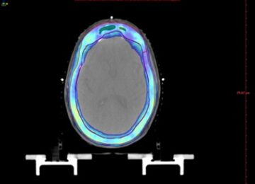

Figure 1. The 95% dose wash in PTV_Calvarium in the axial (a) and sagittal plane (b)


Figure 2. The 50% dose wash in PTV_Calvarium in the axial (a) and sagittal plane (b)
Table 3. Individual OAR doses
| Patient
No. |
OAR doses in Gy | ||||||
| Brain
(Dmax, Dmean) |
Brain-
stem (Dmax) |
Optic
Chiasm (Dmax) |
Optic
Nerve Right (Dmax) |
Optic
Nerve Left (Dmax) |
Eye Right
(Dmax Dmean) |
Eye
Left (Dmax) Dmean) |
|
| 1 | 32.49
21.30 |
30.33 | 27.51 | 26.23 | 26.66 | 18.36
11.44 |
19.26
12.21 |
| 2 | 32.48
21.37 |
31.28 | 25.47 | 28.39 | 27.69 | 30.00
20.19 |
28.24
16.41 |
| 3 | 32.41
19.65 |
26.49 | 31.30 | 29.97 | 29.84 | 26.61
18.95 |
27.35
17.69 |
| 4 | 33.46
21.05 |
30.47 | 29.06 | 26.17 | 29.17 | 25.96
12.18 |
27.78
14.09 |
| 5 | 33.50
21.11 |
32.15 | 26.06 | 27.53 | 25.37 | 27.81
10.72 |
28.64
11.18 |
| 6 | 34.61
18.56 |
26.47 | 25.73 | 33.44 | 31.62 | 33.37
16.70 |
31.23
13.37 |
4. Discussion
The calvarium is the convexity of the roof of the skull that encases the brain parenchyma. It includes the paired frontal, parietal, temporal (squamous part), and the interparietal part of the occipital bone. These are composed of two layers of inflexible cortical bone, namely the inner and outer tables, which enclose a layer of soft cancellous bone, the diploë. The average thickness of the skull bones is between 2 and 6 mm. It is surrounded by endosteal and periosteal sheaths, which are innervated by meningeal, nociceptive fibers as well as sympathetic and parasympathetic fibers (7). The skull base, on the other hand, is composed of flat bones, namely the ethmoid, sphenoid, occipital, paired frontal, and paired temporal bones, which separate the intracranial compartment from the extracranial head and neck (8). It forms the floor of the cranial cavity and is among the most complex anatomical structures (9). Owing to the said area being densely packed with critical neurovascular structures, radio- therapy often emerges as a feasible and safe alternative to surgery for addressing neoplastic lesions of the base. The calvarium or the roof of the skull, though may not share the complexity of the base from a surgical point of view, is a formidable challenge as far as radiotherapy is concerned by virtue of its distinctive convex geometry worsened by the skeletal distortion caused by the metastasis (4)(10).
Brain sparing palliative radiotherapy has been at- tempted in the past, such as that by Wolden et al in cases of diffuse skull metastasis in neuroblastoma, using conventionally planned electron-photon fields to doses between 21 and 30 Gy with varied fractionation schedules. They found that the brain-sparing pho- ton/electron technique delivers a higher dose to the skull bones and a lower mean dose to the brain com- pared with whole-brain palliative RT (11). An evaluation of whole-skull radiotherapy for metastasis has also been conducted by G.S. Gautam et al., who com- pared IMRT vs VMAT and found greater uniformity in dose distribution, less dose spillage to the underlying brain, and lower OAR doses with VMAT (12). Kotecha et al., on the other hand, advocate the use of Gamma Knife Surgery for skull metastasis because of excellent local control, rapid dose fall-off, limited dose to adjacent normal tissue, the opportunity for significant dose escalation, and the ability to treat irrespective of prior radiotherapy (4). However, this is not a feasible option for diffuse metastases. In short, there is no consensus on the most appropriate radiotherapy technique for treating diffuse calvarial metastases due to the limited reporting, but it is not unwise to extrapolate from experience treating the scalp.
A number of techniques have been explored in literature for scalp radiotherapy such as (a) combined photon–electron technique with shifting junctions, where lateral electron fields were matched with lateral photon fields(13), (b) photon and electron fields over- lapped by 3–4 mm to improve dose uniformity, again shifting the junction midway through the treatment (14), (c) photon shell technique with four 90° photon arcs around the head with central blocks for sensitive structures(15), (d) Mould-based high dose rate (HDR) brachytherapy, (e) IMRT, VMAT, Helical Tomotherapy and (f) Protons.
Among the various techniques, 3D-conformal radiotherapy was associated with a higher integral dose, but OARs inadvertently received the treatment dose. IMRT and VMAT were found to have better conformity to the PTV and lower OAR doses, especially when OARs were present in the concavity of the PTV, for example, the brainstem in the concavity of the nasopharynx or the brain in the concavity of the scalp (16). Wojcicka et al., upon evaluating different treatment techniques in extensive scalp irradiation (17), found IMRT to provide the best PTV coverage and the most homogeneous dose within the target. The HDR technique could spare the brain well (particularly in terms of maximum doses), but failed in terms of optical structures. IMRT irradiated a larger volume to a lower dose, whereas the 3D-CRT plan irradiated larger volumes to higher doses, resulting in more than 100% hotspots within the brain. The pho- ton-electron technique, although producing low doses in optical structures, was the least conformal and most cumbersome to implement.
VMAT for scalp lesions is more advantageous, with lower planning technical demands, faster daily setup and delivery, and simpler dose calculations. In addition, it offers excellent dose conformity and homogeneity. The main disadvantage of VMAT how- ever is the increased percentage of low-dose radiation received by the underlying brain (18). Scalp irradiation performed by the same group as above used four half-field arcs and one vertex arc. The mean dose to the brain in the composite plan was 1892 cGy, which is similar to the mean Dmean brain observed in our study, which was 20.50 Gy. However, it must be noted that Lozano et al have treated patients to a dose of up to 60 Gy with conventional fractionation and used a 1 cm bolus. In another study, available in abstract form by Dibs et al., on scalp VMAT using a 3D- milled bolus, in which the whole scalp was irradiated in 40% of cases, the median mean brain dose was 15.4 Gy, which is similar to our results. Their median eye Dmax is 10.9 Gy, which is quite low compared to the mean Dmax Eye in our study (27 Gy) and can be at- tributed to the more superficial location of their PTV (19). The VMAT FF plans, generated by Inui et al. for the treatment of scalp angiosarcomas, used 4 to 7 partial-to-full, non-coplanar arcs. Similar mean brain doses were achieved, to the order of 17.11 ± 5.25 Gy; however, doses to the remaining OARs were lower, as expected (20). In another recent study comparing IMRT with RapidArc for the treatment of skull lesions, doses ranged from 28 Gy to 54Gy. In cases pre- scribed 28 to 30 Gy (4/10), the doses achieved in their RapidArc plans versus our study were as follows: mean Dmean brain 19.48 Gy vs 20.50 Gy, mean Dmax brainstem 28.04 Gy vs 29.53 Gy, and mean Dmax optic chiasm 20.97 Gy vs 27.52 Gy (12).
Owing to the lack of consensus on the most appropriate dose fractionation for BSCI, we used a dose of 30 Gy in 10 fractions based on the results of RTOG 97-14 (21), which compared multi-fraction versus single-fraction palliative radiotherapy for pain- ful bone metastasis. A similar dose fractionation was used by Vikram and Chu in their 1979 study on radiotherapy for skull base metastasis, which ranged from 24 to 36 Gy (3 Gy per fraction) using 2-dimensional techniques, with nearly 80% of patients reporting significant and sustained symptomatic relief (6). In a study by Gautam et al., doses prescribed for skull metastasis ranged from 28 to 54 Gy at conventional fractionation. In a retrospective analysis by Onoe et al. on fractionated EBRT for skull base metastases, a median dose of 30 Gy over 10 fractions was used. They found that among the 29 of 41 patients for whom neurological outcomes were evaluated, 41% had significant improvement in symptoms, 41% mild improvement, and 17% had no change (22). A case series (n=3) from TMH Mumbai published in 2009 that evaluated brain sparing whole skull/scalp radiotherapy, for ex- tensive skull/scalp involvement, with Helical Tomotherapy, used a dose of 23.4 Gy in 13 fractions for one case of Neuroblastoma and 45 Gy in 25 fractions for 2 cases of Non-Hodgkins Lymphoma, reported excel- lent target coverage, significant brain sparing and con- formal avoidance of OARs (23).
BSCI is a technically feasible and safe modality of radiotherapy for palliating patients with multiple calvarial lesions with no or limited brain parenchymal extension, with fair to good performance status, and an anticipated survival of over 3 months. However, data on its effectiveness remain limited. The available literature, including ours, is mainly retrospective and dosimetric. Patient-related outcomes have been re- ported only in studies involving a limited number of calvarial metastases or lesions confined to the skull base, rather than the calvarium per se. Another limitation of our study is the small sample size, whereby all six cases happened to be primary breast cancers, questioning its applicability to other primaries. How- ever, in other studies, BSCI has been used for skull metastases arising from a range of solid tumors and hematolymphoid malignancies, with breast cancer emerging as the most common site.
5. Conclusion
VMAT-based BSCI is a feasible modality for treating diffuse calvarial metastasis as it provides a favorable dose distribution both in terms of conformity and homogeneity while sparing the brain and other critical structures. The need of the hour, however, are larger, prospective, even multi-institutional studies to generate more robust data to establish protocols for calvarial metastasis management, and this work could pave the way for the same.
ABBREVIATIONS
3DCRT – 3-Dimensional Conformal Radiotherapy
CI – Conformity Index
CT – Computerised Tomography
Dmax – Maximum Dose
Dmean – Mean Dose
EUD – Equivalent Uniform Dose
HDR – High Dose Rate
HI – Heterogeneity Index
HI – Homogeneity Index
HTCI – Healthy Tissue Conformity Index
ICRU – International Commission on Radiation Units and Measurements
IMRT – Intensity Modulated Radiation Therapy
LCF – Lesion Coverage Factor OAR – Organ At Risk
PIV – Prescription Isodose Volume
PTV – Planning Target Volume
QUANTEC – Quantitative Analyses of Normal Tissue Effects
TV – Target Volume
TV1 – Volume of the target that receives the prescription dose
VMAT – Volumetric Modulated Arc Therapy
VR1 – Total volume of the prescription isodose
STATEMENTS
Authors contributions: Conception of the study TD; acquisition of data AS; analysis and interpretation of data AS, AS*, AS** JD; drafting the manuscript AS; finalizing the manuscript AS, AS*, TD.
Consent for publication: As the corresponding author, I confirm that the manuscript has been read and approved for submission by all named authors.”
Conflicts of Interest: The authors have no conflicts of interest to declare.
Funding: None.
Ethics: Being purely a retrospective dosimetric analysis of radiotherapy plans for previously treated patients, with no such references linking them to this study, ethics committee approval was not sought.
References
- Posner JB, Chernik Intracranial metastases from systemic cancer. Adv Neurol. 1978;19:579-592.
- Mitsuya K, Nakasu Y, Horiguchi S, et Metastatic skull tumors: MRI features and a new conventional classification. J Neurooncol. 2011;104(1):239-245. doi: 10.1007/s11060-010-0465-5.
- Bradley WG, Neurology in clinical practice. Butterworth-Heinemann; 2004.
- Kotecha R, Angelov L, Barnett GH, et Calvarial and skull base metastases: expanding the clinical utility of Gamma Knife surgery. J Neurosurg. 2014;121 Suppl:91-101. doi: 10.3171/2014.7.GKS141272.
- Laigle-Donadey F, Taillibert S, Martin-Duverneuil N, Hildebrand J, Delattre Skull-base metastases. J Neurooncol. 2005;75(1):63-69. doi: 10.1007/s11060-004-8099-0.
- Vikram B, Chu Radiation therapy for metastases to the base of the skull. Radiology. 1979;130(2):465-468. doi: 10.1148/130.2.465.
- Harrison RA, Nam JY, Weathers SP, DeMonte Intracranial dural, calvarial, and skull base metastases. Handb Clin Neurol. 2018;149:205-225. doi: 10.1016/B978-0-12-811161-1.00014-1.
- Borges Skull Base Bone Lesions I. In: Head and Neck Imaging. Elsevier; 2018. doi: 10.1016/B978-0-323-48563-0.00015-5.
- Gao H, Liu C, Zhang Neuro-endoscope for skull base tumors. Clin Neurol Neurosurg. 2018;170:102-105. doi: 10.1016/j.cli- neuro.2018.05.009.
- Caivano R, Fiorentino A, Pedicini P, Califano G, Fusco A radiotherapy technique for palliative total scalp irradiation. Ann Palliat Med. 2015;4(1):35-38. doi: 10.3978/j.issn.2224-5820.2015.01.02.
- Wolden SL, Barker CA, Kushner BH, et Brain-sparing radiotherapy for neuroblastoma skull metastases. Pediatr Blood Cancer. 2008;50(6):1163-1168. doi: 10.1002/pbc.21384.
- Gautam GS, Rathore VB, Jain RR, Kushwaha P, Singh RS, et Brain Saving Radiotherapy in Skull Metastases using Volumetric Modulated Arc Technique: A Dosimetric Analysis. IOSR J Dent Med Sci. 2022;21(4):26-34. doi: 10.9790/0853-2104052634.
- Akazawa C. Treatment of the scalp using photon and electron beams. Med Dosim. 1989;14(2):129-131. doi: 10.1016/0958- 3947(89)90184-2.
- Tung SS, Shiu AS, Starkschall G, Morrison WH, Hogstrom Dosimetric evaluation of total scalp irradiation using a lateral electron-photon technique. Int J Radiat Oncol Biol Phys. 1993;27(1):153-160. doi: 10.1016/0360-3016(93)90433-v.
- Kinard JD, Zwicker RD, Schmidt-Ullrich RK, Kaufman N, Pieters Short communication: Total craniofacial photon shell tech- nique for radiotherapy of extensive angiosarcomas of the head. Br J Radiol. 1996;69(820):351-355. doi: 10.1259/0007-1285-69- 820-351.
- Zhang RL, Dunkerley N, Wright K, et Superior outcomes of Volumetric Modulated Arc Therapy (VMAT) in the palliation of skull metastases of multiple myeloma: a case report. Int J Radiol Radiat Ther. 2021;8(4):139-142. doi: 10.15406/ijrrt.2021.08.00306.
- Wojcicka JB, Lasher DE, McAfee SS, Fortier Dosimetric comparison of three different treatment techniques in extensive scalp lesion irradiation. Radiother Oncol. 2009;91(2):255-260. doi: 10.1016/j.radonc.2008.09.022.
- Lozano F, Perez N, Iglesias A, et Volumetric arc therapy for total scalp irradiation: case report for a recurrent basal cell carcinoma of the scalp. Ecancermedicalscience. 2017;11:737. doi: 10.3332/ecancer.2017.737.
- Dibs K, Konieczkowski DJ, Grecula JC, Mitchell DL, Baliga S, Gogineni E, et Scalp Volumetric Modulated Arc Therapy Using 3D Milled Bolus: Dosimetry, Toxicity and Outcome. Int J Radiat Oncol Biol Phys. 2023;117(2 Suppl):e292-e293. doi: 10.1016/j.ijrobp.2023.06.1289.
- Inui S, Ueda Y, Ohira S, et Novel strategy with the automatic non-coplanar volumetric-modulated arc therapy for angiosarcoma of the scalp. Radiat Oncol. 2020;15(1):175. doi: 10.1186/s13014-020-01614-3.
- Hartsell WF, Scott CB, Bruner DW, et Randomized trial of short- versus long-course radiotherapy for palliation of painful bone metastases. J Natl Cancer Inst. 2005;97(11):798-804. doi: 10.1093/jnci/dji139.
- Onoe T, Kawashiro S, Sumita K, Ogawa H, Harada H, Asakura H, et Fractionated External Beam Radiation Therapy for Skull Base Metastases With Cranial Nerve Involvement. Int J Radiat Oncol Biol Phys. 2015;93(3):E469. doi: 10.1016/j.ijrobp.2015.07.1747.
- Mallik S, Master Z, Gupta T, Laskar S, Muckaden Brain Sparing Whole Skull/Scalp Radiotherapy: Unique Application of Helical Tomotherapy. Int J Radiat Oncol Biol Phys. 2009;75(3 Suppl):S729. doi: 10.1016/j.ijrobp.2009.07.1663.