

Author(s) :
Taha Ismail Mahmoud Hewala1, Sherif Elzawawy 2 , Sabbah I. Hammoury3, Walaa Ibrahim3
1 Department of Radiation Science, Alexandria University, Egypt
2 Department of Clinical Oncology and Nuclear Medicine, Faculty of Medicine, Alexandria University, Egypt
3 Department of Radiation Oncology, Ayadi Almostakbal Oncology Hospital, Alexandria, Egypt
Corresponding author: Taha Ismail Mahmoud Hewala, Email: tahahewala@hotmail.com
Publication History: Received - 27 June 2025, Revised - 13 October, Accepted - 30 December 2025, Published Online - 30 December 2025.
Copyright: © 2025 The author(s). Published by Casa Cărții de Știință.
User License: Creative Commons Attribution – NonCommercial (CC BY-NC)

 PDF
PDF
 Share
Share
Highlights
• Total Skin Electron Beam Irradiation (TSEBI) with a fixed platform effectively delivered a homogeneous distribution of radiation doses across the majority of the body surface for patients with mycosis fungoides.
• Specific body areas were identified as receiving significantly lower doses due to shielding or positioning, most notably the under the breast fold in under the left arm in men and on the shoulder in tall patients.
• A rotating platform instead of a fixed one could improve dose uniformity, and TLDs should be used routinely to monitor individual dose distribution and ensure treatment accuracy.
Abstract
Background: The aim of the study was to evaluate radiation dose distribution at different body sites for mycosis fungoides patients treated by total skin electron beam irradiation (TSEBI) using a fixed platform.
Materials and methods: We included patients with Mycosis Fungoides who underwent TSEBI from January 2020 to December 2022. Eighteen TLD chips were used to measure the dose distribution at 18 points on the whole-body skin for each patient. All treatment fields were delivered with a Varian VitalBeam linear accelerator (A Siemens Healthineers Company) at a collimator angle of 0°, using an electron applicator of 25×25 cm2 and an SSD of 4 meters.
Results: We included 10 patients (4 women and 6 men) with similar age and body weight. TLD located above the shoulder received a lower mean dose (83.90 ± 1.98 cGy). The TLD lying under the arm received a lower dose (74.07 ± 5.77 cGy). TLDs no 4 (left inner forearm) and no. 11 (right outer forearm) received lower doses (87.52 ± 1.29 cGy and 86.63 ± 1.27 cGy, respectively). The points lying near the beam center (TLDs no. 2, 5, and 9) received higher doses (101.63 ± 1.23 cGy, 105.61 ± 1.04 cGy, and 101.80 ± 1.24 cGy, respectively) than those lying on the peripheral points (TLDs no. 3 and 10), which received 101.35 ± 0.76 cGy and 101.33 ± 0.91 cGy doses, respectively. For female patients, TLDs under the breast fold received the least dose, 80.54 ± 28 cGy.
Conclusion: The use of TSEBI with a fixed platform enabled a homogeneous distribution of absorbed radiation doses across most of the body surface in skin cancer patients
1. Introduction
Mycosis fungoides (MF) is one of the most common types of cutaneous lymphoma, a cancer that originates in the skin and accounts for about 50% of all skin lymphomas. About 75% of primary cutaneous lymphomas arise from T cells, a type of white blood cell important for immune function, and mycosis fungoides may be present in 2/3 of these T-cell cases (1, 2).
MF occurs at a rate of 6-7 cases per million healthy persons, with marked regional variation. The incidence is higher in blacks and is most common in adults and elderly patients over 50 years old (1).
Before receiving a conclusive diagnosis, many individuals experienced symptoms for years. These were often mistaken for eczema (a condition that causes itchy, inflamed skin), psoriasis (a disease that causes red, scaly patches), or similar conditions. The average time from symptom onset to diagnosis is 3 to 4 years, though in some cases it exceeds decades (1, 3-5).
Total skin electron irradiation (TSEI) has been a well-known therapy for about a century. It is particularly effective in treating primary cutaneous T-cell lymphoma, a cancer arising from skin T cells—most commonly mycosis fungoides. TSEI delivers a consistent dose across the skin surface, targeting superficial irritated plaques (raised lesions) and patches (flat discolored areas). This targeted approach kills superficial cancer cells without harming the underlying tissue (6).
Irradiating the entire skin can be difficult. Over the past few years, various radiotherapy modalities have been investigated. These include (I) large-electron-field techniques, (II) rotational techniques, and (III) techniques involving patient shift during irradiation. With large electron field techniques, the patient stands during irradiation. Within one treatment fraction, the patient’s position changes six times to deliver the dose. The sequence starts with the anterior position, facing the gantry, then moves to the front-lateral right side, the back-lateral left side, the posterior, the back-lateral right side, and finally the front-lateral left side. Positions are rotated every 60 degrees around the patient’s longitudinal axis. The raised hand indicates whether the posture is to the right or left. (7).
In vivo dosimetry is essential for confirming and validating the dose distribution. Thermoluminescent dosimetry is most commonly used to verify doses in TSEI, though other in vivo dosimetric techniques are available. (8, 9). In the current study, thermoluminescent dosimeters were used. Its importance lies in the fact that the amount of light emitted is proportional to the absorbed dose in the irradiated material (8-10).
2. Patients and Methods
We included consecutive patients diagnosed with mycosis fungoides who underwent the Stanford technique with six dual-electron fields at Ayady Al-mostakbl Oncology Hospital in Alexandria, Egypt from January 2020 to December 2022. We excluded the patients under 18 years old. Participation was voluntarily and the patients provided written consent prior to their inclusion, in accordance with the institution’s ethical guidelines.
The Varian (A Siemens Healthineers Company) VitalBeam linear accelerator was used. An electron energy of 6 MeV was applied at a dose rate of 2500 MU/min. Lithium Fluoride (LiF) is an alkali halide widely used in constructing personal dosimeters (TLD-100). In this study, 18 TLD-100 chips were used and distributed across the patient’s body surface. The chips measured 3.1 mm × 3.1 mm × 0.38 mm, as shown in Figure 1. TLDs were annealed before use for dosimetry to improve stability and reduce fading. Annealing was performed at 400°C for 1 hour, followed by fast cooling. TLDs were calibrated before use. Calibration involved determining element correction coefficients (ECC) and plotting a calibration curve.
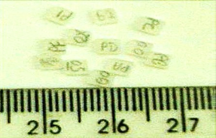
Figure 1. TLD-100 chips with dimensions of 3.1mm×3.1mm×0.38mm
A scattering filter was employed to increase dose homogeneity along the body’s longitudinal axis. It was placed 20 cm from the patient’s body. The patient was standing on a fixed table 25 cm high. TLD Reader (Harshaw 3500) was used to provide cost-effective measurements of the radiation dose absorbed by individual TLD chips.
Eighteen commercial TLD-100 chips were used and distributed over the entire body surface: anterior and posterior sides of the head, breast folds, shoulders, thighs, elbows, forearms, biceps, hips, clavicles, the middle of the groin, and both feet. After chip placement, each patient stands upright in the treatment room with raised arms, so the gantry’s horizontal axis is perpendicular to the body (Figure 2).
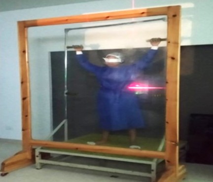
Figure 2. The patient standed in an upright position
The treatment position was changed every 60° around the patient’s longitudinal axis. The treatment position’s right or left orientation is determined by the patient’s raised hand. Every patient standed with angles of (0, 60, 120, 180, 240, 300), so the treatment sites are anterior, right, and left anterior oblique, posterior, right, and left posterior oblique (Figure 3).
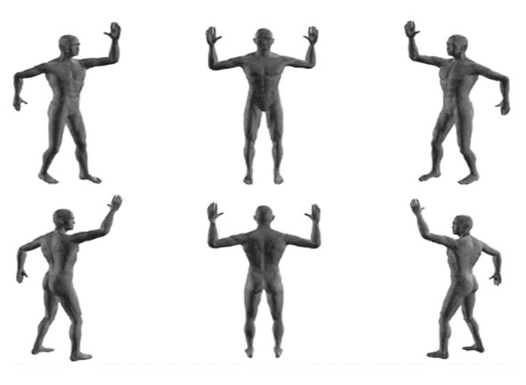
Figure 3. Treatment positions (7)
In the Stanford technique, dual large electron fields are used to guarantee the coverage of the entire patient’s body with electron radiation. For each patient position on the fixed platform, the gantry of the machine rotates at 258.2° to cover the upper body and 281.2° to cover the lower body (Figure 4).
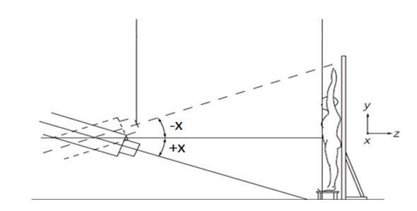
Figure 4. Gantry angles during the Stanford technique (7).
After the treatment session, TLDs were collected from the patient’s body surface. TLDs were read using a Harshaw 3500 TLD reader at temperatures ranging from 55°C to 400°C after 24 hours of irradiation. By obtaining the TLD readings of the irradiated dosimeters in µC, a relationship with the absorbed dose was established, yielding a calibration curve with a linear fit. The calibration curve shows a linear behavior in the studied dose ranges (zero-400 cGy), which means a constant coefficient ratio for dose absorption between all the used TLDs (Figure 5).
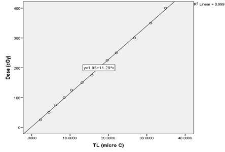
Figure 5. Calibration curve of TLDs
3. Results
Four of the 10 included patients were women and six were men. The patients had a similar mean age (41 for women and 43 for men) and similar weight (74 and 75 kg, respectively). For the female, the mean height was lower (157 cm), compared to median height of men (169 cm). More details are mentioned in Table 1.
Table 1. Patients’ characteristics
| Mean±SD | Female Patients
(n=4) |
Male patients
(n=6) |
|
| Age (years) | 41.5±17.39 | 43.83±14.30 | |
| Weight (KG) | 74±11.80 | 74.83±14.03 | |
| Height (Cm) | 157.25±8.18 | 169.33±10.84 | |
| Stage | I | 75% | 50% |
| II | 25% | 50% | |
The mean absorbed doses measured by TLDs varied across skin locations. The maximum absorbed dose was for the left inner thigh (TLD 6), for all patients The shoulder region received lower doses, especially in tall patients (Table 2).
Table 2. Mean±SD of absorbed doses by TLDs for all patients under the study
| no. | TLD site | Mean ± SD (cGy) | no. | TLD site | Mean ± SD (cGy) | |
| 1 | Forehead | 96.80 ± 1.12 | 10 | Rt. outer humerus | 101.33 ± 0.91 | |
| 2 | Lt. ant. chest | 101.63 ± 1.23 | 11 | Rt. outer forearm | 86.63 ± 1.27 | |
| 3 | Lt. inner humerus | 101.35 ± 0.76 | 12 | Rt. post. thigh | 109.67 ± 1.53 | |
| 4 | Lt. inner forearm | 87.52 ± 1.29 | 13 | Rt. post. knee | 110.74 ± 1.08 | |
| 5 | below the navel | 105.61 ± 1.04 | 14 | Rt. buttocks | 108.83 ± 1.02 | |
| 6 | Lt. inner thigh | 111.72 ± 1.04 | 15 | under breast fold | 80.54 ± 2.8 | |
| 7 | Lt. post. tibia | 106.52 ± 1.45 | 16 | Rt. clavicle | 91.58 ± 1.46 | |
| 8 | Post. head | 100.46 ± 1.39 | 17 | Lt. shoulder | 83.90 ± 1.98 | |
| 9 | Rt. upper back | 101.80 ± 1.24 | 18 | under Lt. arm | 74.07 ± 5.77 |
Abbreviations: rt – right, lt – left, ant. – anterior post.- posterior, SD – standard deviation
For female patients, TLDs under the breast fold (TLD no. 15) have received a lower dose than other sites. For male patients, the minimum absorbed dose was under the left arm (TLD 18). A comparative representation between the two sexes is shown in Figure 6.
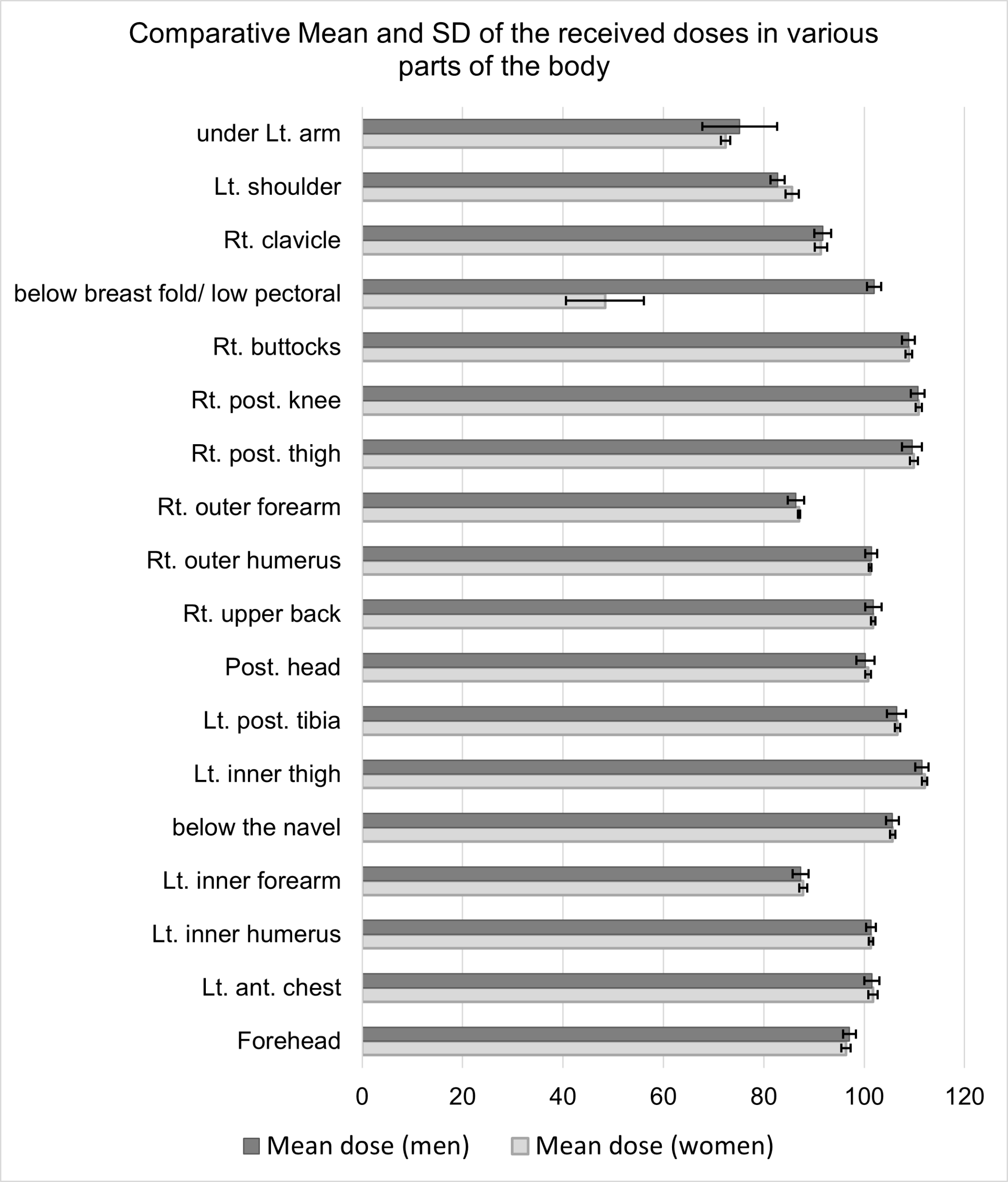
Figure 6. Mean±SD of absorbed doses by TLDs for men and women
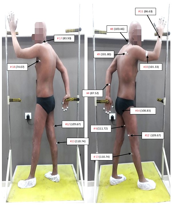
TLD positions and the corresponding measured dose values on the skin surface of one of our patients in this study at the six treatment positions is illustrated in Figure 7.
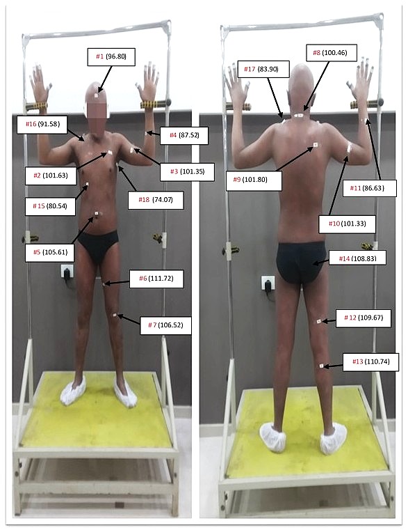
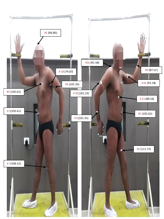
Figure 7. Illustration of TLDs placement and measured values in one of the patients.
4. Discussion
TSEI achieved a good radiation dose distribution over most of the body surface of mycosis fungoides patients, but some areas didn’t receive sufficient doses because they weren’t directly facing the treatment field or were covered by another part of the body. As a consequence, they missed some of the prescribed treatment dose. These low doses were recorded by TLDs placed under the breast folds (in female patients), above the shoulder in tall patients (170-180cm), under both arms, and at the inner and outer sides of both forearms. Also, some areas of the radiation field above the source received lower doses due to gravity, which may cause electrons to travel slightly downward when the source-skin distance is long. At these skin points that received lower radiation doses, the therapeutic effect of radiation may not be enough.
On the other hand, because of the beam diversion effect, the points lying near the beam center received higher doses than those lying on the periphery, as the dose concentration at the beam center was higher than at its edges. Also, due to the falling-off property of electrons under the gravity force, the skin points in the direction of the gravity force received larger radiation doses than those in the opposite direction. Radiation toxicity and inflammation may take place at these skin sites that received higher radiation doses.
Similar to our study, Elsayad et al. compared radiation dose distributions to assess dose variation across anatomic sites. Retrospectively collected data on 85 patients with cutaneous lymphoma or leukemia who underwent TSEBI were reviewed. Patients were irradiated in one of two positions: standing (n = 77) or reclined (n = 8). They concluded that TLD measurements were integral to TSEBT quality assurance. Dose distribution across several anatomical sites correlated significantly with the gender, height, and weight of the treated individuals (11).
Falahati et al. measured dosimetric parameters, including uniformity, surface dose, and maximum depth dose, using TLDs and EBT3 films in total skin electron beam therapy. Stationary and rotary methods were set on the Varian linear accelerator. The source-to-skin distance was set to 400 cm, and the electron beam energy was 6 MeV. The skin dose values were obtained at 21 different points on the phantom surface. The results of dose uniformity in the stationary technique were 10% and 2.6% by TLDs and 6% and 2.3% by EBT3 films along the longitudinal and transverse axes, respectively. Based on their study, stationary techniques were preferred for TSEI. Non-uniformity in dose distribution often occurred at mobile body parts, such as the head, feet, and hands. The dose distribution on the chest, pelvis, and abdomen was much more homogeneous. The results also demonstrated that TLDs should be routinely used in TSEI treatment (12).
Almeida et al. reported that, in some anatomical regions, some dosimeters recorded higher doses than others, which can be explained by their location, due to overlays and irradiation angles (13). Schüttrumpf et al. used the Stanford TSEI technique with six dual-fields. They used TLDs to verify and validate the dose distribution in some patients with localized lesions who were treated with partial skin electron irradiation (PSEI) using large electron fields. They concluded that TLDs enabled individual dose optimization in TSEI and PSEI, enabling a reliable adjustment of the MUs to achieve the prescription dose. They referred to the half-body electron irradiation with the PSEI (9). However, in the current study, the dose distribution was investigated only for TSEI, as no patients were treated with PSEI at the radiotherapy department of Ayady Al-mostakbl Oncology Hospital.
Hensley et al. studied the TSEI technique by irradiating patients in a standing position with two low-energy electron beams at gantry angles of 72 ° and 108 °. They used a 3.9 MeV electron beam after passing through a 6 mm Perspex attenuator and reported that better dose uniformity across the majority of patients’ skin could be achieved with the standing position technique than with the rotational technique (14). Our study differs from that of Hensley and colleagues in several technical parameters. Firstly, the beam energy: the current study used a higher energy (6 MeV), which could achieve better dose distribution at greater distances from the skin surface. Secondly, SSD: the current study used a larger SSD (400 cm), which permitted better coverage of the very tall patients.
Fahimi et al. (15) investigated the uniformity of absorbed dose in the sleeping Stanford technique using dosimetry on the Rando phantom. Their results showed that the dose uniformity achieved in this technique was in the range of (100 ± 25 cGy) and the dose accuracy was 6%. They concluded that the Total Skin Electron Therapy (TSET) technique in sleeping position was very suitable for elderly and disabled patients, and met the required dose uniformity. Furthermore, they recommended using a flattening filter to achieve a more uniform dose distribution.
The main limitations of our study are the relatively small number of patients and its unicentric design. Additionally, some of the TLDs’ positions might have been slightly different among patients, or patients might not have maintained a fixed position during the sessions, or the same position in different sessions.
Our results have practical implications. They suggest that a rotating platform (rather than a fixed one) may allow a more uniform distribution of absorbed radiation dose across the involved skin. In case of a fixed platform, special attention should be given to the skin area under the breast in women and to the shoulder region in tall patients. The inner forearm should face the radiation beam directly in all patient positions during all treatment sessions to avoid underdosage. Both arms should be in front of the supports, and the supports must be secured with low-density material (e.g., self-adhesive tape). The output of the selected electron energy should be calibrated at least twice weekly to avoid increasing the dose delivered in each radiation field in TSEI. TLDs should be routinely used in TSEI treatment to ensure doses are within the reference treatment range and maximize patient benefit.
Abbreviations
Ant. – Anterior
cGy – Centigray
ECC- Element correction coefficients
LiF – Lithium Fluoride
Lt – Left
MF – Mycosis fungoides
MU/min – Monitor units per minute
Mus – Monitor units
post. – Posterior
PSEI- Partial skin electron irradiation
Rt – Right
SD – Standard Deviation
SSD – Source-to-skin distance
TLD – Thermoluminescent dosimeter / Thermoluminescent dosimetry
TSEBI – Total skin electron beam irradiation
TSEBT – Total skin electron beam therapy
TSEI – Total skin electron irradiation
TSET – Total Skin Electron Therapy
Statements
Authors’ contribution: All authors conceived and planned the analysis. TIMH contributed to the interpretation of the results. SE selected the skin cancer patients and treated them with radiotherapy. SIH and WI took the lead in writing the manuscript. TIMH revised and made the final approval. All authors provided critical feedback and helped shape the research, analysis, and manuscript.
Consent for publication: All authors read and approved the final version of the document.
Funding: No funds, grants, or other support were received during the preparation of this manuscript.
Competing Interests: The authors have no relevant financial or non-financial interests to disclose.
Ethics: Patients voluntarily participated in this observational study and provided informed consent for using their images.
References
- Hristov AC, Tejasvi T, Wilcox RA. Mycosis fungoides and Sézary syndrome: 2019 update on diagnosis, risk-stratification, and management. Am J Hematol. 2019;94(9):1027-41.
- Jawed SI, Myskowski PL, Horwitz S, Moskowitz A, Querfeld C. Primary cutaneous T-cell lymphoma (mycosis fungoides and Sézary syndrome): part I. Diagnosis: clinical and histopathologic features and new molecular and biologic markers. J Am Acad Dermatol. 2014;70(2):205.e1-16.
- Arulogun SO, Prince HM, Ng J, Lade S, Ryan GF, Blewitt O, et al. Long-term outcomes of patients with advanced-stage cutaneous T-cell lymphoma and large cell transformation. Blood. 2008;112(8):3082-7.
- Demierre MF, Kim YH, Zackheim HS. Prognosis, clinical outcomes, and quality of life issues in cutaneous T-cell lymphoma. Hematol Oncol Clin North Am. 2003;17(6):1485-507.
- Van Doorn R, Van Haselen CW, van Voorst Vader PC, Geerts ML, Heule F, de Rie M, et al. Mycosis fungoides: disease evolution and prognosis of 309 Dutch patients. Arch Dermatol. 2000;136(4):504-10.
- Smits K, Quint KD, Vermeer MH, Daniëls LA, Willemze R, Jansen PM, et al. Total skin electron beam therapy for cutaneous T-cell lymphomas in the Netherlands: A retrospective analysis of treatment outcomes and selection for high or low dose schedule. Clin Transl Radiat Oncol. 2022;33:77-82.
- Piotrowski T, Milecki P, Skórska M, Fundowicz D. Total skin electron irradiation techniques: a review. Postepy Dermatol Alergol. 2013;30(1):50-5.
- Nolasco AV, Faria LO. The use of the TLD-100 for quality assurance in Total Body Irradiation (TBI). In: Eskola H, Väisänen O, Viik J, Hyttinen J, editors. EMBEC & NBC 2017. IFMBE Proceedings, vol 65. Singapore: Springer; 2018. p. 431-4.
- Schüttrumpf L, Neumaier K, Maihoefer C, Niyazi M, Ganswindt U, Li M, et al. Dose optimization of total or partial skin electron irradiation by thermoluminescent dosimetry. Strahlenther Onkol. 2018;194(5):444-53.
- Rivera T. Thermoluminescence in medical dosimetry. Appl Radiat Isot. 2012;71:30-4.
- Elsayad K, Moustakis C, Simonsen M, Bäcker D, Haverkamp U, Eich HT. In-vivo dosimetric analysis in total skin electron beam therapy. Phys Imaging Radiat Oncol. 2018;6:61-5.
- Falahati L, Nedaie HA, Esfahani M, Banaee N. Dosimetric evaluation of electron total skin irradiation using Gafchromic films and thermoluminescent dosimetry. J Can Res Ther. 2019;15(7):S115-22.
- Almeida SB, Villani D, Sakuraba RK, Rezende ACP, Campos LL. Dosimetric evaluation and comparison of TL responses of LiF:Mg,Ti and μLiF:Mg,Ti in the clinical electron beams dosimetry applied to total skin irradiation (TSEB) treatments. Radiat Meas. 2019;125:15-8.
- Hensley FW, Major G, Edel C, Hauswald H, Bischof M. Technical and dosimetric aspects of the total skin electron beam technique implemented at Heidelberg University Hospital. Rep Pract Oncol Radiother. 2013;19(2):135-43.
- Fahimi MS, Geraily G, Aghili M, Toolee H. Evaluation of Dose Distribution in Optimized Stanford Total Skin Electron Therapy (TSET) Technique in Rando Anthropomorphic Phantom using EBT3 Gafchromatic Films. J Biomed Phys Eng. 2021;11(4):425-34.