


Author(s) :
Sophie Duncan1, Felicity Hudson1,2,3, Michaela Beavan1, Mark Lee1,3, Andrew Wallis1, Sankar Arumugam1,2,3
1 Liverpool and Macarthur Cancer Therapy Centres, Sydney, NSW, Australia
2 Ingham Institute of Applied Medical Research, Sydney, NSW, Australia
3 South West Sydney Clinical School, University of New South Wales, Sydney, NSW, Australia
Corresponding author: Sophie Duncan, Email: sophie.duncan@health.nsw.gov.au
Publication History: Received - , Revised - , Accepted - , Published Online - 29 July 2024.
Copyright: © The author(s). Published by Casa Cărții de Știință.
User License: Creative Commons Attribution – NonCommercial (CC BY-NC)

 PDF
PDF
 Share
Share
Abstract
Background: Pre-simulation Assessment Sessions (PASS) can be utilised to assess respiratory motion in patients receiving stereotactic ablative body radiotherapy (SABR). PASS is an assessment process that uses cine x-ray images to determine whether expiration breath-hold (EBH) or abdominal compression (AC) can be effectively utilised to manage diaphragm motion, prior to computed tomography (CT) simulation. This study aimed to determine the effectiveness of PASS for eligible patients based on diaphragm motion in free breathing (FB) compared to using MMSs.
Material & Methods: Retrospective data on diaphragm motion in FB and elected MMS was collected for 73 patients. Eligible patients were treated between 2018-2022 using SABR for abdominal and lower lobe lung tumours. In the PASS process, the diaphragm motion seen on cine x-ray images was measured through three cycles of FB versus the elected MMS. Differences in FB and MMS diaphragm motion was found for each patient using Wilcoxon Matched Pairs Test.
Results: Of the 73 patients, 28 were treated with EBH, 34 with AC, 2 with alternate strategies and 11 were treated using FB as they were not suitable for a MMS. There was a statistically significant difference between the mean of the amplitude of the diaphragm motion when comparing FB and EBH and FB and AC (p<0.05). There were no associations found between the PASS success rate for any MMS and BMI or age.
Conclusion: PASS is a useful tool which can be used to shape the future of radiotherapy by selecting the patient specific MMS for the reduction of tumour motion during SABR treatments. This study will be used to further investigate the dosimetric effects of MMS on internal margin reductions and normal tissue sparing.
1. Introduction
Stereotactic ablative body radiotherapy (SABR) delivers a high dose of radiation to a relatively small tumour in a limited number of treatment fractions (1), leading to local control rates and quality of life indicators equivalent to that of surgery (2, 3). Its precise targeting and high dose of radiation is capable of ablating tumours directly, and the highly conformal nature allows for increased sparing of adjacent organs at risk (OARs) and healthy tissue (1). Immobilisation instability, patient anxiety, intrafraction movement due to prolonged treatment times and tumour changes are challenges which are faced when trying to achieve optimal accuracy during radiation therapy (RT) treatment (4). Three-dimensional computed tomography (3DCT) images show a snapshot of tumour position during the patients’ breathing cycle, however this may not reflect the true tumour postion and can vary intra- and inter- fractionally. Patients with concerns regarding respiratory motion currently utilise 4-dimensional computed tomography (4DCT) to evaluate tumour movement at all stages of the breathing cycle, to create an individualised internal target volume (ITV). Tumours within the adrenal gland (5), kidney (6), upper gastrointestinal (GI), such as pancreas and hepatobiliary (1), and lower lobe of lung (7) may move with respiration due to their proximity to the diaphragm, therefore, motion management techniques for these patients during RT is becoming increasingly important.
The role of motion management strategies (MMS) in SABR has increased due to its ability to facilitate the delivery of hypofractionated ablative doses whilst reducing normal tissue doses(2). MMS, along with the use of image guidance, are measures used to limit and monitor tumour position variance from respiratory motion and improve treatment accuracy. The active motion management include inspiration breath hold (IBH), expiration breath hold (EBH), abdominal compression (AC), gated and real-time tumour tracking treatment delivery (8). In a passive motion management approach, an ITV is derived from a 4DCT data set which is used to account for tumour motion in a free breathing (FB) state (8). At our centre, deep inspiration breath hold (DIBH) and EBH are performed using the Active Breathing Coordinator (ABC; El- ekta Limited, Crawley, United Kingdom). This provides non-invasive internal immobilisation for anatomy affected by respiratory motion, using efficient assisted breath hold techniques (9). For abdominal compression, we use the Omni-V Stereotactic Patient Positioning System (Bionix, USA) equipment with a respiratory belt to apply pressure on the abdomen, forcing shallow breathing and thereby limiting the motion of the diaphragm and liver compared to FB. Currently, EBH is the gold standard for reducing respiratory motion and providing superior results, however, patient suitability/tolerability can be a limiting factor (10). This evolution of respiratory motion management has allowed for the individualisation of ITVs based on motion in the planning computed tomography (CT) scan (3-dimensional/4-dimensional) when used with the elected MMS (11). Excessive dose to surrounding organs is generally the dose-limiting factor in RT; it also helps avoid tumour un- derdosage which can result in decreased local control (2). Reduction of motion, and therefore reduction of ITVs, limits the amount of surrounding tissue and OARs being irradiated, potentially reducing radiation toxicities (10).
Pre-simulation Assessment Session (PASS) is a structured process used in our centre to determine which patient specific immobilisation best reduces respiratory motion prior to the acquisition of their planning CT or magnetic resonance imaging (MRI). Our centre performs PASS for all patients who are undergoing SABR for the tumours in the adrenal gland, kidney, pancreas, liver and lower lobe of the lung. The session is tailored for each patient and assesses their tolerability and reproducibility of the different MMS for treatment. In this process the position of tumour surrogates, such as im- planted fiducials markers and the diaphragm, are monitored using cine x-ray images with the kV imager available on linear accelerators, to quantify the motion in FB against the MMS that is tolerable to the patient to decide the suitability of MMS that is specific to the patient. Following this process patients have their simulation appointment where, based on the MMS chosen for respiratory motion reduction, a 3DCT with/without MRI (EBH) or 3DCT and 4DCT with/without 4DMRI (AC/FB) are acquired for further assessment of motion for target delineation.
The primary aim of this study was to determine the effectiveness of PASS process for eligible patients based on diaphragm motion in FB versus when using a MMS. This study hypothesises that the diaphragm motion with MMS will be significantly reduced compared to FB. Results from this study have the potential to validate PASS’ role in successfully selecting a reproducible MMS for individual patients to limit respiratory motion and therefore potentially lead to reduced ITVs and dose to surrounding OARs based on subsequent simulation CT.
2. Material and Methods
2.1. PASS Workflow
PASS is a process during which patient-specific motion management techniques are evaluated and selected based on the patient’s tolerability, as well as the effectiveness of reducing and reproducibly managing tumour motion. All patients underwent PASS process completed on an Elekta Versa HD Linear Accelerator with the XVI Imaging System (Elekta Limited, Crawley, United Kingdom). Sessions were run by Radiation Therapists (RTTs) prior to simulation, occasionally with the pres- ence of a Radiation Oncologist (RO). All patients were positioned head-first, supine with arms above head, in a whole body vacbag +/- MMS. Patient eligibility for each respective MMS is outlined in Table 1. Eligibility for EBH with ABC was assessed as the centres’ gold standard. All EBH assessments were conducted at a threshold of 0.1L on expiration. Initially, prior to patients’ PASS, a 45-minute ABC coaching session was arranged to assess tolerability and eligibility of EBH. However, due to the COVID-19 Pandemic, from 2020 onwards this was cancelled as a way of limiting patient time in hospitals and movement within the community. If not eligible for EBH, AC eligibility was assessed using the Omni-V with the Respiratory Belt. Patients were only assessed in the EBH group if they were not pre-emptively ineligible. Patients were only assessed for AC if they failed EBH and didn’t have other comorbidities which made them ineligible for AC, e.g. abdominal hernia.
Table 1. Eligibility criteria for Motion Management Systems
| EBH | AC |
|---|---|
| No dentures | Able to safely get on/off Omni-V device |
| Tolerate equipment (Maintain seal around mouth snorkel) | Able to tolerate pressure of AC belt |
| Understand/follow breathing instructions in English | No pre-existing conditions that may preclude use of AC belt – e.g., abdominal hernia, tenderness at surgical site |
| Maintain Breath hold for a minimum of 15 seconds | Motion reduction of diaphragm >5mm from FB scan |
| Able to reproduce breath hold (BH) threshold for a minimum of x3 BH’s | Able to tolerate prolonged setup position time (~45 minutes) |
| Consistent and reproducible diaphragm positioning on repeat cine x-rays, measured as <2-3mm at each breath hold | |
| Motion reduction of diaphragm >5mm from FB scan | |
| Able to tolerate prolonged setup treatment time (~45 minutes) |
(EBH = exhalation breath hold, AC = abdominal compression, FB = free breathing)
Due to poor contrast on kilovoltage (kv) images, tracking of abdominal tumours is disad- vantaged(12). Previous studies have found the diaphragm to be an accurate surrogate for abdominal and lung tumour/organ motion(12-14). In accordance with this, our process used the apex of the diaphragm as a surrogate for tumour motion which was assessed via cine x-ray images with 110 frames in 20 seconds. The cine x-ray images were acquired using Motion View in XVI Software, which acquires continuous kV images. Figure 1 illustrates the decision tree used to determine the appropriate MMS through the PASS workflow. All patients underwent one to two cine x-ray image acquisitions in FB, which were used to evaluate the maximum amplitude of the respiratory motion of the diaphragm over three breathing cycles. Patients who were eligible for EBH underwent three further image acquisitions whilst holding their breath with the assistance of the ABC system. The amount of displacement and the reproducibility of the diaphragm position with each breath hold, as well as the motion and potential drift of the diaphragm during the breath hold were assessed on the cine image acquisitions. If patients were not eligible for EBH, diaphragm motion of three breathing cycles for AC was collected, with the Respiratory Belt compression below the diaphragm, as much as tolerable. All x-ray images were acquired, in anterior-posterior direction with the F0 filter and 27cm x 27cm imaging field size at approximately junction of ribs, at a tabletop of 10-15cm with laterality dependant on site. The motion of the apex of the diaphragm was measured using the grid, ruler and draw function on XVI.
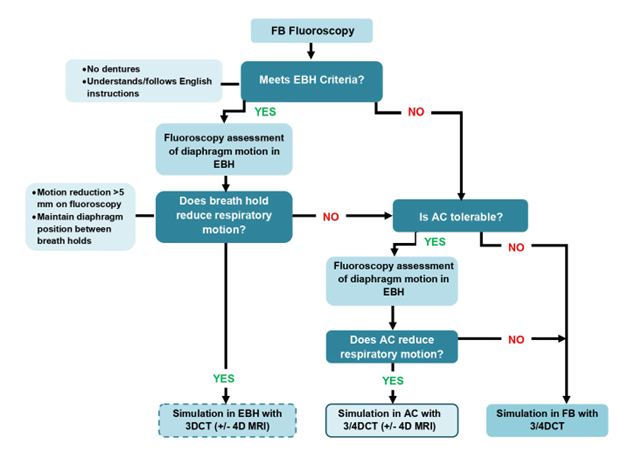
Figure 1. Flowchart of Pre-simulation Assessment Session
(FB = free breathing, EBH = exhalation breath hold, AC = abdominal compression)
2.2. Study Methodology
2.2.1 Patient characteristics
Following South Western Sydney Local Health District Quality Improvement Project approval, retrospective data from patients who underwent a PASS between September 2018 and September 2020 was obtained from MOSAIQ Record and Verify System (Elekta Inc, California, USA). These patients were treated with SABR for tumours located in the liver, pancreas, lower lobe of the lung or adrenal gland. Patients were eligible for the study if they had completed a PASS assessment and had diaphragm motion data, averaged over three breathing cycles, available on the FB and chosen MMS. Table 2 summarises the characteristics of patient cohort considered for this study. Data on patients’ ability to successfully complete all fractions of their treatment following PASS was also ac- quired from MOSAIQ Record and Verify System.
Table 2. Patient characteristics
| Variable | Count (n=73) | |
| N | % | |
| Gender | ||
| Male | 53 | 72.6 |
| Female | 20 | 27.4 |
| Age, years | ||
| 40-49 | 2 | 2.7 |
| 50-59 | 6 | 8.2 |
| 60-69 | 29 | 39.7 |
| 70-79 | 25 | 34.2 |
| 80+ | 11 | 15.1 |
| Primary Tumour Site | ||
| Adrenal | 7 | 9.6 |
| Liver | 57 | 78.1 |
| Lung | 2 | 2.7 |
| Pancreas | 7 | 9.6 |
| MMS | ||
| FB | 11 | 15.1 |
| EBH | 28 | 38.4 |
| AC | 34 | 46.6 |
| BMI† | ||
| <25 | 22 | 30.6 |
| ≥25 | 50 | 69.4 |
| Replans due to MMS | ||
| AC | 2 | 2.7 |
| EBH | 0 | 0 |
| MMS Compliance issues | ||
| AC | 0 | 0 |
| EBH | 1 | 1.4 |
| Cancelled fractions due to MMS | ||
| AC | 2 | 2.7 |
| EBH | 0 | 0 |
| Incomplete treatment due to MMS | ||
| AC | 0 | 0 |
| EBH | 0 | 0 |
(MMS = motion management system, FB = free breathing, EBH = exhalation breath hold, AC = ab- dominal compression, BMI = body mass index) † One patient did not have height information availa- ble and therefore their BMI was not evaluated
2.2.2. Analysis – Changes in motion
Records of average diaphragm motion in FB and the chosen MMS was collected. This was recorded at the time of the patients’ PASS and organised into an inhouse designed Excel (Microsoft, Washington USA) spreadsheet. The mean motion of the diaphragm in FB for all patients and EBH or AC, depending on MMS decided on, was determined. Changes in the mean motion and standard deviation of the dia- phragm in FB versus EBH and AC were evaluated using a Wilcoxon Matched Pairs Test. Differences of p≤0.05 were considered statistically significant. All statistical analyses were performed using RStudio (version 4.2.0).
2.2.3. Analysis – Role of Age and Body Mass Index (BMI) in MMS Eligibility
Patients’ age and BMI at the time of their PASS was also collected from MOSAIQ Record and Verify System. The mean age of patients was 69.4 years old (yo), therefore patients were separated into 2 groups of less than 70yo or 70yo or over. The mean BMI for patients was ~28, hence patients were split into 2 groups of less than 25 (healthy or underweight range) or 25 and over (overweight or obese range)(15). A Fisher’s Exact Test was used to determine whether a statistically significant association existed between a patients’ age or BMI and their eligibility for a MMS.
3. Results
3.1. MMS Selection
Retrospective data for 77 patients was sourced, however only 73 of these patients were evaluated for the study; 2 patients were not included as there was minimal motion detected in FB, therefore no other MMS was assessed, and 2 patients treated in DIBH have not been evaluated in this study as there was too little data available to evaluate statistical significance. Of these 73 patients, 11 patients were treated in FB as they were unable to tolerate the EBH equipment (5) or AC equipment (4), had dentures (1), no MMS was reproducible (1), couldn’t understand or follow instructions (1) or the change in diaphragm motion was <5mm or larger when using a MMS versus FB (3). Patients who failed MMSs were being treated for tumours located in the liver (8), adrenal (1) and pancreas (2). 85% of patients assessed were assigned a MMS which reduced respiratory motion. The mean diaphragm motion was 19.3mm, 2mm and 8.4mm for FB (n=73), EBH (n=28) and AC (n=34) respectively.
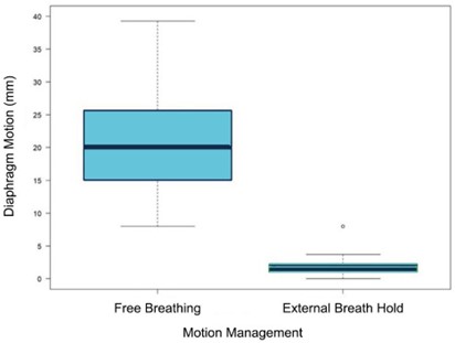
Figure 2. Differences in respiratory motion in free breathing vs exhalation breath hold (EBH) (mm) for patients eligible for EBH
3.2. Respiratory motion changes in FB vs EBH
Figure 2 demonstrates the difference in respiratory motion in FB versus EBH in patients eligible for EBH. The mean respiratory motion of the diaphragm in FB (8–42mm, SD ±8.5mm) and EBH (0–8mm, SD ±1.6mm) was 20.9mm and 2mm, respectively. A significant difference between the motion for these MMSs was observed (p<0.05). The mean reduction of respiratory motion for these patients was 18.9mm.
3.3. Respiratory motion changes in FB vs AC
Figure 3 demonstrates the difference in respiratory motion in FB versus AC in eligible patients. The mean respiratory motion of the diaphragm in FB (6.7–35mm, SD ±5.9mm) and AC (1.7-19.3mm, SD ±4mm) was 17.7mm and 8.4mm, respectively. Significant reduction in motion was observed when using AC (p<0.05). The mean reduction of respiratory motion for these patients was 9.5mm.
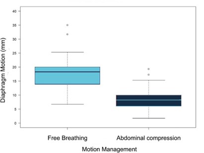
Figure 3. Differences in respiratory motion in free breathing vs abdominal compression (AC) (mm) for patients eligible for AC
3.4. Effects of Age on success of MMS
The relationship between patients’ age and their likelihood of being eligible for a MMS was assessed for EBH (n=47) and AC (n=41). No statistically significant association between age and EBH (p=1) or AC success (p=1) was observed.
3.5. Effects of BMI on success of MMS
The relationship between patients BMI and their likelihood of being eligible for a MMS was assessed for EBH (n=47) and AC (n=40). One patient had no height data and therefore was not assessed. No statistically significant relationship between patients’ BMI and EBH (p=0.1986) or AC success (p=1) was observed.
3.6. Treatment Outcomes
Treatment outcomes are shown in Table 1. Replans were required during treatment for 2 patients due to the use of AC (5.9%); one patient had a fall and was unable to tolerate the AC belt further, whilst another clinically deteriorated and was replanned to a simpler treatment technique. One EBH patient (3.6%) required recoaching on correct breathing technique during treatment, however was still treated. Two AC (5.9%) patients required a treatment session to be cancelled and rescheduled; one patient was cancelled due to an issue with the compression belt leaking where a new belt had to be acquired, the second was unable to tolerate the equipment due to pain. Both patients continued on to complete all treatments. Table 1 outlines the amount of patients which required replans during treatment following PASS (AC=5.9%, EBH=0%), the amount with compliance issues which were MMS related (AC=0%, EBH=3.6%) and how many patients had fractions cancelled (AC=5.9%, EBH=0%) or did not complete treatment due to the use of a MMS (AC=0%, EBH=0%). Two FB patients had inconsistent breathing on treatment and therefore additional CBCTs were needed, and one FB patient required a replan due to large tissue changes.
4. Discussion
The importance of motion management in the delivery of hypofractionated ablative doses, such as SABR, to improve dosimetric coverage and reduce normal tissue dose has been proven across a number of studies (3, 4, 9, 16). The primary aim of this study sought to determine the effectiveness of PASS for eligible patients based on diaphragm motion in FB versus when using a MMS. We have reviewed the feasibility of our PASS process in 73 patients and demonstrated that 85% of these patients were suitable for a MMS which resulted in a reduction in respiratory motion. These patients had minimal complications with <12% of patients in each MMS group requiring replans, compliance issues, cancellations or incompletion of treatment due to MMS.
Despite our departments ability to allocate a suitable MMS for patients receiving SABR to lower lobe lung and abdominal tumours using PASS, patient stresses or changes in performance status can result in difficulties on treatment. In total, 7 patients who were classed as suitable for AC at their PASS required replans, however only 2 of these were MMS related. All other patients planned with AC who needed a replan were due to soft tissue changes or stent insertion between PASS/simulation and treat- ment. Two patients allocated EBH at PASS required replans but both were non-MMS related; PTV/tissue changes. In total one patient had compliance issues with the EBH equipment, specifically the snorkel mouthpiece, however was still treated after radiation therapists recoached the patient. Only two patients suitable for AC had fractions cancelled during treatment due to MMSs, other patients included in this study had fractions cancelled due to non-MMS related issues such as patients being too unwell to attend treatment, which was resolved and these patients also completed treatment at a later date without further incidences. In total, 6.5% of patients did not complete treatment however none of these cases were due to MMSs; 3.2% of the treatment cancellations were a result of significant clinical deterioration with/without palliative treatment instead and the remaining was due to a change in imaging results and therefore no RT was needed. It is also important to note that patients nominated to be treated in FB also faced obstacles on treatment which were due to compliance issues or required replans, demonstrating the day-to-day issues faced by RTTs during treatment. Overall, we can see that MMS equipment is the biggest issue faced on treatment following PASS, however this still affects a very small amount of patients in this study and majority of patients completed treatment with excellent compliance and no incidences.
Hardcastle et al. similarly evaluated the distribution of use of voluntary EBH (VEBH), AC, FB gated and FB (17); this study had success rates of 67.6% for VEBH, 20% for AC, 6.9% for FB gated and 5.5% for FB. We found that approximately 36% of patients were pre-emptively ineligible for EBH due to having dentures, not being able to follow instructions or not being able to tolerate the ABC equipment. Therefore, the higher success rates for VEBH demonstrated by Hardcastle et al. could be due to the fact that it was voluntary and was not using the ABC equipment and patients potentially found it easier to comply with. However, this techniques’ lack of integration of breath hold equip- ment with the treatment system means it is potentially less reliable and/or consistent when tracking the breathing cycle and could result in a greater chance of treatment errors such as geographical misses. Hardcastle et al. had a very similar process and selection criteria for determining patients’ eligibility for MMSs, proving that processes like PASS can be used across multiple departments. Similar to using the liver dome/diaphragm as a surrogate for respiratory motion in our study, Eccles et al. evaluated the reproducibility of liver position using ABC equipment (18). Of the 34 patients in this study, 21 were screened to be suitable for ABC (62%). This study found that interfraction craniocaudal reproducibility was up to 7.9mm, indicating that day-to-day reproducibility of the diaphragm may differ (18), highlighting the need for a rigorous selection process, such as PASS, for patients receiving higher dose per fraction treatments such as SABR. This also suggests that departments should implement more image guidance throughout patients’ treatment. In another study completed by Mampuya et al, 53% of patients evaluated were eli- gible for AC, demonstrating similar numbers to this study (47%) (19). This study also demonstrated the limitations of AC by highlighting that the remaining 47% of patients, despite having respiratory motion >8mm in FB, were ineligible for AC due to medical reasons, such as gallstones, abdominal aneurysm/surgery, dementia or chronic obstructive pulmonary disease or the amplitude of tumour motion wasn’t significantly reduced (19). Likewise, in this study we had three patients who could not have AC assessed due to tenderness post-surgery, four patients not able to tolerate the equipment and five patients’ respiratory motion was not significantly reduced or remained the same when using the equipment.
A literature review conducted by Darréon et al, found that sites including the diaphragm, lower lobe of the lung, liver, pancreas, kidney and oesophagus have significant respiratory motion, with motion amplitude ≥ 14mm (11). Lu et al. compared 3 consecutive CT scans with patients in IBH to evaluate the changes in the centroid of the liver to reflect intrafraction motion. This study revealed that 7% of shifts in the anterior-posterior direction and 11% of the shifts in the superior-inferior direction exceeded 5mm (20). Since this is outside of our standard 5mm PTV expansion, this demonstrates how respiratory motion can be a major cause of geographical misses in radiotherapy. PASS’s personalised assessment of patients and strict eligibility criteria has the potential to identify patients who would fall into this category and may result in a decreased risk of geographical miss. A standard PTV expansion at our centre would generally be ITV + 3-5mm. For and for EBH, PTV is defined by a 5mm expansion from GTV with no ITV due to the limited respiratory motion.. The PASS process and resulting respiratory motion reduction has allowed us to select the appropriate MMS to individualise ITV/GTV margins and therefore potentially reduce PTV margins. Lu et al. also found that, due to intrafractional changes in liver position across the 3 CT scans, 41% of patients plans’ were not meeting the clinical dose objectives; gross tumour volumes (GTV) volumes receiving 100% of the prescription ranged from 76% to 100% (20). Geographical misses such as this can result in suboptimal local control rates (2) and increased dose to volume of liver and GI structures (3). Abbass et al. concluded that through a review of literature, when treating hepatic tumours, normal liver dose can be significantly reduced by using individualised margins with respiratory motion management (21). EBH is our preferred method due to its’ stability and reproducibility of diaphragm position. Furthermore, Taniguchi et al. found that a dosimetric analysis of OAR doses on end-expiration and end-inspiration phases on 4DCT of 18 patients’ for pancreas SABR, revealed the duodenum dose to be higher in IBH compared to EBH due to greater overlap with the duodenum and PTV due to organ position changes (21, 22). Furthermore, Qi et al. found that the use of AC for patients receiving lower lobe lung tumour SABR decreased GTV excursion by 54.69% in the superior-inferior direction but did not change in the left-right direction (23). However, the GTV excursion increased by 5.56% in anterior-posterior direction when using AC for these patients (23); this highlights a limitation of our PASS process as we do not evaluate anterior- posterior or the left-right movement.
Despite the success rates of PASS for motion reduction, there are still some limitations of this study. These include the use of the diaphragm as a surrogate for tumour motion; although studies have found a correlation between the diaphragm motion and tumour/organ motion in the superior-inferior and anterior-posterior direction there are some discrepancies in the left/right direction (12, 13). Therefore, this error should be considered in the treatment margins through the primary use of 4DCT at simulation for target delineation. The increased time on the machines and financial implications required for PASS appointments versus the decreased simulation time due to not needing to repeat simulation scans has not been assessed. Extra training and education sessions were required for staff understanding of the process. Issues with reproducibility for AC can also be resolved with adequate training of staff, radiation therapists setting the patient up again and the department is now testing new equipment with radiopaque markers for compression belt placement. The impact of COVID-19 on our processes also could have potentially led to a decrease in the success rates of EBH in this study. To further streamline success and improve PASS processes, there is currently a screening sheet for patients being created within the department to highlight patients who are ineligible for MMSs prior to completing PASS. For EBH, the screening tool includes evaluating patients’ ability to hold their breath in exhale for minimum 20 seconds, understand and follow breath hold instructions, tolerate RT positioning for at least 45 minutes, tolerate equipment and whether they have dentures or missing teeth. For AC, the screening tool will determine if a patient has a history of abdominal hernias, a stoma bag or a pre-existing compromised lung function and if the patient is able to tolerate compression of the abdominal region for at least 45 minutes.
5. Conclusion
This study has determined PASS to be an effective process for assigning eligible patients as demonstrated by 85% of patients being assigned the appropriate MMS prior to their simulation scans; specifically, by examining the diaphragm motion in FB versus when using a MMS. Individualisation of MMSs through the PASS process leads to significant reduction in respiratory motion as a result. Patients also had minimal issues on treatment related to MMSs, with the large majority completing treatment successfully. No associations were found between the PASS success rate for any MMS and BMI or age. Moving forward, the outcome of this study can lead into future evaluation of the dosimetric impacts as a result of utilising PASS.
Abbreviations
3DCT – 3-dimensional computed tomography 4DCT – 4-dimensional computed tomography AC – abdominal compression
BMI – body mass index
CT – computed tomography
DIBH – deep inspiration breath hold EBH – expiration breath hold
FB – free breathing GI – gastrointestinal
GTV – gross tumour volume IBH – inspiration breath hold ITV – internal target volume kV – kilovoltage
MMS – motion management system MRI – magnetic resonance imaging OAR – organ at risk
PASS – pre-simulation assessment session RO – radiation oncologist
RT – radiation therapy RTT – radiation therapist
SABR – stereotactic ablative body radiotherapy VEBH – voluntary expiration breath hold
yo – years old
Statements
Authors Contributions: SD took the lead in writing the manuscript. MB did the statistical analysis and interpretation. All remaining authors contributed in the final approval of the manuscript and helped shape the research and analysis.
Consent for Publication: As the corresponding author, I confirm that the manuscript has been read and approved for submission by all named authors.
Conflicts of Interest: The authors declare no conflict of interest.
Funding Sources: None
Statement of Ethics: This manuscript does not contain any studies carried out by the authors on humans or animals and did not require ethics approval.
References
- Tchelebi L, Zaorsky N, et al. Stereotactic Body Radiation Therapy in the Management of Upper GI Malignancies. Biomedicines. 2018;6(1).
- Cole AJ, Hanna GG, et Motion management for radical radiotherapy in non-small cell lung cancer. Clin Oncol (R Coll Radiol). 2014;26(2):67-80.
- Gargett M, Haddad C, et Clinical impact of removing respiratory motion during liver SABR. Radiat Oncol. 2019;14(1):93.
- Wu VWC, Ng APL, et al. Intrafractional motion management in external beam radiotherapy. J Xray Sci Technol. 2019;27(6):1071-86.
- Franzese C, Nicosia L, et Stereotactic body radiation therapy for adrenal gland metastases: outcome and predictive factors from a multicenter analysis. Clin Exp Metastasis. 2021;38(6):511-8.
- Siva S, Louie AV, et Pooled analysis of stereotactic ablative radiotherapy for primary renal cell carcinoma: A report from the International Radiosurgery Oncology Consortium for Kidney (IROCK). Cancer. 2018;124(5):934-42.
- Li Y, Wu W, et Using 4DCBCT simulation and guidance to evaluate inter-fractional tumor variance during SABR for lung tumor within the lower lobe. Sci Rep. 2021;11(1):19976.
- Li H, Dong L, et AAPM Task Group Report 290: Respiratory motion management for particle therapy. Med Phys. 2022;49(4):e50-e81.
- 2023 [Available from: https://www.elekta.com/products/radiation-therapy/#sec-motion.
- Galayini M, Hazem M, et Implementing a Pre-simulation Assessment Session for abdominal SABR: respiratory motion management. Radiotherapy and Oncology. 2019;141.
- Darreon J, Bouilhol G, et Respiratory motion management for external radiotherapy treatment. Cancer Radiother. 2022;26(1-2):50-8.
- Veldman JK, van Kesteren Z, et Accuracy of abdominal organ motion estimation in radiotherapy using the right hemidiaphragm top as a surrogate during prolonged breath-holds quantified with MRI. Med Phys. 2023;50(6):3299-310.
- Yang J, Cai J, et al. Is diaphragm motion a good surrogate for liver tumor motion? Int J Radiat Oncol Biol Phys. 2014;90(4):952-8.
- Zhou D, Quan H, et A feasibility study of intrafractional tumor motion estimation based on 4D-CBCT using dia- phragm as surrogate. J Appl Clin Med Phys. 2018;19(5):525-31.
- Health Body Mass Index (BMI) calculator 2023 [Available from: https://www.health.nsw.gov.au/heal/Pages/bmi.aspx.
- Molitoris JK, Diwanji T, et Advances in the use of motion management and image guidance in radiation therapy treatment for lung cancer. J Thorac Dis. 2018;10(Suppl 21):S2437-S50.
- Hardcastle N, Gaudreault M, et Selection of motion management in liver stereotactic body radiotherapy and its impact on treatment time. Phys Imaging Radiat Oncol. 2023;25:100407.
- Eccles C, Brock KK, et Reproducibility of liver position using active breathing coordinator for liver cancer radio- therapy. Int J Radiat Oncol Biol Phys. 2006;64(3):751-9.
- Mampuya WA, Nakamura M, et Interfraction variation in lung tumor position with abdominal compression during stereotactic body radiotherapy. Med Phys. 2013;40(9):091718.
- Lu L, Ouyang Z, et Dosimetric assessment of patient-specific breath-hold reproducibility on liver motion for SBRT planning. J Appl Clin Med Phys. 2020;21(7):77-83.
- Abbas H, Chang B, et Motion management in gastrointestinal cancers. J Gastrointest Oncol. 2014;5(3):223-35.
- Taniguchi CM, Murphy JD, et Dosimetric analysis of organs at risk during expiratory gating in stereotactic body radiation therapy for pancreatic cancer. Int J Radiat Oncol Biol Phys. 2013;85(4):1090-5.
- Qi Y, Li J, et al. Effect of abdominal compression on target movement and extension of the external boundary of peripheral lung tumours treated with stereotactic radiotherapy based on four-dimensional computed tomography. Radiat Oncol. 2021;16(1):173


