


Author(s) :
Remus Șereș1, Dragoș Goadă1, Andreea – Iulia Pricopie2, Andrada Deac1, Corina Bocșa4, Bogdan Petruț1,3
1“Prof. Dr. Ion Chiricuţă” Oncology Institute, Cluj-Napoca, Romania
2 Department of Biochemistry and Chemistry of environmental factors,” George Emil Palade” University of Medicine, Pharmacy, Science and Technology, Târgu-Mureș, România
3Department of Urology, Faculty of Medicine, “Iuliu Haţieganu” University of Medicine and Pharmacy, Romania
42nd Internal Medicine Department, “Iuliu Hațieganu” University of Medicine and Pharmacy Cluj-Napoca
Corresponding author: Remus Șereș , Email: seres.remus@yahoo.com
Publication History: Received - , Revised - , Accepted - , Published Online - July 2022.
Copyright: © The author(s). Published by Casa Cărții de Știință.
User License: Creative Commons Attribution – NonCommercial (CC BY-NC)

 PDF
PDF
 Share
Share
Highlights
-
This case report describes a 40-year-old patient with malignant melanoma presenting with synchronous urinary bladder and gluteal muscle metastases, an exceptionally rare dissemination pattern.
-
Histopathology and immunohistochemistry (S100, HMB-45, Melan-A positive) confirm metastatic melanoma infiltrating the full thickness of the bladder wall, while PET-CT identifies metabolically active skeletal muscle and nodal disease.
-
Molecular testing reveals a BRAF V600E mutation, guiding the initiation of targeted therapy after progression on cytotoxic chemotherapy.
-
Treatment with Dabrafenib induces durable disease control with prolonged progression-free survival and preserved performance status, highlighting the impact of BRAF-targeted therapy in aggressive metastatic melanoma.
Abstract
Malignant melanoma has been known to metastasize; several cases in the literature have reported its metastasis to the urinary bladder. Metastasis to the gluteal muscle, however, is quite unusual. We report a case of malignant melanoma metastatic lesions in the urinary bladder and gluteal muscle, with a very good response to targeted therapy despite the aggressive behavior of this disease.
1. Introduction:
Malignant melanoma is the 17th most common cancer worldwide, and is responsible for nearly 73% of skin cancer-related deaths due to its aggressive behavior (1). Melanomainduced metastatic lesions of the bladder are extremely rare, with few reported cases (2). Skeletal muscle metastasis is also uncommon, possibly due to the large percentage of asymptomatic cases and the challenges faced in diagnosing it, as there are no specific imaging techniques for differentiating between carcinomas, sarcomas, and other muscular disorders (3).
This study presents a case of melanoma with concomitant urinary bladder and gluteal muscle metastasis. To our knowledge, there is no similar case in the literature with this pattern of dissemination. Given the rarity of the case, we performed a literature review to obtain further information.
2. Case presentation:
A 40-year-old male with no relevant medical, familial, or psychosocial history presented to the Department of Urology of a regional hospital in August 2016. The patient had been experiencing dysuria and hematuria for a month. A computed tomography (CT) scan of the abdomen and pelvis revealed a tumoral mass invading the anterior and lateral wall of the urinary bladder with no involvement of the seminal vesicles, prostate, or ureters. A cystoscopy was performed, but the patient experienced rapid worsening of symptoms. Due to the depth of malignant invasion of the bladder wall, the surgical team performed radical cystoprostatectomy with pelvic lymphadenectomy and urinary deviation, a procedure known as bilateral percutaneous ureterostomy.
The histopathological examination confirmed the diagnosis of metastasis from a malignant melanoma, which had infiltrated all layers of the bladder wall, and was associated with mucosal ulceration, serous involvement, and infiltration of the adjacent striated muscle. The mitosis index was 8 mitoses/mm2. Tumor necrosis represented approximately 30% of the tumor volume. Nine lymph nodes were resected and examined, and no tumor deposits were found. Immunostains were positive for S100, HMB-45, and Melan-A and negative for CK7, CK20, and CK8/18, BRAF V600 mutant status.
The skin of the patient was also examined, revealing a right paravertebral regressed nevus that may have represented the origin of the melanoma, which began as a prolonged ulcerative area 4 years prior.
The performed ultrasound (US) and CT scans of the abdominal and pelvic regions showed suspicious gluteal and hepatic lesions with extensive collateral vascular circulation and marked splenomegaly. These findings may have been related to a previously diagnosed portal cavernoma (Fig.1,2).
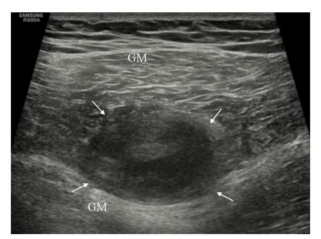
Fig.1 Right gluteal metastasis revealed by ultrasound
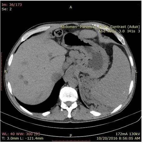
Fig. 2 Possible liver metastasis revealed by CT scan
Upon performing a fluorodeoxyglucose (FDG) positron emission tomography (PET) scan in October 2016, multiple metabolically active lesions were found: two in the left axillary region, measuring 25/20 mm and 55/35 mm respectively, with a maximum standardized uptake value (SUVmax) of 15.68, five retroperitoneal lesions, and two lesions in the right gluteal muscle, measuring 42/33 mm and 31/21 mm respectively with SUVmax of 14.34 (Fig. 3).
Unfortunately, immunotherapy and BRAFtargeted therapy were not available in Romania when treatment began.
Given the fulminant progression of the disease, systemic chemotherapy with Dacarbazin was initiated in October 2016. After 4 cycles of Dacarbazin, the patient started experiencing pain in the right lower limb. This was attributed to progression in the inguinal lymph nodes. In February 2017, the treatment was switched to Carboplatin and Paclitaxel. After two cycles, clinical progression was noted in the inguinal, axillary, and supraclavicular lymph nodes.

Fig. 3 Metastasis in axillary, retroperitoneal lymph node, and gluteal muscle revealed by PET scan
A molecular analysis of the tumor demonstrated a BRAF V600E mutation. In March 2017, treatment with Dabrafenib (Tafinlar) was initiated. Unfortunately, at that time only Dabrafenib was reimbursed by the patient’s insurance and a MEK inhibitor (e.g. Trametinib) could not be given.
The patient was followed up with monthly clinical evaluations and lab tests. His clinical status improved and the analgesics were discontinued. The treatment with Dabrafenib was well tolerated, with minimal hematological toxicity.
Thoracic, abdominal, and pelvic CT scans were done every 6 months, and documented persistent stable disease overall. At the January 2022 assessment the axillary, mediastinal, and abdominal lymph nodes were lower than 1 cm and no other lesions were noted.
As of last follow-up, in June 2022, the patient continued to be asymptomatic, with a maintained performance status (PS) of 1. The CT scans continued to show stable disease. He is currently continuing treatment with Dabrafenib.
3. Discussion:
Bladder invasion, either caused by metastatic melanoma or by melanoma developing in the bladder wall, are rare conditions which can be diagnosed almost exclusively by cystoscopy. The diagnostic is established by immunohistochemical analysis with S-100, HMB-45, and Melan-A specific antigens (4). Symptomatic bladder lesions can be resected by transurethral surgery. Partial or radical cystectomy are more aggressive approaches, and are usually performed for solitary or localized metastatic disease (5). Due to the aggressivity of this tumor, the patient was surgically treated with radical cystectomy and prostatectomy.
Compared to other similar cases reported in the literature and treated with transurethral resection (TUR)/cystectomy and that had synchronous metastases, our patient has a good PS and demonstrated a progression free survival (PFS) of almost 48 months (Table 1).
Table 1. Treatment of melanoma cases with bladder metasases
| Reference | Sex/ Age | Synchronous metastasis | Treatment | Survival |
|---|---|---|---|---|
| Bartone (6) | F/70 | Lymph nodes | Partial cystectomy | Died within 2 months |
| Dasgupta (7) | F/35 | Axillary lesions | TUR | Died within 4 months |
| Meyer (8) | F/60 | Lungs | TUR | Died 2 months after resec tion |
| Silverstein (9) | M/56 | Lymph nodes | Intra-tumor BCG vac cine followed by par tial cystectomy | Alive at 8 month follow-up |
| Stein (10) | M/50 | Absent | TUR/chemotherapy | Alive at 2 year follow-up |
| Arapantoni Dadioti (11) | F/28 | Brain, lungs Lymph nodes | TUR | Died 2 months after resec tion |
| Efesoy (12) | F/60 | Lungs | TUR | Died 7 months after resec tion |
| Nair (13) | M/54 | Widespread in cluding ureteral and renal pelvic | TUR and laser abla tion of ureteral and re nal pelvic tumors | Died within 3 months of TURBT |
| Shkula (14) | M/60 | Widespread | TUR | Alive at 1 year follow-up |
An uncommon feature of this case is the metastasis to the gluteal muscle. Metastasis to the skeletal muscle occurs only in 0.8% of patients with malignant melanoma.
Literature reports on muscle metastasis from malignant melanoma are scarce. In a retrospective cohort study that included 305 patients diagnosed with malignant melanoma, only 4 patients with skeletal muscle metastases were identified (15).
Other reports described muscle metastases from melanoma in the quadriceps, temporalis, and sartorius muscles. (16-18). Calvert and Goforth reported the presence of metastatic melanoma in the skeletal muscles of patients examined post-mortem (19). Portilla et al. reported an isolated rectus abdominis metastasis from melanoma (20). Hering et al. reported on a series of 15 patients with skeletal muscle metastases, of which only 2 of the primary tumors were melanomas. The melanomas metastasized to the gracilis and vastus lateralis, respectively (21).
It is critical to emphasize the importance of investigating the BRAF status for therapeutic purposes. Patients with tumors with BRAF mutations that received targeted therapies had better OS compared to patients treated with chemotherapy or patients that did not receive treatment (15) (Table 2).
Table 2. Skeletal metastasis from melanoma and survival depending of BRAF status
| Sex/age | Metastasis site | BRAF status | Survival (months) |
|---|---|---|---|
| F/78 | Toe | Wild type | 11 |
| F/56 | Gluteus muscle | Wild type | 4 |
| F/53 | Wing of ilium | Mutated | 29 |
| M/45 | Spine | Wild type | 2 |
| F/76 | Temporal bone | Wild type | 2 |
| F/38 | Knee | Mutated | 24 |
The management of metastatic melanoma is strongly dependent on the tumor’s location, stage, and genetic profile. Before the introduction of immunotherapy, cytotoxic chemotherapy such as Dacarbazine and its active metabolite, Temozolomide, were the first agents used in advanced-stage melanoma.
In the immunotherapy era, chemotherapy was abandoned, as it confers only modest clinical benefit.
Inhibited T-cell functionality is a hallmark of cancer, including malignant melanoma. On their surface, the tumor cells express inhibitory molecules that down-regulate CD4+ and CD8+ lymphocyte activity, such as cytotoxic Tlymphocyte-associated antigen-4 (CTLA- 4) and Programmed Death Protein 1/ Programmed Death Protein Ligand 1 (PD-1/PD-L1). The development of specific immune checkpoint antibodies targeting PD-1, PD-L1, and CTLA-4 revolutionized the treatment of advanced-stage melanoma. The response rate and OS in metastatic melanoma were significantly increased after treatment with checkpoint inhibitors was introduced (18). Patients treated with checkpoint inhibitors have to be closely monitored because of potential autoimmune side effects.
BRAF mutations are present in approximately 40-60% of melanoma tumors. BRAFtargeted inhibitors like Dabrafenib and Vemurafenib increase the OS and PFS for patients with BRAF V600E mutations.
The clinical activity and tolerability of BRAF inhibitors administered to BRAF mutant melanoma patients was first demonstrated in the BREAK-2 phase 2 trial. In the BREAK-2 trial, 84% of patients had prior systemic treatment, and the median follow-up was 13 months. The 5- year PFS was found to be 11%.
In the BREAK-3 trial, that randomized Dabrafenib and Dacarbazine, the five-year OS was 24% for Dabrafenib and 22% for Dacarbazine. Patients in the Dacarbazine arm were allowed to switch to Dabrafenib when their disease progressed. 59% did the switch, explaining the similar 5-year OS between the two arms (24).
Lactate dehydrogenase (LDH) is a useful biomarker for assessing melanoma prognosis. The 5-year PFS in patients with normal LDH was 16% vs. 4% in those with elevated LDH in several phase 2 and 3 clinical trials (24). Long-term treatment with Dabrafenib seems to be well-tolerated. Based on the results of the COMBI-d and COMBI-v trials, the combination of Dabrafenib with Trametinib is currently the standard of care for first-line treatment of patients with sensitive BRAF mutation, leading to long-term benefit in approximately one third of patients (25). Dabrafenib is used alone only when unacceptable toxicity to Trametinib occurs.
Based on the results of the above studies, Dabrafenib should be used in combination with Trametinib. In our case, Dabrafenib was used alone, because Trametinib was not available.
4. Conclusions:
We report a case of aggressive malignant melanoma with an unusual metastatic pattern involving the bladder and skeletal muscle. Remarkably, our patient demonstrated a longterm response to Dabrafenib despite the fact that the drug was started seven months after initial diagnosis and prior progression with chemotherapy. The patient continues to demonstrate stable disease more than 5 years after introducing targeted therapy.
Abbreviations:
CT – computed tomography
US – Ultrasound
FDG-PET – Fluorodeoxyglucose – positron emission tomography
SUV Ibm Max – maximum standardized uptake value lean body mass
MEK – mitogen-activated proteine kinase
CTLA – 4 – Cytotoxic T Lymphocyte Associated protein 4
PR – partial response
PS – performance status
ECOG – Eastern Cooperative Group
PFS – Progression Free Survival
PD – 1/PD-L1 – Programmed Death Protein 1/Programmed Death Protein-Ligand 1
TUR – transurethral resection
M – male, F – female
OS – overall survival
Statements:
Authors’ contributions: All co-authors equally contributed to the manuscript.
Consent for publication: As the corresponding author, I confirm that the manuscript has been read and approved for submission by all co-authors.
Conflict of interest: All authors declare having no competing interests associated with this publication.
Funding Sources: This research did not receive any specific grant from funding agencies in the public, commercial, or not-for-profit sector.
Informed Consent: Written informed consent was obtained from the patient for publication of this case report and any accompanying images. A copy of the written consent is available for review.
Ethical Approval: The treatment strategy was approved by the “Institute of Oncology Cluj-Napoca” tumor board.
References:
- Global Cancer Observatory. Melanoma of skin [Internet]. 2020 [cited 2021 Nov 27]. Available from: https://gco.iarc.fr/today/data/factsheets/cancers/16-Melanoma-of-skin-fact-sheet.pdf
- Kumar RA, Batra N, Frankel J, et al. Melanoma metastasis to the bladder: A case report. Urology Case Reports. 2022;40:101941
- Paterson A, Sut M, Kaul A. Metastatic malignant melanoma of the urinary bladder: Case report and literature review. Central European Journal of Urology. 2012;65:232-234
- Patil RV, Woldu SL, Lucas E, et al. Metastatic melanoma to the bladder: case report and review of the literature. Urol Case Rep. 2017;11:33-36
- Viswanathan N, Khanna A. Skeletal muscle metastasis from malignant melanoma. British Journal of Plastic Surgery. 2005;58:855-858
- BartoneFF. Metastatic melanoma of the bladder. Journal of Urology. 1964;91:151–155
- Dasgupta T, Brasfield R. Metastatic melanoma. A clinicopathological study. Cancer. 1964;17:1323-1339.
- Meyers ML, Balch CM. Diagnosis and treatment of metastatic melanoma Cutaneous melanoma, 3rd ed St. Louis: Quality Medical Publishing. 1998;325–372.
- Silverstein MJ, DeKernion J, Morton DL. Malignant melanoma metastatic to the bladder. Regression following intratumor injection of BCG vaccine. Journal of the American Medical Association. 1974;229(6):688
- Stein BS, Kendall AR. Malignant melanoma of the genitourinary tract. J Urol.1984;132:859-868
- Arapantoni- Dadioti P, Panayiotides J, Kalkandi P, et al. Metastasis of malignant melanoma to a transitional cell cercinoma of the urinary bladder. European Journal of SurgicalOncology. 1995;21
- Efesoy O,Cayan S.Bladdermetastasis of malignant melanoma: a case report and review of literature. Med Oncol. 2011;28:667-669
- Nair BC, Williams NC, Cui C, Summers D, et al.Conjunctival Melanoma: Bladder and Upper Urinary Tract Metastases . Journal of ClinicalOncology. 2011;29(9)
- Shukla A,Wingate JT,Baker KC, et al. HexaminolevulinateBlue-LightCystoscopy in a Patientwith Metastatic Melanoma of the Bladder. Journal of Endourology Case Reports. 2016;2:68-70
- Nieves Gómez-Leóna, Vilma Pacheco-Barciab, Ana I. Ballesterosb, Javier Fragac, Ramon Colomerb, and Alfonsa Friera. Skeletal muscle and solitary bone metastases from malignant melanoma: multimodality imaging and oncological outcome. Melanoma Research 2018, 28:562–570
- Mercer NS, Devaraj VS. Intramuscular metastatic melanoma with an unknown primary. Br J Plast Surg. 1990;43(3):367-368
- Calvert J, Pigg TS. A case of melanotic sarcoma. Trans Pathol Soc Lond. 1898;49:297
- Goforth JL. Malignant melanoma of the vulva. Surg Gynaecol Obstet. 1926;43:322
- Portilla G, Cruz A, Juan N, et al. Isolated rectus abdominis metastasis from melanoma, an extremely rare case. Int J Surg Case Rep. 2016;26:121-123
- Moss ALH, Rees MJW. Metastatic malignant melanoma in muscle. Br J Plast Surg. 1984;37:250
- Herring CL, Harrelson JM, Scully SP. Metastatic carcinoma to skeletal muscle a report of 15 patients. Clin Orthop. 1998;355:272-281
- Jenkins RW, Fisher DE. Treatment of Advanced Melanoma in 2020 and Beyond. J Invest Dermatol. 2021;141(1):23-31
- Tanda ET, VanniI,Boutros A, et al.Current State of Target Treatment in BRAF MutatedMelanoma. Front Mol Biosci. 2020;7:154
- Hauschild A, Ascierto PA, Schadendorf D, Grob JJ, Ribas A, Kiecker F, Dutriaux C, Demidov LV, Lebbé C, Rutkowski P, Blank CU, Gutzmer R, Millward M, Kefford R, Haas T, D’Amelio A Jr, Gasal E, Mookerjee B, Chapman PB. Long-term outcomes in patients with BRAF V600-mutant metastatic melanoma receiving dabrafenib monotherapy: Analysis from phase 2 and 3 clinical trials. Eur J Cancer. 2020. 125: 114-120.
- Robert C, Grob JJ, Stroyakovskiy D, Karaszewska B, Hauschild A, Levchenko E, Chiarion Sileni V, Schachter J, Garbe C, Bondarenko I, Gogas H, Mandalá M, Haanen JBAG, Lebbé C, Mackiewicz A, Rutkowski P, Nathan PD, Ribas A, Davies MA, Flaherty KT, Burgess P, Tan M, Gasal E, Voi M, Schadendorf D, Long GV. Five-Year Outcomes with Dabrafenib plus Trametinib in Metastatic Melanoma. N Engl J Med. 2019. 381(7): 626-636.