


Author(s) :
Andrew Naus1, Norbert Banyi2, Roy Ma3
1West Point Grey Academy, Vancouver, Canada
2University of British Columbia, Faculty of Medicine, Vancouver, Canada
3BC Cancer, Department of Radiation Oncology
Corresponding author: Norbert Banyi, Email: nbanyi@student.ubc.ca
Publication History: Received - , Revised - , Accepted - , Published Online - 1 October.
Copyright: © The author(s). Published by Casa Cărții de Știință.
User License: Creative Commons Attribution – NonCommercial (CC BY-NC)

 PDF
PDF
 Share
Share
Highlights
-
Eye-sparing radiotherapy has replaced routine enucleation: For small–medium uveal melanomas, plaque brachytherapy (PB) offers survival comparable to enucleation and remains the most widely used modality, while SRS/SRT and proton therapy expand options for challenging cases.
-
Proton beam irradiation delivers highly conformal dose with strong control rates, limited by access: Leveraging the Bragg peak, PBI achieves >90% local control and ~70–85% 5-year OS in published series, but global availability and infrastructure cost are key constraints.
-
Canada’s unique PBI experience at TRIUMF (Vancouver): As the only Canadian proton center, TRIUMF treated >200 OM patients (1995–2017), reporting ~91% tumor control, ~82% 5-year survival, and ~80% eye preservation; emerging combinations (e.g., ATM inhibition) aim to reduce radioresistance and potentially lower dose.
Abstract
Ocular melanoma (OM) originates from melanocytes in the eye, predominantly in the uvea, particularly the choroid. The yearly incidence is around six cases per million. OM is not primarily driven by ultraviolet exposure like skin melanoma, but is usually caused by mutations in GNAQ or GNA11. Symptoms like blurry vision and visual field defects appear late. Diagnosis is often made via eye exams, specialized ultrasound, and rarely biopsy. This narrative review describes the radiation treatment modalities of OM and highlights the landscape of proton beam irradiation in Canada.
Historically, enucleation was the standard of care for OM. However, current strategies consider tumor size, location, patient age, visual potential, and metastatic presence. Primary treatments include radiation therapy and surgery. Radiation therapy includes plaque brachytherapy (PB), proton beam irradiation (PBI), stereotactic radiosurgery (SRS), and stereotactic radiotherapy (SRT). Surgery includes endoresection, exoresection, and enucleation. Tebentafusp-tebn has been FDA-approved for metastatic cases.
PB, the most common radiation therapy for OM, involves radioisotopes delivering radiation into the tumor. Comparable survival rates between PB and enucleation for medium choroidal melanoma have made PB the standard of care. PB has certain limitations, mainly surgical complications. PBI uses a particle accelerator for focused, high-energy proton radiation, yielding high tumor control and survival rates, though the availability of proton facilities is a significant limitation.
Vancouver is the only center in Canada for PBI, administered not in a healthcare facility but at TRIUMF (Tri-University Meson Facility). TRIUMF, the world’s largest cyclotron particle accelerator, in partnership with BC Cancer and UBC Department of Ophthalmology and Eye Care Center, has treated over 200 ocular melanoma patients between 1995 and 2017, achieving a 91% tumor control rate and 82% five-year survival rate. Emerging combination therapies like Ataxia Telangiectasia Mutated (ATM) protein kinase inhibition before PBI show potential, possibly reducing radiation dose and resistance.
1. Introduction
1.1.Epidemiology
Known primarily as “skin cancer,” melanoma is a tumor arising from melanocytes located at various anatomic locations, including skin, mucosal membrane (nasal mucosa, oropharyngeal, pulmonary, gastrointestinal, vaginal, anal/rectal, urinary tract), ocular region (uvea, conjunctiva, eyelid, orbit), and rarely from unknown primary sites.
The annual incidence of eye melanoma in the United States is about six cases per one million persons (approximately 2500 new diagnoses annually) (1). Among ocular melanomas, 83% arise from the uvea, 5% from the conjunctiva, and 10% from other sites (2). The most common site for uveal melanoma is the choroid. In a study of 8033 patients with uveal melanoma by Shields et al., the tumor was located in the iris in 285 (4%), ciliary body in 492 (6%), and choroid in 7256 (90%) cases (3). Melanoma of the uveal tract is a lifethreatening tumor that also jeopardizes the function of the eye. While deaths caused by the disease only account for a few cancer deaths a year, approximately one-quarter of affected patients eventually die from metastasis due to the lack of systemic treatment (1). While skin melanoma has been linked with ultraviolet exposure and harbors a BRAF or NRAS mutation, the impact of ultraviolet rays is significantly less in the development of ocular melanoma, which is usually initiated by a mutation in GNAQ or GNA11 (4, 5).
1.2.Clinical presentation and diagnosis
Ocular melanoma is often asymptomatic for many years before any symptoms arise. From a clinical and practical perspective, the most common symptoms are blurry vision and visual field defects (blind spots), while in our experience, most of the other symptoms described in the literature are exceptionally rarely seen in clinical practice.
Without presenting symptoms, an optometrist or general ophthalmologist discovers most ocular melanomas via routine eye examinations. Still, an expert ocular oncologist ultimately confirms it, most often without needing a pathologic diagnosis (6). A uveal nevus is the most common simulating lesion in the differential diagnosis of uveal melanoma, and these two entities are challenging to distinguish on clinical examination. Preexisting choroidal nevi are where most choroidal melanomas are believed to arise, although few of them transform into melanoma (<1:8000) (7). Ocular examination through specialized eye ultrasound, fluorescein angiography, or optical coherence tomography (OCT) are techniques for diagnosing ocular melanoma (8). Fine needle aspiration biopsy is used with caution to avoid seeding. Still, it is necessary to confirm the diagnosis in complex cases and through genetic profiling to determine the underlying genetic abnormalities of each tumor, assessing its metastatic risk and prognosis (9).
Studies suggest that in 50% of patients, ocular melanoma will metastasize (10). The rate of metastasis varies with the anatomical location of the primary tumor within the eye, with iris melanoma having the lowest rate of metastases, as compared to ciliary bodies and choroidal melanomas (10). In patients with uveal melanoma, the clinical and histopathologic features of the primary tumor that are associated with poor prognosis include increased patient age, the largest basal diameter of the tumor, ciliary body involvement, extra scleral tumor extension, epithelioid cell type, and vasculogenic mimicry patterns (11). Patients who present with primary uveal melanoma and synchronous distant metastases are rare (less than four percent of all patients diagnosed with uveal melanoma).
1.3.Risk factors
Several risk factors, including light eyes, the Caucasian population, and certain skin conditions such as cutaneous nevi, dysplastic nevus syndrome, iris nevi, and BAP1 mutation, have been identified (1). The somatic or germline mutation of BAP1 predisposes patients to develop uveal melanoma, malignant mesothelioma, cutaneous melanomas, basal cell carcinoma, and renal cell carcinoma, requiring molecular testing and referral to hereditary cancer programs (12). A large study of iris and posterior melanomas showed a metastatic rate of 15% at 5 years and 25% at ten years (11). The most common metastasis site is the liver (60-89%), followed by the lungs (24-29%), skin and soft tissue (11-12%), bone (8-17%), and lymph nodes (11%) (11,13). Identifying metastases prompts whole-blood genotyping testing for HLA-A*02:1, affecting therapy selection (14).
2. Results and discussion
2.1.Treatment options for ocular melanoma
Historically, the main treatment option for primary tumors in patients with ocular melanoma was enucleation. Many factors, including the size and location of the tumor, presence of extraocular extension, visual potential, patient age and preference, and presence or absence of metastases, currently guide its management, which remains a multidisciplinary challenge. Radiotherapy tends to be the preferred treatment for small and medium tumors since the Collaborative Ocular Melanoma Study (COMS) showed that survival rates between enucleation and plaque radiotherapy are comparable in patients with medium choroidal melanomas (15). At the same time, enucleation is usually performed for larger and more advanced melanomas, poor visual function, recurrent tumors, multifocal iris melanoma, and diffuse iris melanoma (13). There are currently very few treatments approved for metastatic ocular melanoma and despite the advances in the treatment of primary tumors, the rate of mortality once metastases are diagnosed is unchanged in decades (16). Several options have been reported without significant or lasting improvement, including cryotherapy, chemotherapy, and immunotherapy. Most recently, in 2022, the US Food and Drug Administration (FDA) approved tebentafusp-tebn (Kimmtrak) for the treatment of metastatic uveal melanoma or that which cannot be surgically removed, in patients with HLA_A* 02:01 (16).
2.1.1. Observation: For asymptomatic patients with small uveal melanocytic tumors (<12 mm in diameter and <2 to 3 mm in height), initial management is often observation for evidence of growth, with follow-up at two- to four-month intervals rather than immediate intervention. Imaging modalities commonly used in monitoring a small suspicious uveal melanocytic tumor include fundus photography, ultrasonography, and fundus autofluorescence to identify evidence of tumor growth, subretinal fluid, orange lipofuscin pigmentation, and other risk factors for malignant transformation (17).
2.1.2. Surgery: The main surgical procedure used for ocular melanomas is enucleation, and represented historically, the standard of care. Although eye preservation is essential and highly desirable, enucleation remains the only option for large tumors unsuitable for radiation. Indications for enucleation include large tumors with thickness >10 or 12 mm and/or a basal diameter >18 mm (18), poor visual potential and extraocular growth, proximity to the optic disc, and extensive involvement of iris, angle, or ciliary body (19).
Local resection is sometimes performed for small iris tumors only and almost never for the other anatomical locations within the eye. Tumor resection can be based either on an external approach, known as exoresection, or an ab-interno approach, known as endoresection. Even in such cases, postoperative vision is suboptimal, as an incomplete iris leaks light and vision looks distorted.
2.1.3 Radiation therapy: The current standard of care treatment for primary uveal melanoma is radiation therapy and two main types of radiation therapy exist. Since uveal melanomas are relatively radioresistant, and the eye is a closed and small space, they must be treated with high-dose focused radiation. Radiation therapy can be delivered to the eye in various ways. It can be done with a surgically implanted radioactive brachytherapy plaque containing various isotopes such as iodine-125, ruthenium-106, or palladium-103. Alternately, external beams can deliver the radiation, either in the form of charged particles such as proton or helium ions or photons, using stereotactic techniques with a Gamma knife or a Linear Accelerator.
In this review, we offer a brief and practical comparison of each technique’s characteristics, advantages, disadvantages, and complications, focusing on proton beam irradiation, where we incorporate aspects learned during 22 years of treatment of ocular melanoma at TRIUMF, Canada.
3. Radiation therapy techniques used for ocular melanoma
All cells, whether normal or malignant, have some radio sensitivity secondary to two cellular factors: rapid cell division and the cell’s innate ability to repair DNA damage. Ionizing radiation damages the DNA of malignant cells by causing double-strand breaks, most commonly causing the cell to misrepair (20). The extent of the damage becomes most evident during mitosis, wherein the damaged cells lose the ability to self-replicate.
Another damage caused by the radiation encompasses alterations in the growth of signal transduction pathways, apoptosis, and the general regulation of the cell cycle. Cancer cells divide rapidly and often and have immature DNA repair mechanisms compared to normal cells. Melanoma has a relatively low radio sensitivity compared to many cancers but is far more radiosensitive than normal tissue (21).
A summary of radiation therapy modalities can be seen in Table 1.
Table 1: Summary comparison of radiation therapy methods
| Method | Plaque Brachytherapy (PB) | Stereotactic Radiosurgery (SRS) | Proton Beam Irradiation (PBI) |
|---|---|---|---|
| Availability | High | High | Low |
| Cost | Low | Moderate | High |
| Indications | Small – medium, away from optic nerve |
Larger (>7mm thickness), near optic nerve |
Larger (>7mm thickness), near optic nerve |
| Complication of concern |
Radiation induced glaucoma |
Radiation induced glaucoma |
Radiation induced glaucoma |
| Contraindications | Tumor close to optic nerve, large tumor thickness in comparison to base. |
Tumor volume > 1/3 of globe volume |
Tumor volume > 1/3 of globe volume |
3.1.Plaque Brachytherapy (PB)
Plaque brachytherapy is the most common form of radiation therapy used in treating uveal melanoma worldwide and utilizes low-energy radioisotopes that constantly emit radiation photons due to radio decay, delivered through the eye wall into the tumor. Plaque radiotherapy with an apex dose of 70–100 Gy (22) is one of the most commonly used treatment modalities for posterior uveal melanoma. Initial head-tohead comparison of enucleation and PB was performed through COMS, the largest randomized controlled trial in ocular oncology, with >2,000 enrolled patients (23). The results revealed no survival differences at 5, 10, and 12 years between plaque brachytherapy and enucleation in patients with medium choroidal melanoma, making PB the treatment of choice in suitable tumors. Because the saucer-shaped plaque is sutured surgically to the sclera for the duration of treatment, very high-energy radiation is emitted near the source. There is a rapid energy loss away from the source, translating into a high degree of efficacy and little damage beyond the tumor target. Several radioisotopes have been used, including cobalt-60 (60Co), iodine-125 (125I), iridium-192 (192Ir), palladium-103 (103Pd), and ruthenium-106/rhodium-106 (106Ru/106Rh) (25). The most commonly used isotope in the United States is 125I, a gamma emitter with deeper tissue penetrance that allows for treating larger tumors. However,125I is rarely used in Europe and South America, where Ruthenium-106, a beta emitter, is most often used (although it is ineffective in large melanomas due to its lower penetrance). The choice of radioisotope is based less on their efficacies and toxicities, which are relatively similar clinically but based on availability. Iodine-125 and palladium-103 are often used in North America due to the large repositories of these radioisotopes and the consistent supply chain. Similarly, the available quarry of ruthenium in Europe makes it the dominant radioisotope for plaque brachytherapy in Europe and South America (24). With intraoperative ultrasonography for plaque localization, the local recurrence rates, a risk factor for metastasis with plaque brachytherapy, have decreased substantially and are now similar to charged-particle radiation therapy. Consensus opinion guidelines for using radioactive plaque therapy have been published by the American Brachytherapy Society (ABS), but research is ongoing into the optimal dosimetry parameters for uveal melanoma (25). Despite comparison studies between various radioisotopes, clinically, the radioisotope is selected based on relative convenience and availability of the isotope to each treatment facility.
As PB is the standard of care for ocular melanoma, there are constant efforts to improve the technique, especially plaque placement, use of newer radioisotopes, and identification of the best optimal dose for the tumor apex, without or in combination with the inhibition of the Ataxia telangiectasia mutated (ATM) protein kinase (26). Pulverization of gold nanoparticles on the tumor before brachytherapy can absorb photons of energy, preventing the photons from passing through the tumor, and has been studied to reduce the dose of radioisotopes (27). Improvement in radiological techniques may allow for ultra-high field MRI instead of ultrasound for three-dimensional visualization of the tumor, restricted by the availability of a seven-tesla magnet required for this procedure (19). Alternatively, modern techniques, such as 3D printing of the eye and orbit, have also been studied to enhance treatment planning further (28). The main limitation for PB remains related to surgery for plaque placement. Surgical complications include double vision due to muscular injury, dry eye due to conjunctival peritomy, eyelid swelling, and the risk of anesthesia, which makes especially elderly patients poor candidates for this therapy.
3.2 Stereotactic Radiosurgery (SRS)
Stereotactic radiosurgery is an advanced type of external beam radiotherapy used mainly for larger tumors that can be visualized by CT scan or MRI, which are used to localize the tumor within the eye. The primary type of ionizing radiation utilized is photon-based (gamma or Xray waves), characterized by slightly lower energy and increased scattering.
3.2.1 Gamma knife: A gamma knife involves immobilization within a cylindrical headpiece anchored to the patient’s skull with multiple beams directed at the tumor from multiple directions simultaneously, with a potential for high amounts of radiation to be delivered at once. It appears to cause a significant slowing of the blood flow through the central retinal artery and a significant increase in vascular resistance of the smaller retinal arteries, which may explain one of its main complications: the development of therapyrelated secondary glaucoma, reported as high as 47% in some series (29). Due to its multibeam ability to attack the tumor from multiple angles, several authors have also been able to de-escalate the radiation dose and still obtain an excellent killing effect.
3.2.2 Cyberknife: Like gamma knife, cyberknife is a photon-based radiotherapy that involves the external application of multiple high-intensity beams from multiple directions in rapid succession without fixation to the skull. The head is immobilized with a custom-made aquaplast mask. It offers the ability to treat larger eye tumors not amenable to brachytherapy or local resection. The availability of a cyberknife also may influence the treatment decision regarding its use, as it may be the only treatment option available in some countries (stereotactic linear particle accelerator, Slovakia). Cyberknife therapy has a higher incidence of secondary glaucoma following treatment in comparison to other standard methods of uveal melanoma radiation (30).
3.3.Proton Beam Irradiation (PBI)
Proton Beam Irradiation, an alternate method of external beam irradiation, uses heavy charged particles such as helium ions or protons. Although helium therapy was successful and showed promising results, it was discarded due to expensive operating costs. Proton beam therapy utilizes a particle accelerator to release high-energy protons from an atomic nucleus. Because of their relatively large size, protons are not scattered within the cellular nucleus upon arrival at the target cell. This allows for a much-focused beam of radiation with minimal effect on the tissue surrounding the tumor. Protons also have the advantage of undergoing a rapid reduction of energy upon arrival at their target, known as the Bragg effect, which implies that high-energy protons will stop at their target, cause damage to their target, and not pass through the target to cause an effect on tissue past the tumor (normal tissue). Protons are thus able to cause more DNA damage with less total radiation energy.
PBI is an effective and safe treatment for ocular melanoma, with excellent local tumor control >90% and a 5‑year overall survival of 70‑85% (31, 32). PBI could be preferred to brachytherapy for tumors with a location that may challenge plaque positioning or have a risk of suboptimal immobilization of the plaque (for instance, posterior pole), in the treatment of larger tumors with a height of more than 5 mm, or tumors with a narrow base (32) The main limitation of PBI is the availability of protons and the facilities that produce them. Developing a proton accelerator is cumbersome and extremely expensive, and PBI is available only in a few centers worldwide (19). For decades, there were only two in the United States (Boston and California) and a few in Europe, making treatment for most patients impractical. Second, successful proton therapy of an eye requires placing a radiopaque target on the eye’s surface to aim the proton beam, which requires a surgical procedure ahead of treatment, typically under general anesthesia. However, the placement of tantalum markers used to mark the tumor and safety margins is surgically easier than plaque placement, and computer adjustments are possible after insertion. Third, the proton accelerator is external to the eye, which is a highly mobile organ. Because the beam is very focused, there is a real possibility of missing the target tumor with consequent treatment failure. Despite the limitations, continuous development can improve the technique to achieve better outcomes (33).
At the beginning of 2023, 89 proton therapy centers operated worldwide and 41 in the United States (34). Vancouver remains the only center in Canada for PBI, administered not in a healthcare facility but at TRIUMF (TRI-UNI-VERSITY MESON FACILITY), the world’s largest cyclotron particle accelerator (520Mev, 18 meter-long diameter vacuum tank). Between 1995 and 2017, one of TRIUMF’s beamlines used for cutting-edge research in nuclear physics was also used for cancer treatment of ocular melanoma at the Proton Therapy Research Center, where in conjunction with BC Cancer and the UBC Department of Ophthalmology and Eye Care Center, more than 200 patients were treated, many under the direct care of one of the authors (Dr. Ma). Approximately 40 patients with eye melanoma were diagnosed locally, and a quarter of them underwent PBI at TRIUMF, while the remaining had brachytherapy. Patients aged 14 to 86 had customized equipment to ensure their treatment’s comfort and safety. The painless treatment lasted 90 seconds and was administered four times over four days, similar to protocols used by other centers (35). Local tumor control was 91% effective, with a five-year survival rate of 82%, with the affected eye saved in 80% of cases (32). The local experience confirmed the importance of PB and PBI radiation treatments, which were considered complementary for achieving the best outcomes and required personalized treatment from multidisciplinary treatment selection to implementation and followup. Figures 1-4 depict the facilities and patient set-up for administering this therapy, and figure 5 the pathology of an ocular melanoma treated by enucleation in Vancouver.
For the treatment of larger tumors and tumors close to the optic nerve, plaque brachytherapy is not used. Instead, SRS or PBI has been used for many years as alternatives to enucleation. While SRS has been demonstrated to have higher rates of post-treatment glaucoma, vitreous hemorrhage, and significant visual loss, overall rates of eye retention and survival are similar between SRS and PBI (36, 37). PBI remains the less accessible option of the two due to its prohibitive cost.
Among the most exciting combination therapies is inhibiting ATM protein kinase before radiation therapy to reduce the required radiation dose. PBI may be the first to incorporate ATM inhibition and decrease the ocular melanoma relative resistance before initiating PBI.
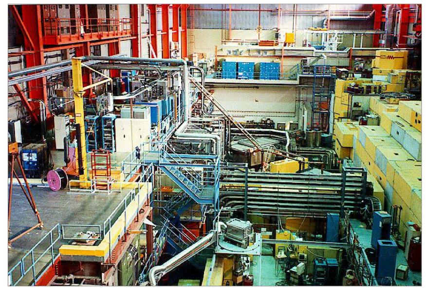
Figure 1: TRIUMF: The only Canadian Center for Proton Beam Therapy Treatments: Ocular Melanoma Treatments From 1995 to 2017: Not a Hospital Facility
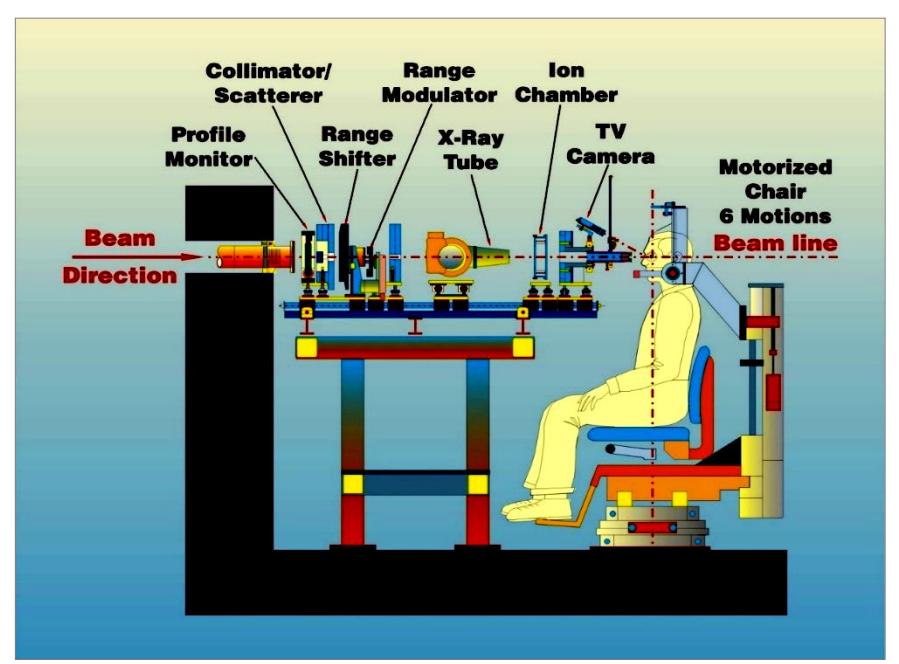
Figure 2: The Beam Preparation and Patient Equipment at TRIUMF, Canada The proton beam is centered using a profile monitor onto a collimator scatterer, which has a .8 mm Pb foil that uniformly spreads out the beam over about 30 mm diameter. The range shifter and range modulator are placed near the scatterer. The dose is measured using an air ion chamber and the patient is seated in a chair with 6 degrees of freedom.
Figure 3: Treatment Set Up and Patient Treatment at TRIUMF, Vancouver, Canada
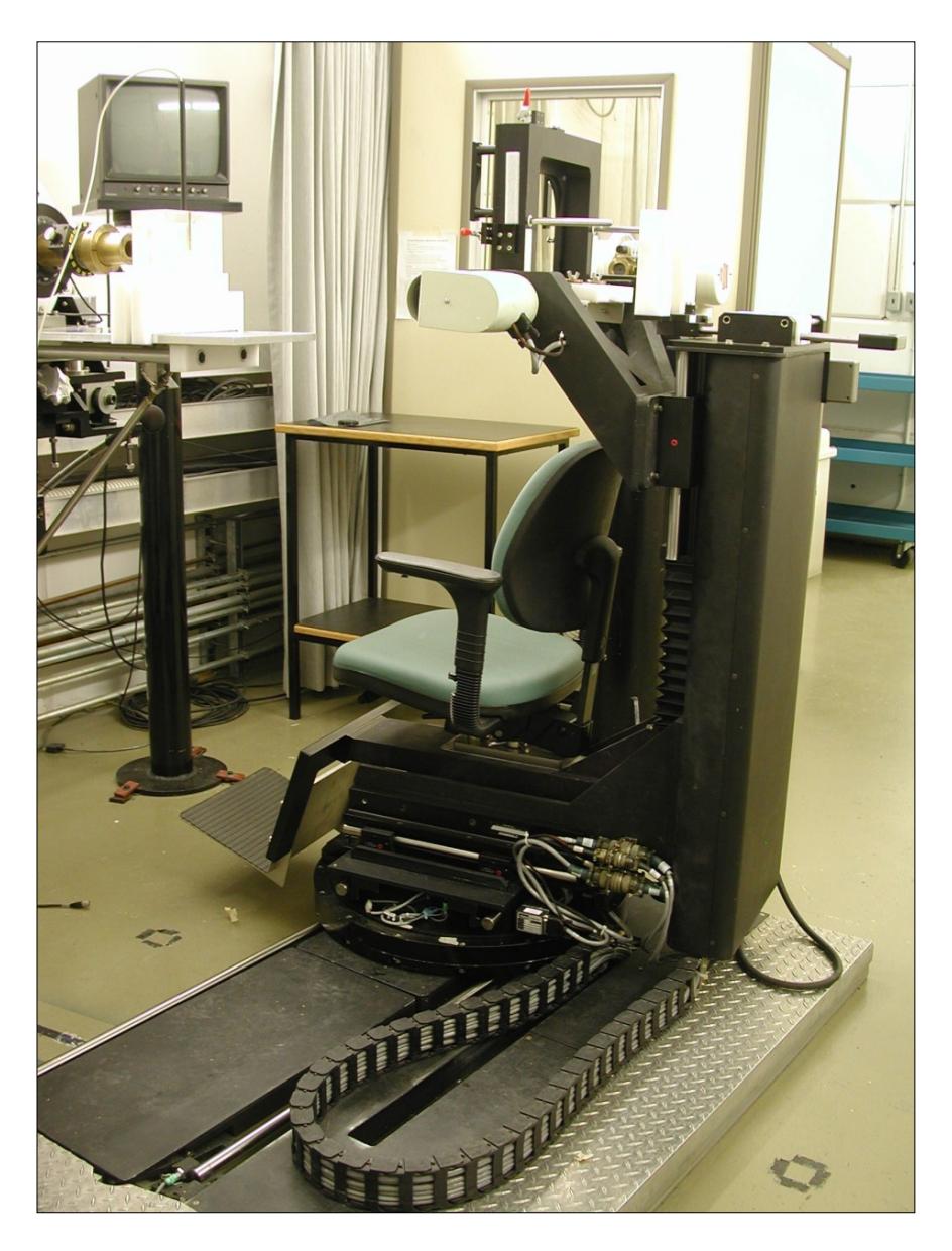
Patient Chair and Set-Up for Treatment: The chair has six degrees of freedom and can be rotated around a vertical axis, typically plus/minus 15 degrees to assist the positioning of the patient
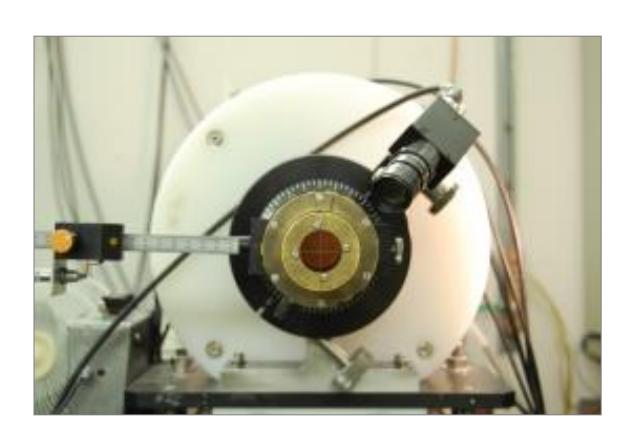
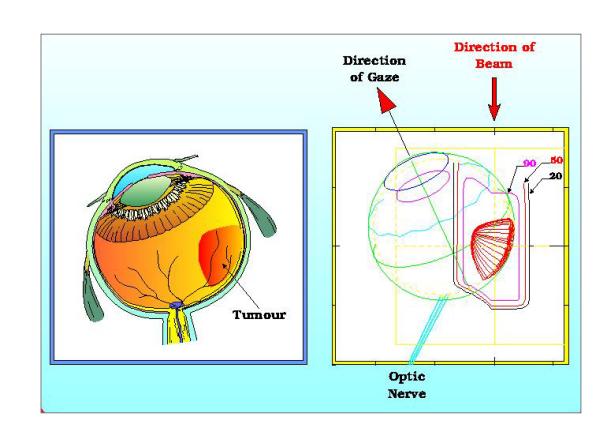
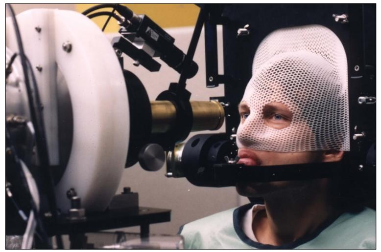
Before Initiation of Treatment: multiple gaze angles are assessed, and the most optimal azimuthal and polar angles are chosen to treat the tumor, but spare anterior chamber, optic nerve, and lacrimal gland.
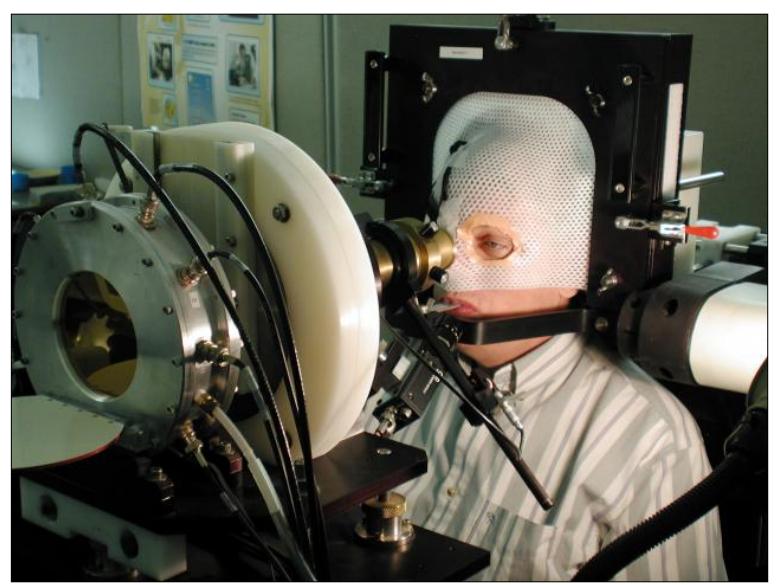
Patient in Treatment Position (treatment time approximately 1 minute, 50Gypr in 4# in 4 days)
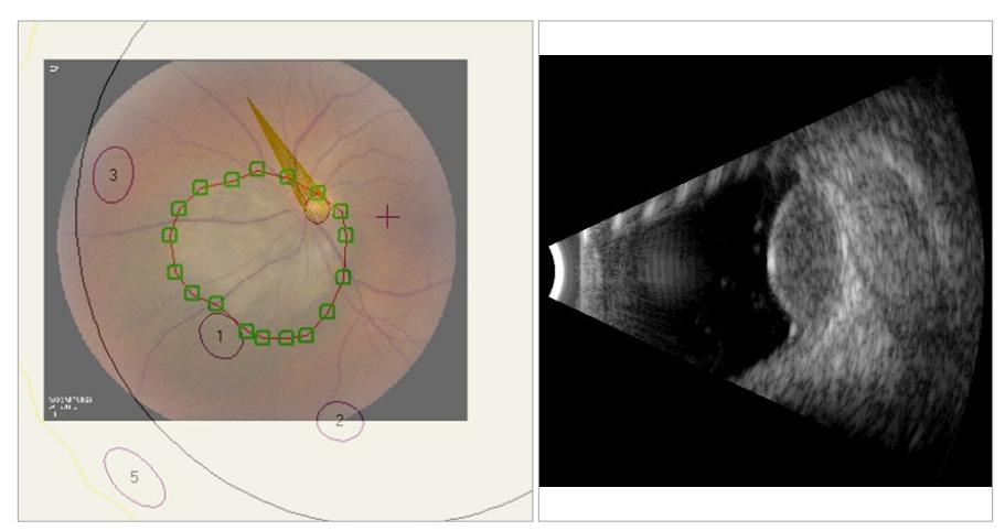
Figure 4. Treatment Planning: The tumor is drawn onto the globe using EYEPLAN software, aided by ultrasound photographs of the fundus.
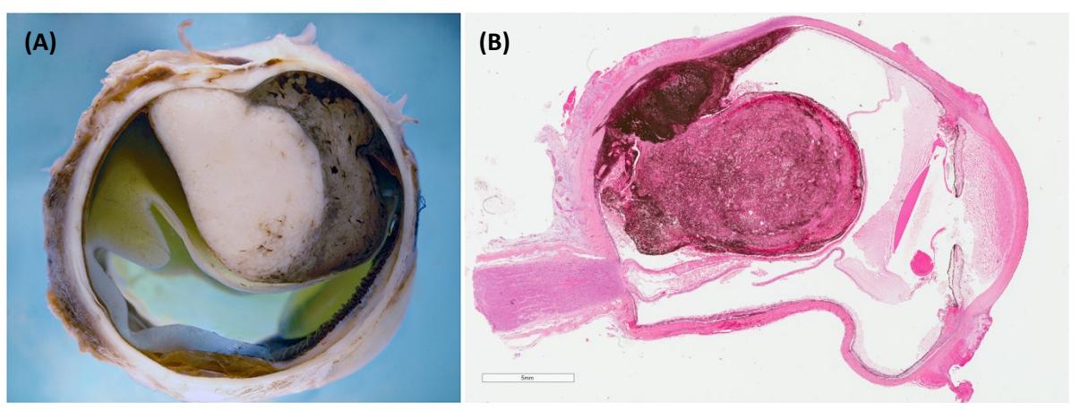
Figure 5: Ocular Melanoma: (A) macroscopic and (B) microscopic view from an enucleation procedure. (Courtesy of Dr. Rasmussen, Ophthalmologic Pathologist, Vancouver General Hospital).
4. Conclusion
OM remains a rare disease with treatment specific to its unusual location. Therefore, it may be one type of cancer many oncologists may never directly encounter. For those, as well as for acknowledging the dedication, work, and results of the Canadian group at BC Cancer and TRIUMF, we decided to write this narrative review, which may spark the readers’ curiosity to look for future reads on the topics of OM or PBI.
Abbreviations:
BRAF – v-Raf Murine Sarcoma Viral Oncogene Homolog B
NRAS – Neuroblastoma RAS Viral Oncogene Homolog
GNAQ – Guanine Nucleotide Binding Protein (G Protein), Q Polypeptide
GNA11 – Guanine Nucleotide Binding Protein (G Protein), Alpha 11
OCT – Optical Coherence Tomography
BAP1 – BRCA1 Associated Protein-1
HLA-A02-1 – Human Leukocyte Antigen A02-1
COMS – Collaborative Ocular Melanoma Study
FDA – Food and Drug Administration
TRIUMF – Tri-University Meson Facility
PB – Plaque Brachytherapy
Gy – Gray (unit of ionizing radiation dose)
ABS – American Brachytherapy Society
ATM – Ataxia Telangiectasia Mutated
MRI – Magnetic Resonance Imaging
PBI – Proton Beam Irradiation
PBT – Proton Beam Therapy
CT – Computed Tomography
UBC – University of British Columbia
MeV – Mega Electron Volt
Statements:
Authors’ contributions: AN had the idea for this review. AN, NB, and RM conceived the paper. AN took the lead in writing the manuscript; NB and RM also contributed to the writing of the manuscript, provided critical feedback and made the final approval.
Consent for publication: “As the corresponding author, I confirm that the manuscript has been read and approved for submisson by all named authors”.
Conflict of interests: The authors declare no conflict of interest. Funding sources: There are no funding sources for this work.
Statement of ethics: No ethics approval was required for this narrative review.
References:
-
Jager MJ, Shields CL, Cebulla CM, Abdel-Rahman MH, Grossniklaus HE, Stern MH, Carvajal RD, Belfort RN, Jia R, Shields JA, Damato BE. Uveal melanoma. Nat Rev Dis Primers. 2020 Apr 9;6(1):24. doi: 10.1038/s41572-020-0158-0. Erratum in: Nat Rev Dis Primers. 2022 Jan 17;8(1):4. PMID: 32273508.
-
Jovanovic P, Mihajlovic M, Djordjevic-Jocic J, Vlajkovic S, Cekic S, Stefanovic V. Ocular melanoma: an overview of the current status. Int J Clin Exp Pathol. 2013 Jun 15;6(7):1230-44. PMID: 23826405; PMCID: PMC3693189.
-
Shields CL, Kaliki S, Shah SU, et al. Iris melanoma: features and prognosis in 317 children and adults. J AAPOS. 2012;16(1):10–6. https://doi.org/10.1016/j.jaapos.2011.10.012.
-
Van Raamsdonk CD, Bezrookove V, Green G, et al. Frequent somatic mutations of GNAQ in uveal melanoma and blue naevi. Nature. 2009;457(7229):599–602. https://doi.org/10.1038/nature07586.
-
Van Raamsdonk CD, Griewank KG, Crosby MB, et al. Mutations in GNA11 in uveal melanoma. N Engl J Med. 2010;363(23):2191–9. https://doi.org/10.1056/NEJMoa1000584.
-
Amaro A, Gangemi R, Piaggio F, Angelini G, Barisione G, Ferrini S and Pfeffer U: The biology of uveal melanoma. Cancer Metastasis Rev 36: 109-140, 2017
-
Krantz BA, Dave N, Komatsubara KM, MarrBP and CarvajalRD: Uveal melanoma: Epidemiology, etiology, and treatment of primary disease. Clin Ophthalmol 11: 279-289, 2017
-
Kaliki S and Shields CL: Uveal melanoma: Relatively rare but deadly cancer. Eye (Lond) 31: 241-257, 2017.
-
Frizziero L, Midena E, Trainiti S, Londei D, Bonaldi L, Bini S and Parrozzani R: Uveal melanoma biopsy: A review. Cancers (Basel) 11: 1075, 2019
-
Carvajal RD, Schwartz GK, Tezel T, Marr B, Francis JH, Nathan PD. Metastatic disease from uveal melanoma: treatment options and future prospects. Br J Ophthalmol. 2017 Jan;101(1):38-44. doi: 10.1136/bjophthalmol-2016-309034. Epub 2016 Aug 29. PMID: 27574175; PMCID: PMC5256122.
-
Kaliki S, Shields CL, Shields JA. Uveal melanoma: estimating prognosis. Indian J Ophthalmol. 2015 Feb;63(2):93-102. doi: 10.4103/0301-4738.154367. PMID: 25827538; PMCID: PMC4399142.
-
Pilarski R, Carlo MI, Cebulla C, et al. BAP1 Tumor Predisposition Syndrome. 2016 Oct 13 [Updated 2022 Mar 24]. In: Adam MP, Mirzaa GM, Pagon RA, et al., editors. GeneReviews® [Internet]. Seattle (WA): University of Washington, Seattle; 1993-2023. Available from: https://www.ncbi.nlm.nih.gov/books/NBK390611/
-
Kaliki, S., Shields, C. Uveal melanoma: relatively rare but deadly cancer. Eye 31, 241–257 (2017). https://doi.org/10.1038/eye.2016.275
-
Howlett S, Carter TJ, Shaw HM, Nathan PD. Tebentafusp: a first-in-class treatment for metastatic uveal melanoma. Ther Adv Med Oncol. 2023 Mar 21;15:17588359231160140. doi: 10.1177/17588359231160140. PMID: 36970111; PMCID: PMC10031621.
-
Hawkins BS; Collaborative Ocular Melanoma Study Group. The Collaborative Ocular Melanoma Study (COMS) randomized trial of pre-enucleation radiation of large choroidal melanoma: IV. Ten-year mortality findings and prognostic factors. COMS report number 24. Am J Ophthalmol. 2004 Dec;138(6):936-51. doi: 10.1016/j.ajo.2004.07.006. PMID: 15629284.
-
Chen LN, Carvajal RD. Tebentafusp for the treatment of HLA-A*02:01-positive adult patients with unresectable or metastatic uveal melanoma. Expert Rev Anticancer Ther. 2022 Oct;22(10):1017-1027. doi: 10.1080/14737140.2022.2124971. Epub 2022 Sep 19. PMID: 36102132; PMCID: PMC10184536.
-
PDQ Adult Treatment Editorial Board. Intraocular (Uveal) Melanoma Treatment (PDQ®): Health Professional Version. 2023 May 12. In: PDQ Cancer Information Summaries [Internet]. Bethesda (MD): National Cancer Institute (US); 2002-. Available from:https://www.ncbi.nlm.nih.gov/books/NBK66047/
-
Singh, M., Durairaj, P. & Yeung, J. Uveal Melanoma: A Review of the Literature. Oncol Ther 6, 87–104 (2018). https://doi.org/10.1007/s40487-018-0056-8
-
Foti, P.V., Travali, M., Farina, R. et al. Diagnostic methods and therapeutic options of uveal melanoma with emphasis on MR imaging—Part II: treatment indications and complications. Insights Imaging 12, 67 (2021). https://doi.org/10.1186/s13244-021-01001-w
-
Azzam EI, Jay‑Gerin JP, Pain D. Ionizing radiation‑induced metabolic oxidative stress and prolonged cell injury. Cancer Lett. 2012;327(1‑2):48‑60
-
Groenewald C, Konstantinidis L, Damato B. Effects of radiotherapy on uveal melanomas and adjacent tissues. Eye (Lond). 2013 Feb;27(2):163-71. doi: 10.1038/eye.2012.249. Epub 2012 Nov 30. PMID: 23196647; PMCID: PMC3574249.
-
Karimi S., Arabi A., Siavashpour Z., Shahraki T., Ansari I. Efficacy and complications of ruthenium-106 brachytherapy for uveal melanoma: A systematic review and meta-analysis. J. Contemp. Brachytherapy. 2021;13:358–364. doi: 10.5114/jcb.2021.106191
-
Margo C.E. The Collaborative Ocular Melanoma Study: An Overview. Cancer Control. 2004;11:304–309. doi: 10.1177/107327480401100504
-
Reichstein DA, Brock AL. Radiation therapy for uveal melanoma: a review of treatment methods available in 2021. Curr Opin Ophthalmol. 2021 May 1;32(3):183-190. doi: 10.1097/ICU.0000000000000761. PMID: 33770014.
-
American Brachytherapy Society Ophthalmic Oncology Task Force. Electronic address: paulfinger@eyecancer.com; ABS – OOTF Committee. The American Brachytherapy Society consensus guidelines for plaque brachytherapy of uveal melanoma and retinoblastoma. Brachytherapy. 2014 Jan-Feb;13(1):1-14. doi: 10.1016/j.brachy.2013.11.008. Epub 2013 Dec 24. PMID: 24373763
-
Hussain RN, Coupland SE, Khzouz J, Kalirai H, Parsons JL. Inhibition of ATM Increases the Radiosensitivity of Uveal Melanoma Cells to Photons and Protons. Cancers (Basel). 2020 May 28;12(6):1388. doi: 10.3390/cancers12061388. PMID: 32481544; PMCID: PMC7352388
-
Asadi S, Vaez-zadeh M, Masoudi SF, Rahmani F, Knaup C, Meigooni AS. Gold nanoparticle-based brachytherapy enhancement in choroidal melanoma using a full Monte Carlo model of the human eye. J Appl Clin Med Phys. 2015 Sep 8;16(5):344–357. doi: 10.1120/jacmp.v16i5.5568. PMID: 26699318; PMCID: PMC5690168.
-
Subashi E, Jacobs C, Hood R, et al. A design process for a 3D printed patient specific applicator for HDR brachytherapy of the orbit. 3D Print Med 2020; 6:15. doi: 10.1186/s41205-020-00068-3.
-
Sarici A.M., Pazarli H. Gamma-knife-based stereotactic radiosurgery for medium- and large-sized posterior uveal melanoma. Graefe’s Arch. Clin. Exp. Ophthalmol. 2012;251:285–294. doi: 10.1007/s00417-012-2144-z.
-
Zemba M, Dumitrescu OM, Gheorghe AG, Radu M, Ionescu MA, Vatafu A, Dinu V. Ocular Complications of Radiotherapy in Uveal Melanoma. Cancers (Basel). 2023 Jan 4;15(2):333. doi: 10.3390/cancers15020333. PMID: 36672282; PMCID: PMC9856287
-
Verma V., Mehta M. Clinical Outcomes of Proton Radiotherapy for Uveal Melanoma. Clin. Oncol. 2016;28:e17–e27. doi: 10.1016/j.clon.2016.01.034
-
Weber B., Paton K., Ma R., Pickles T. Outcomes of Proton Beam Radiotherapy for Large Non-Peripapillary Choroidal and Ciliary Body Melanoma at TRIUMF and the BC Cancer Agency. Ocul. Oncol. Pathol. 2015;2:29–35. doi: 10.1159/000433546.
-
Egger E, Schalenbourg A, Zografos L, Bercher L, Boehringer T, Chamot L, et al. Maximizing local tumor control and survival after proton beam radiotherapy of uveal melanoma. International Journal of Radiation Oncology*Biology* Physics. 2001;51(1):138–47. doi:10.1016/s0360-3016(01)01560-7
-
Administrator. Facilities in operation [Internet]. [cited 2023 May 29]. Available from: https://www.ptcog.site/index.php/ facilities-in-operation-public
-
Dogrusöz M., Jager M.J., Damato B. Uveal Melanoma Treatment and Prognostication. Asia-Pac. J. Ophthalmol. 2017;6:305. doi: 10.22608/apo.201734.
-
Van Beek JGM, Ramdas WD, Angi M, van Rij CM, Naus NC, Kacperek A, et al. Local tumour control and radiation side effects for fractionated stereotactic photon beam radiotherapy compared to proton beam radiotherapy in uveal melanoma. Radiotherapy and Oncology. 2021;157:219–24. doi:10.1016/j.radonc.2021.01.030
-
Sikuade MJ, Salvi S, Rundle PA, Errington DG, Kacperek A, Rennie IG. Outcomes of treatment with stereotactic radiosurgery or proton beam therapy for choroidal melanoma. Eye. 2015;29(9):1194–8. doi:10.1038/eye.2015.109


The proton beam is centered using a profile monitor onto a collimator scatterer, which has a .8 mm Pb foil that uniformly spreads out the beam over about 30 mm diameter. The range shifter and range modulator are placed near the scatterer. The dose is measured using an air ion chamber and the patient is seated in a chair with 6 degrees of freedom.
