


Author(s) :
Bogdan Silviu Ungureanu1, Victor Mihai Sacerdoțianu1, Dan Nicolae Florescu1, Lucian Mihai Florescu2, Ion Rogoveanu1, Tudorel Ciurea1, Dan Ionuț Gheonea1
1 Gastroenterology Department, University of Medicine and Pharmacy of Craiova, Romania
2 Radiology Department, University of Medicine and Pharmacy of Craiova, Romania
Corresponding author: Bogdan Silviu Ungureanu, Email: boboungureanu@gmail.com
Publication History: Received - , Revised - , Accepted - , Published Online - 2021.
Copyright: © The author(s). Published by Casa Cărții de Știință.
User License: Creative Commons Attribution – NonCommercial (CC BY-NC)

 PDF
PDF
 Share
Share
Highlights
-
A 61-year-old presenting with jaundice, pruritus, abdominal pain, and fever with a cholestatic laboratory profile, in a context where malignant cholestasis is typically attributed to biliary obstruction or liver metastases—yet imaging shows no hepatic lesions or biliary dilatation.
-
Abdominal ultrasound/CT identifies a left renal mass (~6 cm) with perirenal fat infiltration; liver imaging remains negative for metastases or intrahepatic obstruction, aligning with a Stauffer syndrome variant (paraneoplastic hepatic dysfunction associated with RCC).
-
The patient undergoes radical nephrectomy with negative margins; pathology confirms clear cell renal cell carcinoma with perirenal fat invasion and no vascular involvement, consistent with RCC-associated paraneoplastic hepatic dysfunction (classically linked to inflammatory mediators such as IL-6 in mechanistic discussions).
Abstract
Cholestatic jaundice is usually linked to a malignant disease if it is secondary to a mechanical obstruction of the bile duct or due to hepatic metastasis. As a paraneoplastic syndrome, cholestasis has been described in lymphoproliferative disorders, in prostate cancer, and as the Stauffer syndrome with non-metastatic dysfunction in patients with renal cell carcinoma (RCC). We present the case of a 61 year old patient with a paraneoplastic manifestation with cholestatic jaundice due to RCC with kidney and lung metastases. Clinical characteristics of patients with RCC vary and sometimes manifest in a peculiar way. Paraneoplastic syndrome in RCC may present cholestatic jaundice, and with the exclusion of more frequent causes, it should be taken into account in the differential diagnosis for icteric liver disease. This rare but possible situation requires prompt recognition, as treatment correlates with disease spread and therefore will influence patients’ overall survival rates.
1. Introduction:
Cholestatic jaundice is usually linked to a malignant disease if it is secondary to a bile duct mechanical obstruction or to hepatic metastasis. As a paraneoplastic syndrome, cholestasis has been described in lymphoproliferative disorders [1], prostate cancer [2] and as the Stauffer syndrome with non-metastatic dysfunction in patients with renal cell carcinoma (RCC) [3]. Also known as vanishing bile duct syndrome, it should be taken into consideration if a malignant disease has been previously diagnosed.
2. Case report:
A 61 year old patient was admitted into our Gastroenterology Department for jaundice, pruritus, upper abdominal pain and fever. Symptomatology began almost a month previously with nausea, low appetite and fatigability. The symptoms considerably worsened during this period. During our medical interview, we found that the patient also had a type II diabetes and also underwent surgery for a stage II sigmoid adenocarcinoma 5 years before. He denied any other medical problems. He also denied using alcohol, tobacco or illicit drugs.
The physical examination was significant for an intense jaundice, tenderness in the upper abdominal quadrants and a hepatosplenomegaly. The vitals were completely normal. The laboratory results were significant for inflammatory markers, severe anemia and cholestasis. Acetaminophen levels, hepatitis A, B and C serologies, ceruloplasmin, α-fetoprotein level and antinuclear antibody profile were all within normal limits. Also, blood and urine cultures were negative.
A colonoscopy was performed and no lesions were found. An abdominal ultrasound showed hepatosplenomegaly, with the liver’s right lobe measuring 16 cm and the spleen approximately 17 cm. The gallbladder had small quantities of sediment. Further examination revealed on the left kidney a large hypoechoic mass of approximately 6 x 5 x 5 cm. The patient underwent a CT scan which confirmed the presence of a heterogeneously enhanced left renal tumor (Figure 1). No distant metastasis was present. The kidney was working properly, had no visible dilations, no vessel thrombosis, although the surrounding perirenal fat appeared infiltrated. The liver showed no intrahepatic dilations and no masses.
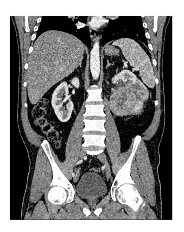
Fig 1. Left renal tumour on CT scan
Subsequently, given the fact that no signs of hepatic metastases were found and other possible cholestatic disorders were excluded, we directed the patient to the Oncology Department with the diagnosis of paraneoplastic syndrome due to renal cancer. A left radical nephrectomy was performed with free tumor surgical margins. Pathology revealed a clear cell renal carcinoma, invading the perirenal fat, with no vascular involvement.
3. Discussion:
Cancer related cholestatic jaundice is usually related to the main bile duct obstruction, widespread liver metastasis or secondary to a paraneoplastic syndrome [4]. Several malignant disorders such as bronchial adenosarcoma, leyomyosarcoma, malignant hystiocytoma, prostate cancer and RCC have been described as causes of cholestatic jaundice without liver metastases [5, 6, 7]. Renal cell associated paraneoplastic syndromes are relatively frequent, as almost 10 to 40% of patients diagnosed with RCC deviate from the classic triad of palpable mass, hematuria and flank pain [8]. An uncommon intrahepatic cholestatic syndrome (Stauffer’s Syndrome) was originally described in 1961 by M.H. Stauffer and it usually manifests as anicteric cholestasis in the absence of hepatic metastases [3]. Several cases have been published so far, that associate hepatosplenomegaly, fever, weight loss, with elevated liver enzymes, high erythrocyte sedimentation rate and abnormal liver function tests with no other obvious cause of cholestasis than renal cancer [9,10]. The underlying mechanism of paraneoplastic cholestasis is not clear yet. However, several theories suggested that it may be caused by the tumor cells that secrete hepatotoxic substances which generate an autoimmune cross reaction by activating T cells and producing antibodies against liver antigens and, in a final stages, may also lead to hepatic injury. Also, interleukin-6 has been incriminated due to its capacity to stimulate hepatic protein production [8]. The only successful treatment consists in a nephrectomy. Subsequent elevation of hepatic enzymes nephrectomy usually suggests a local or distal relapse [11].
In our case the patient presented with both cholestatic and systemic symptoms. Jaundice, pruritus, dark urine supported the cholestasis diagnosis, while other more general symptoms such as weight loss, fatigability might have been associated with the existing diabetes. The differential diagnostic of fever was: infection, inflammatory disease or malignancy.
All direct mechanical causes directly obstructing the biliary tree were excludedand no other causes of intrahepatic cholestasis were found. Common causes of hepatitis such as alcohol or viral hepatitis were ruled out and we also excluded acetaminophen intoxication, infectious disease, primary biliary cirrhosis or other immunological disease as all the laboratory tests were normal. On the other hand, imaging investigations showed several hypoechoic masses on the left kidney which suggested a renal cancer. Although alkaline phosphatase and γ-glutamiltransferase levels were elevated at presentation, no clinical hyperbilirubinemia was present. Additionally, no other laboratory abnormalities were observed, and following nephrectomy, the patient’s status improved with liver serum enzymes decreasing significantly.
Interleukin 6 (IL-6) is considered an autocrine growth factor with effect on liver cells, and is part of the inflammatory inflammation response to tissue injury. The Il-6 increase in Stauffer’s syndrome IL-6 may represent an acute inflammatory reaction caused by the liver infiltration by immune cells [12,13].
RCC is well known for its variety of paraneoplastic symptoms and has been labeled “the internist tumor”. Together with hypercalcemia, polycythemia, coagulopathy, amyloidosis, vasculopathies, neuromyopathies, non-metastatic hepatic disfunction has been described as a paraneoplastic syndrome in the context of the initial diagnostic of renal cancer, or relapse after nephrectomy [8].
4. Conclusion:
Clinical characteristics of patients with RCC vary and sometimes manifest in peculiar ways. RCC may present as cholestatic jaundice, and after excluding more frequent causes, a rare paraneoplastic syndrome should be taken into account in the differential diagnosis of icteric liver disease. This rare but possible association requires prompt recognition, as prognosis correlates with disease stage and, therefore, an early recognition of this syndrome may improve, sometimes, patients’ outcome.
Abbreviations:
IL-6 – Interleukin 6
RCC – renal cell carcinoma
Statements:
Author’s contributions: BSU, VMS and DNF wrote the paper, LMF, IR reviewed the paper, TC and DIG reviewed and approved the final version.
Consent for publication: As the corresponding author, I confirm that the manuscript has been read and approved for submission by all co-authors.
Conflicts of interests: All authors declare having no competing interests asociated with this publication.
Funding resources: This research did not receive any specific grant from funding agencies in the public, commercial, or not-for-profit sector.
Informed Consent: The informed consent was obtained from the patient(s) for publication and any accompanying images.
Statement of Ethics: The accompanying manuscript does not contain any studies carried out by the authors on humans or animals.
Ethical Approval: The treatment strategy/study protocol was approved by a local tumor board/ethics committee.
References:
- Scalabrini DR, Caravelli D, Schianca FC, D’Ambrosio L, Tolomeo F, Boccone P, et al. Complete remission of paraneoplastic vanishing bile duct syndrome after the successful treatment of Hodgkin’s lymphoma: a case report and review of the literature. BMC Res Notes. 2014;7(1):529.
- Kang MK, Park JG, Lee HJ. Cholestatic Jaundice as a paraneoplastic manifestation of prostate cancer aggravated by steroid therapy. Med Princ Pract. 2018;27(2):197-200.
- Stauffer MH. Nephrogenic hepatosplenomegaly. Gastroenterology. 1961;40:694
- Dourakis SP, Sinani C, Deutsch M, Dimitriadou E, Hadziyannis SJ. Cholestatic jaundice as a paraneoplastic manifestation of renal cell carcinoma. Eur J Gastroenterol Hepatol. 1997;9(3):311-4.
- Sharara AI, Panella TJ, Fitz JG. Paraneoplastic hepatopathy associated with soft tissue sarcoma. Gastroenterology. 1992;103(1):330–2.
- Chavarriaga J, Fakih N, Cataño J, Villaquiran C, Rodriguez S, Patino G. Stauffer syndrome, clinical implications and knowledge gaps, does size matter? Case report. BMC Urol. 2020 20(1):105.
- Sharma N, Darr U, Darr A, Sood G. Stauffer Syndrome: A Comprehensive Review of the Icteric Variant of the Syndrome. Cureus. 2019 Oct 30;11(10):e6032.
- Palapattu GS, Kristo B, Rajfer J. Paraneoplastic syndromes in urologic malignancy: the many faces of renal cell carcinoma. Rev Urol. 2002;4(4):163-70
- Kranidiotis GP, Voidonikola PT, Dimopoulos MK, Anastasiou-Nana MI. Stauffer’s syndrome as a prominent manifestation of renal cancer: a case report; Cases J. 2009;2(1):49
- Morla D, Alazemi S, Lichtstein D. Stauffer’s syndrome variant with cholestatic jaundice: a case report. J Gen Intern Med. 2006 Jul;21(7):C11-3
- Fontes-Sousa M, Magalhães H, da Silva FC, Maurício MJStauffer’s syndrome: A comprehensive review and proposed updated diagnostic criteria. Urol Oncol. 2018 Jul;36(7):321-326.
- Blay JY, Rossi JF, Wijdenes J, Menetrier-Caux C, Schemann S, Négrier S, Philip T, Favrot M. Role of interleukin-6 in the paraneoplastic inflammatory syndrome associated with renal-cell carcinoma. Int J Cancer. 1997;72(3):424-30.
- Takamatsu K, Mizuno R, Hattori S, Kono H, Mikami S, Oya M. A case of paraneoplastic liver dysfunction with elevated serum interleukin-6 in clinically localized renal cell carcinoma. Int Cancer Conf J. 2015 Jun 28;5(1):66-67.
Table 1. Laboratory Values
| Relapse | First Episode | ||
| Hemoglobin (Normal 13 – 16)
MCV |
6.3
85.5 |
10.1
87.2 |
g/dl
|
| Hematocrit (Normal 40-48) | 19.6 | 37 | % |
| Platelets | 226.000 | 245.000 | mmc |
| Leukocytes | 4.700 | 5.900 | mmc |
| Urea | 39 | 35 | mg/dl |
| Creatinine | 1.09 | 1.07 | mg/dl |
| Glycemia (Normal 70 – 110) | 226 | 150 | mg/dl |
| Colesterol | 203 | 179 | mg/dl |
| AST | 39 | 25 | U/l |
| ALT | 56 | 34 | U/l |
| Total Bilirubin (Normal 0,2 -1,2)
Direct Bilirubin Alkaline Phosphatase (Normal 13-45) γ-glutamiltransferase (Normal 9 – 50) CRP (Normal < 5) ESR (Normal 10/15) Total Protein Albumin Prothrombin Time |
5.19
3.76 667 770 178.9 >160 7.3 2.57 13 |
1.5
0.9 320 270
48/110
10 |
mg/dl –
mg/dl U/l – U/l – mg/dl 1h/2h mg/dl mg/dl s |
MCV, mean corpuscular volume; AST, aspartate aminotransferase; ALT, alanine aminotransferase; CRP, C-reactive protein; ESR, erythrocytes sedimentation rate;