


Author(s) :
Cristina Lungulescu1, Ștefan Răileanu2, Ștefania Dumitru2, Mihaela Dănciulescu3
1 University of Medicine and Pharmacy of Craiova, Doctoral School, Craiova, Romania
2 Department of Oncology, Filantropia Clinical Hospital, Craiova, Romania
3 University of Medicine and Pharmacy of Craiova, Department of Oncology, Craiova, Romania
Corresponding author: Cristina Lungulescu, Email: cristina.lungulescu@yahoo.com
Publication History: Received - , Revised - , Accepted - , Published Online - 2021.
Copyright: © 2021 The author(s). Published by Casa Cărții de Știință.
User License: Creative Commons Attribution – NonCommercial (CC BY-NC)

 PDF
 Share
Highlights
Abstract
Prostate cancer is the second most frequent malignancy worldwide, with an increased incidence over the last two decades. Most patients are asymptomatic at diagnosis due to widespread opportunistic prostate-specific antigen-based screening. Cerebral metastases originating from prostatic tumors are rare, occurring in 0.16% to 0.63% of prostate cancer cases, and generally developing in the setting of widespread disease. We report the case of a 62-year-old male with symptomatic isolated brain metastasis as an early clinical presentation of prostate adenocarcinoma in the setting of a normal PSA and digital rectal examination.
Next-generation sequencing panel revealed a mutation in the PALB2 gene. Germline mutation in genes involved in homologous recombination repair complex, such as BRCA1, BRCA2, and PALB2 increase the incidence and the aggressiveness of multiple cancer types including prostate cancer, yet can provide alternative treatment options using PARP inhibitors after disease progression for patients previously treated with enzalutamide or abiraterone.
1. Introduction:
Prostate cancer is the second most frequent malignancy worldwide (1) and the second leading cause of cancer death among men (2). The incidence of prostate cancer has increased over the last two decades due to enhanced detection capabilities and screening programs. With widespread opportunistic prostate-specific antigen-based (PSA) screening most patients are asymptomatic at diagnosis (3). Patients with PSA level > 4.0 ng/mL are commonly considered candidates for prostate biopsy. Furthermore, early stages can be detected after a routine digital rectal examination (DRE). Histopathological confirmation usually follows a transrectal ultrasound‐guided prostate biopsy (TRUS) performed after a rise in prostate specific antigen, or after transurethral resection of the prostate (TURP) for glandular hyperplasia (1). Prostatic adenocarcinoma accounts for more than 95% of cases, and the pathologic report subsequently determines the Gleason score which is based on architectural characteristics of the cancer cells. The remaining histological types are uncommon, and are not limited to neuroendocrine tumors, small cell carcinomas, sarcomas, or lymphomas (4). Symptoms including urinary frequency, urgency, hesitancy, nocturia, poor urine stream, hematospermia, and terminal hematuria are generally indicative of advanced disease (1). Prostate cancer metastases frequently involve the skeleton (84%), lymph nodes and liver (5). Brain metastasis from prostate cancer are rare, accounting for less than 1% of cases, and occurs in patients with neuroendocrine tumors and scarcely with adenocarcinoma (6). Although invasion of the central nervous system occurs in the setting of extensive metastasis, the increased incidence of brain metastases from prostatic malignancy recorded over the past 20 years is the expression of the patients’ extended survival
(2). Current treatment strategies for brain metastasis involve neurosurgical resection and/or stereotactic radiosurgery and/or whole brain radiotherapy (WBRT). Patients’ outcomes usually correlate with elements such as clinical tumor stage, Gleason score, and PSA level at diagnosis (3).
2. Case Report:
A 62-year-old male with a personal history of benign prostatic hyperplasia, who underwent routine PSA screening since 2017 with normal PSA levels, presented to the Emergency Department for confusion, nausea, agitation and grand mal seizures in March 2020. Upon further assessment a computed tomography (CT) and a magnetic resonance imaging (MRI) scan of the brain revealed a right temporal hemorrhagic intracerebral expansive process (65 mm), with mass effect on the midline structures. The patient underwent craniotomy and resection of the mass lesion in April 2020, with pathologic studies of the tumor in favor of carcinoma metastasis with extensive adjacent hemorrhagic areas, and further diagnosis of brain metastasis from carcinoma of unknown primary. The patient was referred to the Oncology Clinic one month after surgery. Initial clinical exam was normal, with no skin lesions suspicious of melanoma and no history of smoking, with ECOG performance status 0. Contrastenhanced head-chest-abdomen-pelvis CT scan displayed no evidence of a primary tumor. Subsequent immunohistochemical (IHC) exam was performed on the surgically removed brain tumor, with tumor cells positive for PSA and prostate-specific membrane antigen (PSMA), resulting in a diagnosis of brain metastasis of prostate acinar adenocarcinoma with cribriform pattern. The patient was later referred to the Urology Clinic where digital rectal examination revealed no abnormalities. PSA at follow-up visit was 2,6 ng/ml. In June 2020 the patient underwent “wholebrain” external-beam (total dose – 30Gy), under depletive and corticosteroid treatment, with good tolerance. Pelvic MRI scan showed suspicious changes in the peripheral area of the left mid-prostate (Fig.1 and 2) and signal changes in the left seminal vesicle (PI-RADS 4), with no local lymph node involvement, bones or abdominal organs, followed by prostate biopsy in July 2020. Pathological study revealed prostate adenocarcinoma with perineural invasion, Gleason score 8 (4+4). PSA levels remained under the upper limit of normal range (3,86 ng/ml). Molecular analysis revealed a mutation in PALB2 (partner and localizer of BRCA2) gene, supporting deficient homologous recombination (HR) repair status. The patients was started on systemic treatment with first line androgen deprivation therapy (ADT) – LH-RH agonist (triptorelin) and second generation antiandrogen (bicalutamide – 50 mg/qd, 28 days) – the only available first line ADT reimbursed in our country; abiraterone and third generation antiandrogen are second line options.
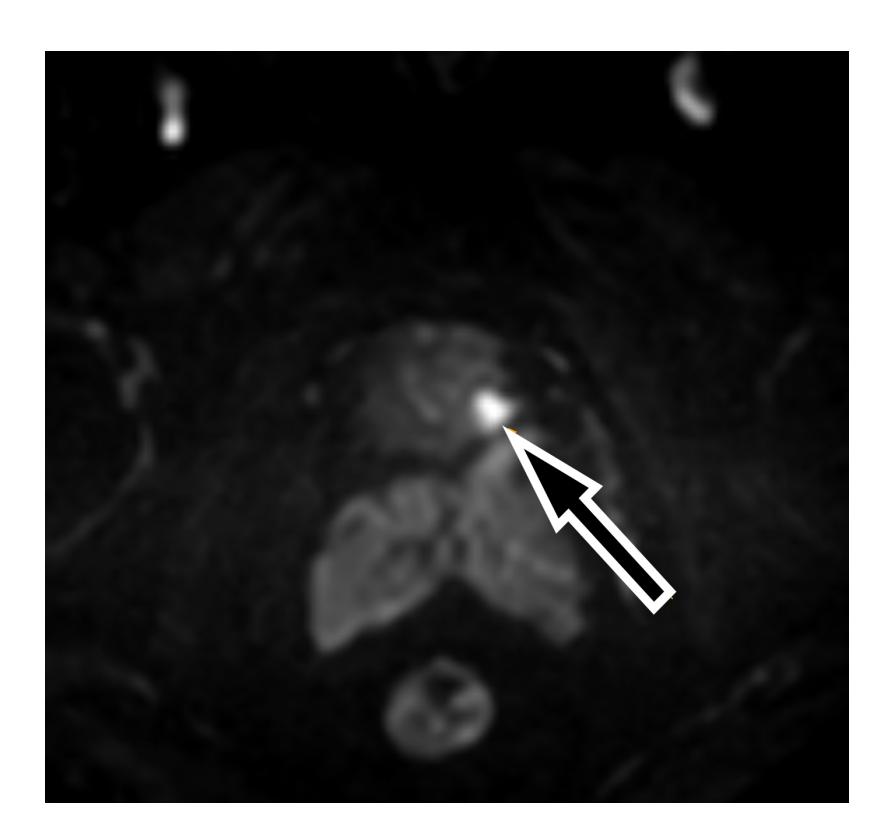
Fig. 1. Pelvic MRI. MRI performed before a first series of biopsies, showed, in its contrast-enhanced dynamic sequence a lesion of the left base (orange arrow) of the prostate.
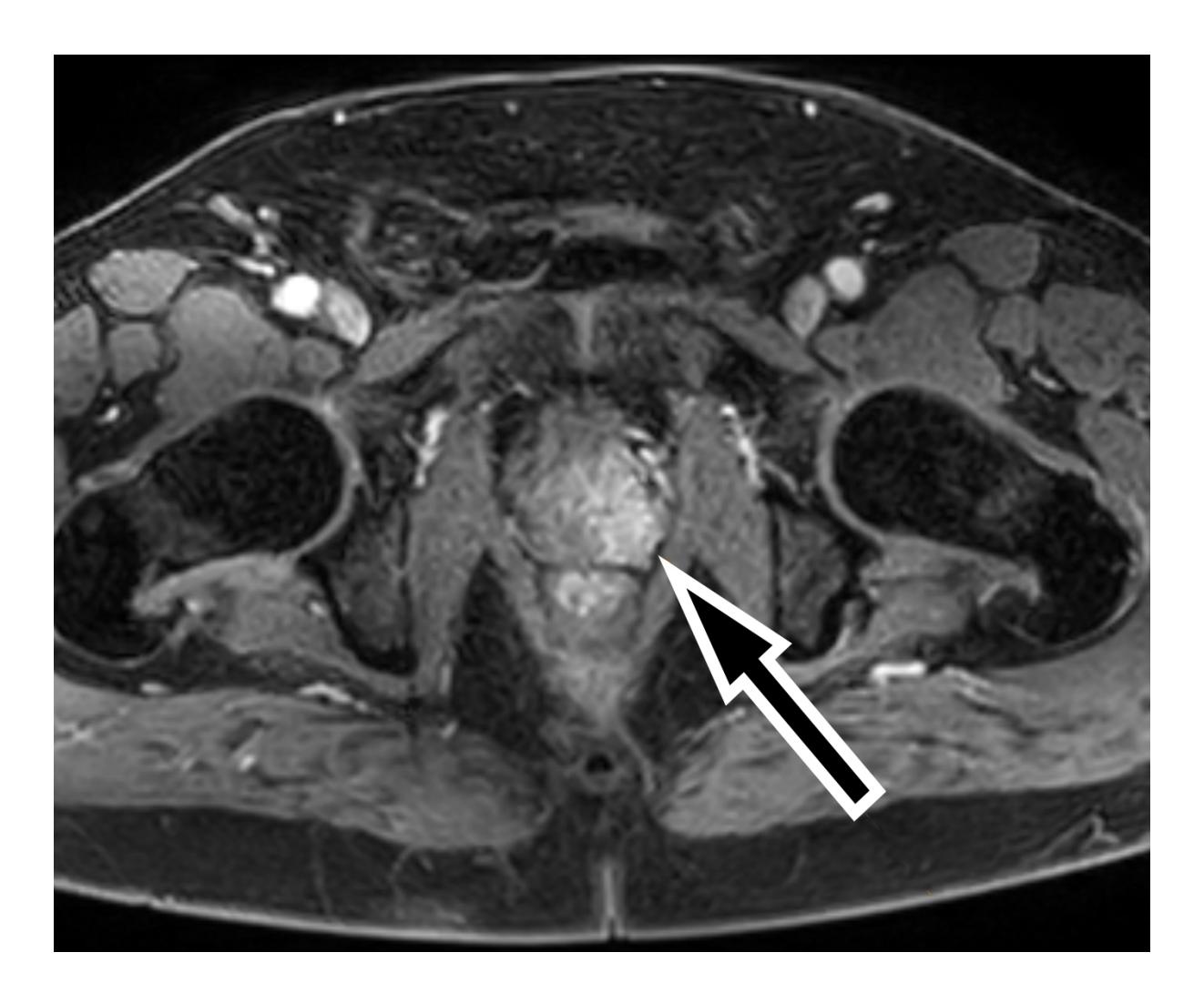
Fig. 2. Pelvic MRI. MRI showing in its contrast-enhanced dynamic sequence a lesion of the left base (orange arrow) of the prostate.
3. Discussion:
Cerebral metastasis represents a serious challenge in oncology and is estimated that approximately 30% of cancer patients will develop central nervous system (CNS) secondary tumors during their lifetime (7). Alarmingly, statistics show that up to 45% of cancer patients acquire brain metastasis annually. Brain metastases develop as a result of hematogenous dissemination or contiguity of secondary bone involvement of the skull. Secondary cerebral tumors far exceed primary nervous system malignancies. The incidence of secondary brain tumors varies in accordance with certain cancer sites, brain metastasis commonly arising from lung cancer, breast cancer and melanoma.
Prostate cancer disseminates in approximately 18% of cases, consistently affecting the bone and lymph nodes (5). CNS involvement originating from prostatic tumors is exceptional, developing in 0.16% to 0.63% of prostate cancer cases, and generally occurring in the setting of widespread disease (6). It is commonly believed that the brain parenchyma is
resistant to the invasion and establishment of prostate malignant cells. Isolated brain metastasis without evidence of other metastatic sites is extraordinary, the M.D. Anderson Experience revealing only 1 such case from 16,280 patients (8), while another study cites 1% of 74,826 patients with metastatic prostate cancer (5). A literature review describes 11 other case reports with isolated brain metastasis from prostatic primary tumor (2).
Neuroendocrine tumors are more commonly associated with SNC secondary tumors. In particular, small cell carcinomas (SCC) from the prostate are believed to behave like SCC the lungs and other organs, displaying a propensity to metastasize to the brain (8). However, the histopathological and IHC results of our patient do not describe any neuroendocrine differentiation.
Furthermore, our case presented with a normal PSA at time of diagnosis, which remained < 4.0 ng/mL throughout follow-up. However, previous studies show that metastatic prostate cancer is not necessarily corelated with high levels of PSA, even with CNS involvement (6). The positive predictive value for prostate cancer is only 30% for the upper limit of normal range of PSA. By contrary, benign prostatic hypertrophy or prostatitis increase PSA levels. Routine PSA screening is currently considered an imperfect test, although a significant number of prostate malignancies are uncovered following this method. The positive predictive value of PSA testing increases subsequent to an abnormal finding on DRE (3). Nonetheless, our case exhibits metastatic prostate cancer with normal DRE and PSA levels. In similar instances, advances in prostate MRI using Prostate Imaging Reporting and Data System (PI-RADS – fivepoint standardized reporting system for abnormalities found on MRI) can help identify lesions and guide the biopsy, with a sensitivity of 0.89 and specificity of 0.73 for prostate cancer (9). Computed tomography scans are less accurate, as described in our case. Although not available in our country, these patients could have benefited from a more specific imaging study, such as prostate-specific membrane antigen (PSMA) PET-CT.
Germline mutation in genes involved in homologous recombination repair complex, such as BRCA1, BRCA2, and PALB2 increase the incidence and the aggressiveness of multiple cancer types including prostate cancer, yet can provide alternative treatment options using poly ADP ribose polymerase (PARP) inhibitors (10). Next-generation sequencing (NGS) panel testing 23 genes was performed from the patient’s saliva using polymerase chain reaction (PCR) amplification. The following genes were evaluated for germinal mutations: ATM, BARD1, BRCA1, BRCA2, BRIP1, CDH1, CHEK2, EPCAM, MLH1, MRE11A, MSH2, MSH6, NBN, PALB2, PMS2, PTEN, RAD50, RAD51C, RAD51D, RINT1, STK11, TP53, XR. The patient tested positive exclusively for PALB2 germinal mutation. In this case, the PALB2 germline mutation may result in beneficial response to PARPinhibitors (namely, olaparib) after disease progression for patients who had previously received treatment with enzalutamide or abiraterone, although this therapy is not currently reimbursed in our country for prostate malignancies.
A review of the cases reported in literature of patients with isolated brain metastases from prostate cancer found that surgical resection, SRS, and WBRT are used as a sole treatment or in various combinations. Survival in cases of solitary metastasis to the brain from prostate cancer following treatment commonly varies between 2 to 20 months (11). Upon disease progression, the patient should benefit from therapies such as abiraterone and cabazitaxel, which are able to cross the blood-brain barrier, as opposed to docetaxel (12).
Abbreviations:
ADT – androgen deprivation therapy
BRCA1 – Breast Cancer gene 1
BRCA2 – Breast Cancer gene 2
CNS – central nervous system
CT – computed tomography
DRE – digital rectal examination
ECOG – Eastern Cooperative Oncology Group
HR – homologous recombination
IHC – immunohistochemistry
LH-RH – luteinizing hormone-releasing hormone
MRI – magnetic resonance imaging
NSG – next-generation sequencing
PALB2 – partner and localizer of BRCA2
PARP – poly ADP ribose polymerase
PCR – polymerase chain reaction
PET-CT – Positron Emission Tomography – Computed Tomography
PI-RADS – prostate imaging–reporting and data system
PSA – prostate-specific antigen
PSMA – prostate-specific membrane antigen
SCC – small cell carcinomas
TRUS – transrectal ultrasound
TURP – transurethral resection of the prostate
WBRT – whole brain radiotherapy
4. Conclusion:
Symptomatic brain metastasis as an early clinical presentation of prostate adenocarcinoma is extremely rare. Although the underlying mechanisms are not currently fully understood, we hypothesize that a mutation in PALB2 gene could account for the aggressive tumor biology, and explain the tendency for cerebral metastasis spread.
Statements:
Authors’ contributions: CL, SD, SR carried out collecting and summarizing the clinical materials of the patient. CL and MD drafted the manuscript.
Consent for publication: As the corresponding author, I confirm that the manuscript has been read and approved for submission by all co-authors.
Conflict of interest: All authors declare having no competing interests associated with this publication.
Funding Sources: This research did not receive any specific grant from funding agencies in the public, commercial, or not-for-profit sector.
Informed Consent: The informed consent was obtained from the patient for publication and any accompanying images.
Statement of Ethics: The accompanying manuscript does not contain any studies carried out by the authors on humans or animals.
Ethical Approval: The treatment strategy was approved by a local tumor board.
References:
- Chmielowski B, Territo M. Manual of clinical oncology. 8th ed. Philadelphia: Wolters Kluwer; 2017; p.566- 80. ISBN: 9781496349576.
- Campagna JP, Feia K. Isolated brain metastasis of prostate carcinoma in the setting of normal prostate specific antigen. Urol Case Rep. 2018 Sep 4;21:67-69.
- Niederhuber JE, Armitage JO, Doroshow JH, Kastan MB, Tepper JE. Abeloff’s Clinical Oncology. 6th ed. Elsevier; 2020; p.1401-15. ISBN: 978-0-323-47674-4.
- The American Cancer Society’s oncology in practice: clinical management. 1st ed. Wiley-Blackwell; 2018; p.344-8. ISBN: 9781118592076.
- Gandaglia G, Abdollah F, Schiffmann J, Trudeau V, Shariat SF, Kim SP, Perrotte P, Montorsi F, Briganti A, Trinh QD, Karakiewicz PI, Sun M. Distribution of metastatic sites in patients with prostate cancer: A populationbased analysis. Prostate. 2014 Feb;74(2):210-6.
- Kanyılmaz G, Aktan M, Yavuz BB, Koç M. Brain metastases from prostate cancer: A single-center experience. Turk J Urol. 2018 Jun 5;45(4):279-283.
- Achrol AS, Rennert RC, Anders C, Soffietti R, Ahluwalia MS, Nayak L, Peters S, Arvold ND, Harsh GR, Steeg PS, Chang SD. Brain metastases. Nat Rev Dis Primers. 2019 Jan 17;5(1):5.
- Tremont-Lukats IW, Bobustuc G, Lagos GK, Lolas K, Kyritsis AP, Puduvalli VK. Brain metastasis from prostate carcinoma: The M. D. Anderson Cancer Center experience. Cancer. 2003 Jul 15;98(2):363-8.
- Turkbey B, Rosenkrantz AB, Haider MA, Padhani AR, Villeirs G, Macura KJ, Tempany CM, Choyke PL, Cornud F, Margolis DJ, Thoeny HC, Verma S, Barentsz J, Weinreb JC. Prostate Imaging Reporting and Data System Version 2.1: 2019 Update of Prostate Imaging Reporting and Data System Version 2. Eur Urol. 2019 Sep;76(3):340-351.
- Tang T, Wang LA, Wang P, Tong D, Liu G, Zhang J, Dai N, Zhang Y, Yuan G, Geary K, Zhang D, Liu Q, Jiang J. Case Report: Co-Existence of BRCA2 and PALB2 Germline Mutations in Familial Prostate Cancer With Solitary Lung Metastasis. Front Oncol. 2020 Oct 26;10:564694.
- Barakat T, Agarwal A, McDonald R, Ganesh V, Vuong S, Borean M, Chow E, Soliman H. Solitary brain metastasis from prostate cancer: a case report. Ann Palliat Med. 2016 Jul;5(3):227-32.
- Caffo O, Veccia A, Russo L, Galligioni E. Brain metastases from prostate cancer: an emerging clinical problem with implications for the future therapeutic scenario. Future Oncol. 2012 Dec;8(12):1585-95.

