


Author(s) :
Andrada Turcas1,2, Bogdan Fetica3, Adrian Trifa4,5,6, Viorica Nagy2
1Oncology Department, University of Medicine and Pharmacy “Iuliu Hatieganu” Cluj-Napoca, Romania
2Radiotherapy Department, Oncology Institute “Prof. Dr. Ion Chiricuta” Cluj-Napoca, Romania
3Pathology Department, Oncology Institute “Prof. Dr. Ion Chiricuta” Cluj-Napoca, Romania
4Department of Genetics, “Victor Babeș” University of Medicine and Pharmacy, Timișoara, Romania
5Department of Genetics, Oncology Institute “Prof. Dr. Ion Chiricuta” Cluj-Napoca, Romania
6Oncohelp Hospital, Timișoara, Romania
Corresponding author: Andrada Turcas, Email: andradaturcas@outlook.com
Publication History: Received - , Revised - , Accepted - , Published Online - 1 April 2023.
Copyright: © The author(s). Published by Casa Cărții de Știință.
User License: Creative Commons Attribution – NonCommercial (CC BY-NC)

 PDF
PDF
 Share
Share
Highlights
• Muir–Torre syndrome, a rare Lynch syndrome variant, can remain unrecognized for decades despite a striking pattern of recurrent sebaceous and non-sebaceous cutaneous tumors.
• Loss of MSH2/MSH6 expression on IHC and identification of a pathogenic germline MSH2 variant provide a definitive molecular diagnosis and immediate implications for cascade testing.
• Delayed recognition carries major clinical consequences, including missed opportunities for intensified surveillance, risk-reducing strategies, and biomarker-driven systemic therapies.
• Multidisciplinary management and structured screening of at-risk relatives remain the cornerstone, while immunoprevention and targeted approaches are emerging future directions for dMMR/MSI-driven hereditary cancers.
Abstract
Muir-Torre syndrome, a subtype of Lynch syndrome, is a rare genetic disorder. We present the case of a female patient with an extended family and personal history diagnosed with numerous benign and malignant tumors of various histology, including some with sebaceous features, beginning at 41. The majority were cutaneous tumours, treated with complete resection, and which frequently recurred. Visceral cancers included endocervical adenocarcinoma, vulvar squamous-cell carcinoma and urothelial carcinoma, treated surgically, followed by systemic oncological treatments and external beam radiotherapy.
Following a 20-year evolution, extensive genetic blood testing revealed a pathogenic variant in the MSH2 gene, c.1861C>T (p.Arg621*), in the heterozygous state. Given this unusual clinical presentation and molecular profile, the patient was ultimately diagnosed with Muir-Torre syndrome. The prognosis was poor, with an inoperable recurrence of the urothelial carcinoma and extensive lymph node metastasis of vulvar squamous cell carcinoma.
1. Introduction
Muir Torre syndrome (MTS) is a rare genetic disease, considered a subtype of hereditary non-polyposis colorectal cancer (HNPCC), also called Lynch syndrome, an autosomal dominant disorder with variable penetrance (1–4). The concerned genes (MSH2, MSH6, MLH1, MLH3, PMS2) are involved in DNA repair mechanisms (Mismatch Repair/ MMR) and their pathogenic variants lead to microsatellite instability (MSI)(5–7). Clinically, this pathology is characterized by the presence of multiple visceral and skin tumours, both malignant and benign, with the typical occurrence of cutaneous sebaceous tumours. The clinical diagnostic criteria include the presence of at least one visceral cancer in a patient under the age of 60, with positive personal and family history of cancer, and the presence of at least one sebaceous tumour. Another distinctive feature is the appearance of skin tumours, especially on the face and scalp, preceding the visceral ones.
So far, approximately 200 such cases have been published in the literature. Table 1 summarises the main diagnostic criteria for Muir-Torre syndrome (8).
Table 1. Muir-Torre Syndrome- diagnostic criteria
Muir-Torre Syndrome – Diagnostic criteria
- Minimum 1 sebaceous tumor
- Adenoma
- Epithelioma
- Carcinoma
- Basal cell carcinoma with sebaceous features
- Minimum 1 visceral malignant tumor
- Age <60 years (disease onset/first tumor)
- Positive family history
- Positive personal history
- Lynch Syndrome-related malignancies
2. Case Presentation
Clinical features and patient history
We present the case of a female patient with a significant family history of cancer, as following: father with colorectal cancer, deceased at the age of 50, one sister with breast cancer and another sister with gastric cancer, and four other relatives diagnosed with different types of cancer, mostly of breast or gynecological origin, all at young ages, the youngest being diagnosed at 20 years of age. Figure 1 shows this family’s cancer history genealogical scheme.
The first symptom occurred at the age of 41, when she presented with a nasal tumour located in the left alar region, which was completely surgically removed and diagnosed as a superinfected keratoacanthoma. She returned 6 years later (age 47) with an endocervical tumour, diagnosed as stage IB1 papillary serous endocervical adenocarcinoma. A total hysterectomy with bilateral salpingooopherectomy was performed, followed by local pelvic radiotherapy with a dose of 50Gy in 25 fractions. Periodic checks were carried out for 5 years, the patient being disease free.
At the age of 53, the patient returned with multiple small tumours at the left alar and cervico-occipital level, which, after excision, were diagnosed as basal cell carcinomas. In the next two years she presented with multiple and various skin tumours, mostly perioral and palmar, pathologically diagnosed as grade 1 keratinized squamous cell carcinoma), all staged at the time of excision as stage I (pT1N0). The patient returned with multiple tumours of the scalp, which turned out to be sebaceous adenomas and carcinomas. Additionally, she presented multiple other skin tumours of various histological subtypes: G1 keratinized squamous cell vulvar carcinoma, (excised pT1b L0V0R0), benign fibrous axillar histiocytoma (excised), submental dermatofibroma (excised, pT1), G1 keratinized squamous cell carcinoma at the palmar level (excised pT1).
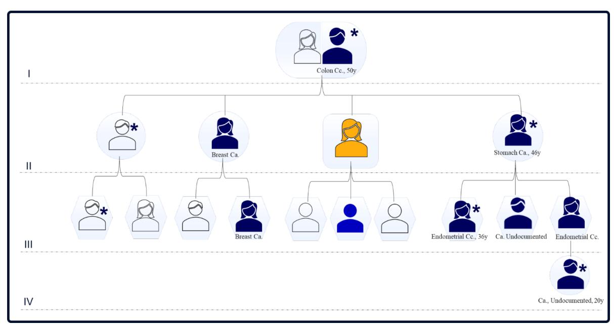
Figure 1. Family tree and cancer history: dark blue= cancer present; yellow= presented patient; blue= mutation identified by genetic testing; *= deceased; y= years-old; CC= carcinoma, Ca=cancer
In the upcoming period, the skin lesions progressed, with repeated presentations for the excision of tumours from the face, anterior thorax and hand. The following histological types were identified: actinic/seborrheic keratosis, squamous carcinoma keratinized G2, molluscum contagiosum and sebaceoma. Throughout this time, the patient did not exhibit any systemic symptoms and there was no alteration of the general condition or quality of life.
At the age of 62, she experienced mild dysuria. Following a computed tomography (CT), an endo-vesical polyp and non-specific nodules in the ascending colon were identified. A colonoscopy was performed, and identified post-irradiation changes, such as scarring and strictures. Following the cystoscopy and biopsy of the endo-vesical polyp, the diagnosis of urothelial carcinoma of the urinary bladder was concluded. The patient underwent a Trans-Urethral Vesical Resection (TURV) procedure and the final diagnosis was G3 papillary urothelial carcinoma, pT1NxMx). Ten bladder instillations with Mitomycin C were administered as adjuvant treatment.
After several months, the patient returned with a mass in the right inguinal region, which was biopsied and subsequently diagnosed as a lymph node metastasis of a non-keratinizing squamous cell carcinoma, of unknown origin. However, given anatomic location, the probability of a metastasis from the previously treated vulvar carcinoma was clinically suspected. A thoraco-abdominal-pelvic CT scan was performed, which identified multiple pelvic, retroperitoneal, and lumbo-aortic adenopathies. The bladder was clear. Systemic treatment with Paclitaxel and Carboplatin (10 cycles) was administered, with only a modest response on the pelvic adenopathies, as assessed by CT imaging. In addition, due to haematological toxicity and peripheral neuropathy, dose de-escalation of the last 3 cycles of chemotherapy was done.
The following year, four months after the completion of the last chemotherapy cycle, the bladder tumour recurred, with extensive invasion of adjacent organs (rectum, pelvic soft tissue). Unfortunately, at that time, patient was no longer a surgical candidate and, palliative treatment and pain control was recommended. Patient’s history and clinical evolution are presented in Figure 2.
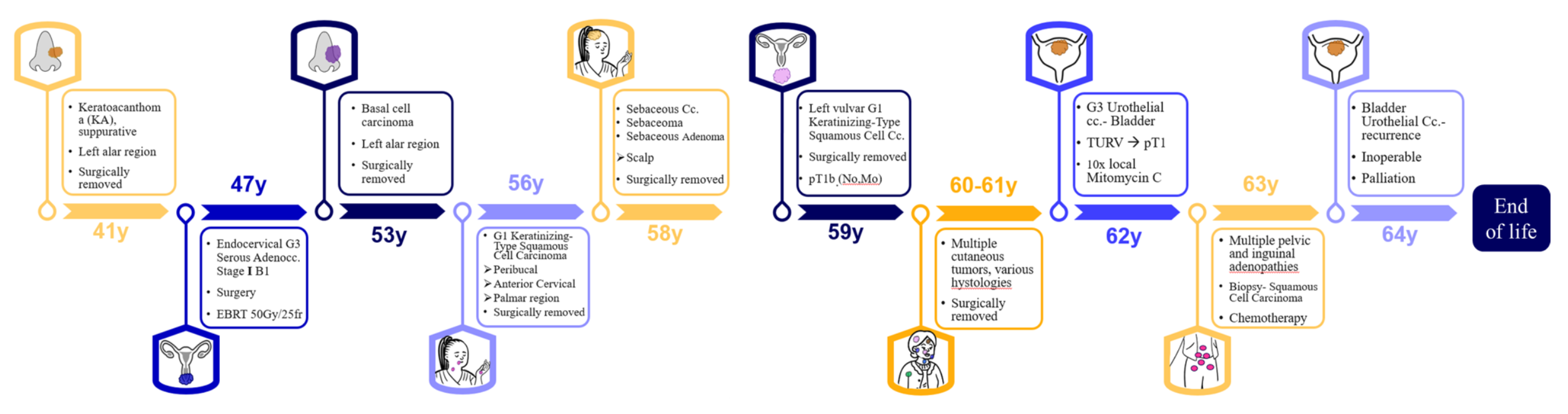
Figure 2. Patient history and evolution; y = years, G1 = grade 1; EBRT = External beam radiotherapy; CC= carcinoma
3. Molecular and genetic testing
Given the significant personal and family history, the suspicion of a hereditary cancer syndrome was raised. Immuno-histochemical testing was performed on the resected bladder tumour specimen, revealing loss of nuclear expression of MSH2 and MSH6 proteins involved in DNA repair (mismatch repair deficient), thus indicating a possible microsatellite instability syndrome. The expression of MLH1 and PMS2 proteins was retained. According to the NCCN guidelines (9) the absence of MSH2 and MSH6 staining suggests either a germline MSH2/EPCAM pathogenic gene variant or rarely a MSH6 pathogenic gene variant. In case of a sporadic cancer, the cause would probably have been of somatic origin.
Also, considering the fulfilment of the Amsterdam and NCCN criteria (9,10) (> 3 family members affected by HNPCC-related cancers, at least one first-degree relative and > 2 affected successive generations, with at least 1 case of colorectal carcinoma under the age of 50 and histologically confirmed lesions), a decision was made to perform extensive genetic testing (83 gene Multi-cancer panel, performed at Invitae, USA panel, using peripheric venous blood). The 83 genes involved in most types of hereditary cancers are presented in Table 2. DNA Sequencing and deletion/duplication of the 83 genes was performed, resulting in the identification a germline mutation pathogenic variant in the MSH2 gene, namely c.1861C>T (p.Arg621*), in heterozygous state. This particular non-sense variant usually leads to a premature translational stop signal in the MSH2 gene. It is expected to result in an absent or disrupted protein product. This variant has been submitted to ClinVar database (variation ID 90804), where 16 entries were identified in several patients suspected of Lynch syndrome or other types of hereditary colo-rectal cancer (11). Given the rich family history and the identified germline pathogenic variant, genetic testing was extended to the patient’s first-degree relatives, with one out of three descendants carrying the same pathogenic variant in the MSH2 gene.
4. Discussions
The presented case is impressive in terms of its complicated history and long evolution, with the final diagnosis discovered more than 20 years after the initial presentation.
The patient fulfilled all required criteria for Muir-Torre syndrome, a rare subtype of Lynch syndrome. She presented with multiple benign and malignant skin tumours, with over ten histological types, including sebaceous tumours (being essential for the clinical diagnosis of the Lynch syndrome subtype). She also had several visceral cancers – endocervical adenocarcinoma, followed by a vulvar squamous cell carcinoma, which also metastasized into the pelvic and inguinal lymph nodes, and finally the urothelial cell carcinoma of the bladder; genitourinary cancers are present in 25% of Muir Torre syndrome cases(12–15).
Also, the strong family history further reinforces this diagnosis, with multiple relatives across at least four generations affected by different malignancies, including first degree relatives (16). Guided by the Amsterdam criteria for genetic testing in the case of suspected HNPCC syndrome (10), we decided to perform an extensive germline testing of both the patient and the first degree descendants. The genetic testing in this case brought essential information to both the patient, as well as the family, identifying a pathogenic germline variant in the MSH2 gene, thus confirming the germline origin of the MMR deficiency observed in patient’s tumour (5).
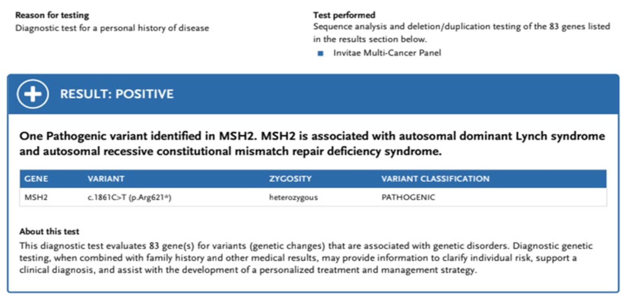
Figure 3. Genetic testing result- germline mutation in MSH2 gene identified.
With such a complex presentation, there were several therapeutic challenges. Notably, the stage IB vulvar carcinoma might have benefited from ipsi- or bilateral inguinal lymph node dissection, and the option of external beam radiotherapy could have been discussed, given the long time since the initial pelvic irradiation (more than 10 years). More so, the urothelial cancer was a high grade, pT1 tumour, which is classified as a high-risk tumour that might have benefited from a more radical surgical treatment (cystectomy), intravesical Bacillus Calmette-Guerin (BCG) or immunotherapy, especially in the context of MSI-high tumours(17). Also, clinical trial screening and enrolment or off-label medication prescription could have been possible and might have offered her a chance to a targeted treatment(18). Additionally, the option of using PET-CT for follow-up in the context of such frequently re-occurring tumours could have been beneficial.
In terms of identified pathogenic variant in healthy relatives, the level of vigilance should be increased. Genetic counselling and sustained screening for the prevention and early detection of cancers for which they may be at risk should be recommended, according to the latest NCCN guidelines(9). Specifically, we made several recommendations to the patient’s descendant in which we detected the MSH2 pathogenic variant, regarding the follow-up and surveillance: yearly clinical examinations, colorectal screening by periodic colonoscopy (every 1-2 years), gynecological examination, with pelvic ultrasound and aspiration biopsy every year, from age 30 to 35 years, gastric cancer screening by periodic upper gastro-intestinal endoscopy (every 2-4 years), and regular, frequent skin examination(19). Taking the high risk of both endometrial and ovarian cancer into account, the options of risk-reducing surgical interventions (bilateral prophylactic salpingooophorectomy and hysterectomy) were also addressed. Given the family history of breast cancer, which represents an independent risk factor for this type of cancer, yearly breast surveillance was also recommended, starting earlier than in the standard risk population (20,21). More, oral chemoprophylaxis with retinoids (isotretinoin) for cutaneous tumours prevention could be an option for these patients. Other options include subcutaneous Interferon 2alpha and topic applications of retinoids (21–24). A more recent breakthrough in this area are vaccines for immunoprevention of DNA mismatch repair deficient cancers, which show promising results in initial phases(25).
Table 2. The 83- gene panel tested for this case, comprising genes involved in most types of hereditary cancers
| ALK | APC | ATM | AXIN2 | BAP1 | BARD1 | BLM | BMPR1A |
|---|---|---|---|---|---|---|---|
| BRCA1 | BRCA2 | BRIP1 | CASR | CDC73 | CDH1 | CDK4 | CDKN1B |
| CDKN1C | CDKN2A | CEBPA | CHEK2 | CTNNA1 | DICER1 | DIS3L2 | EGFR |
| EPCAM | FH | FLCN | GATA2 | GPC3 | GREM1 | HOXB13 | HRAS |
| KIT | MAX | MEN1 | MET | MITF | MLH1 | MSH2 | MSH3 |
| MSH6 | MUTYH | NBN | NF1 | NF2 | NTHL1 | PALB2 | PDGFRA |
| PHOX2B | PMS2 | POLD1 | POLE | POT1 | PRKAR1A | PTCH1 | |
| RAD50 | RAD51C | RAD51D | RB1 | RECQL4 | RET | RUNX1 | PTEN |
| SDHAF2 | SDHB | SDHC | SDHD | SMAD4 | SMARCA4 | SDHA | |
| STK11 | SUFU | TERC | TERT | TMEM127 | TP53 | WT1 | |
| SMARCE1 | TSC1 | TSC2 | VHL | WRN | SMARCB1 |
There are approximatively 200 other cases reported in the literature describing this syndrome, thus emphasizing the particularity of this case (26–31). Without strong evidence for specific treatment or prophylaxis options for these patients, active screening and managing tumours in the early stages seems the best available approach. Novel therapies, such as targeted molecules, immunotherapy, or vaccines, might become a solution in the future. However, given the rarity of this disease, clinical trials are difficult to design and patient enrolment could be challenging, requiring international efforts and long periods of inclusion and follow-up (25,32–34).
5. Conclusions
Muir-Torre syndrome, a rare subtype of the Lynch syndrome, is characterized by microsatellite instability, which causes cancer predisposition in patients who carry pathogenic variants in specific mismatch-repair genes. The case we described follows the typical presentation of this disease, with multiple tumours of various histology, both visceral and cutaneous. Patient management is usually multidisciplinary and tumour-specific, with screening and surveillance essential for the patient and their family.
Abbreviations:
CC – Carcinoma
CT – Computed Tomography
DNA – Deoxyribonucleic acid
G – Grade
Gy – Gray
HNPCC – Hereditary Non-Polyposis Colorectal Cancer
MSI – Microsatelite Instability
MMR – Mismatch Repair
MLH – MutL protein homolog
MSH – MutS homolog
NCCN – National Comprehensive Cancer Network
pT – Pathological tumoral stage
PMS – Mismatch repair endonuclease PMS
TURV – Trans-Urethral Vesical Resection
Statements:
Authors’ contributions: AT collected the data and wrote the manuscript; BF, AT, VN participated in patient management, including diagnosis and treatment and manuscript proofing/editing;
Consent for publication: As the corresponding author, I confirm that the manuscript has been read and approved for submission by all co-authors.
Conflict of interest: All authors declare having no competing interests associated with this publication.
Funding Sources: This research did not receive any specific grant from funding agencies in the public, commercial, or not-for-profit sector.
Informed Consent and Ethical Approval: The informed consent was obtained from the patient for publication and any accompanying images.
References:
- Georgiou D, Monje-Garcia L, Miles T, Monahan K, Ryan NA. A Focused Clinical Review of Lynch Syndrome. Cancer Manag Res (Internet). 2023 Jan (cited 2023 Feb 12);15:67–85. Available from: https://pubmed.ncbi.nlm.nih.gov/36699114/
- Rebuzzi F, Ulivi P, Tedaldi G. Genetic Predisposition to Colorectal Cancer: How Many and Which Genes to Test? Int J Mol Sci (Internet). 2023 Jan 21 (cited 2023 Feb 12);24(3):2137. Available from: https://pubmed.ncbi.nlm. nih.gov/36768460/
- Bogani G, Maggiore ULR, Raspagliesi F. Lynch syndrome Muir-Torre variant: implication in gynecologic oncology. J Gynecol Oncol (Internet). 2018 Sep 1 (cited 2023 Feb 12);29(5). Available from: https://pubmed.ncbi.nlm. nih.gov/30049022/
- Ponti G, de Leon MP. Muir-Torre syndrome. Lancet Oncol. 2005 Dec;6(12):980–7.
- de Carvalho N de A, Niitsuma BN, Kozak VN, Costa FD, de Macedo MP, Kupper BEC, et al. Clinical and Molecular Assessment of Patients with Lynch Syndrome and Sarcomas Underpinning the Association with MSH2 Germline Pathogenic Variants. Cancers (Basel) (Internet). 2020 (cited 2023 Feb 12);12(7):1–18. Available from: https://pubmed.ncbi.nlm.nih.gov/32659967/
- Jessup CJ, Redston M, Tilton E, Reimann JDR. Importance of universal mismatch repair protein immunohistochemistry in patients with sebaceous neoplasia as an initial screening tool for Muir-Torre syndrome. Hum Pathol. 2016 Mar 1;49:1–9.
- South CD, Hampel H, Comeras I, Westman JA, Frankel WL, De La Chapelle A. The frequency of Muir-Torre syndrome among Lynch syndrome families. J Natl Cancer Inst. 2008 Feb;100(4):277–81.
- John AM, Schwartz RA. Muir-Torre syndrome (MTS): An update and approach to diagnosis and management. J Am Acad Dermatol. 2016 Mar 1;74(3):558–66.
- Gupta S, Weiss JM, Burke CA, Chen LM, Chung DC, Clayback KM, et al. NCCN Guidelines Version 2.2022 Genetic/Familial High-Risk Assessment: Colorectal Continue. 2022 (cited 2023 Feb 20); Available from: https://www.nccn.org/home/
- Lynch Syndrome (Hereditary Nonpolyposis Colorectal Cancer Syndrome). Diagnostic Pathol Gastrointest. 2016 Jan 1;498–9.
- VCV000090804.26 ClinVar NCBI (Internet). (cited 2023 Feb 20). Available from: https://www.ncbi.nlm.nih. gov/clinvar/variation/90804/?new_evidence=true
- Roberts ME, Riegert-Johnson DL, Thomas BC, Rumilla KM, Thomas CS, Heckman MG, et al. A clinical scoring system to identify patients with sebaceous neoplasms at risk for the Muir-Torre variant of Lynch syndrome. Genet Med. 2014 Sep 11;16(9):711–6.
- Konstantinova AM, Kastnerova L, Michal M, Kolm I, Kazakov D V. Sebaceous Tumors of the Skin: A Study of 145 Lesions From 136 Patients Correlating Pathologic Features and DNA Mismatch Repair Staining Pattern. Am J Dermatopathol. 2021 Mar 1;43(3):174–81.
- Nassour AJ, Jain A, Hui N, Siopis G, Symons J, Woo H. Relative Risk of Bladder and Kidney Cancer in Lynch Syndrome: Systematic Review and Meta-Analysis. Cancers (Basel) (Internet). 2023 Jan 1 (cited 2023 Feb 12);15(2). Available from: https://pubmed.ncbi.nlm.nih.gov/36672455/
- Eiger-Moscovich M, Eagle RC, Shields CL, Racher H, Lally SE, Silkiss RZ, et al. Muir-Torre Syndrome Associated Periocular Sebaceous Neoplasms: Screening Patterns in the Literature and in Clinical Practice. Ocul Oncol Pathol (Internet). 2020 Aug 1 (cited 2023 Feb 12);6(4):226–37. Available from: http://www.ncbi.nlm.nih.gov/ pubmed/33005611
- Burris CKH, Rodriguez ME, Raven ML, Reddy DN, Xu YG, Wiggs JL, et al. Muir-Torre Syndrome: The Importance of a Detailed Family History. Case Rep Ophthalmol (Internet). 2019 May 1 (cited 2023 Feb 12);10(2):180–5. Available from: https://pubmed.ncbi.nlm.nih.gov/31692600/
- Motta R, Cabezas-Camarero S, Torres-Mattos C, Riquelme A, Calle A, Figueroa A, et al. Immunotherapy in microsatellite instability metastatic colorectal cancer: Current status and future perspectives. J Clin Transl Res (Internet). 2021 Aug 8 (cited 2023 Mar 15);7(4):511. Available from: /pmc/articles/PMC8445628/
- Woods AD, Grushchak S, Williams KM, Tan A, Krunic AL. Combination treatment of bilateral periocular sebaceous carcinomas with microsatellite instability with neoadjuvant pembrolizumab and Mohs surgery. Australas J Dermatol. 2022 Nov 1;63(4):e345–9.
- Stoffel EM, Mangu PB, Gruber SB, Hamilton SR, Kalady MF, Lau MWY, et al. Hereditary Colorectal Cancer Syndromes: American Society of Clinical Oncology Clinical Practice Guideline Endorsement of the Familial Risk– Colorectal Cancer: European Society for Medical Oncology Clinical Practice Guidelines. J Clin Oncol (Internet). 2015 Jan 1 (cited 2023 Apr 13);33(2):209. Available from: /pmc/articles/PMC5321096/
- Cohen PR, Kohn SR, Kurzrock R. Association of sebaceous gland tumors and internal malignancy: The muir-torre syndrome. Am J Med (Internet). 1991 Jan 1 (cited 2023 Feb 12);90(1):606–13. Available from: http://www.amjmed.com/article/000293439190637D/fulltext
- Ponti G, Manfredini M, Tomasi A, Pellacani G. Muir–Torre Syndrome and founder mismatch repair gene mutations: A long gone historical genetic challenge. Gene. 2016 Sep 1;589(2):127–32.
- Joshi TP, Farr MA, Lewis DJ. Isotretinoin as chemoprophylaxis for cutaneous malignancies in Muir-Torre syndrome: A novel concept. Dermatol Ther (Internet). 2022 Jul 1 (cited 2023 Feb 12);35(7). Available from: https://pubmed.ncbi.nlm.nih.gov/35477944/
- Spielvogel RL, DeVillez RL, Roberts LC. Oral isotretinoin therapy for familial Muir-Torre syndrome. J Am Acad Dermatol (Internet). 1985 Mar 1 (cited 2023 Feb 12);12(3):475–80. Available from: http://www.jaad.org/article/ S0190962285700667/fulltext
- Mascitti H, De Masson A, Brunet-Possenti F, Bouaziz JD, Laly P, Mourad N, et al. Successful Treatment of Generalized Eruptive Keratoacanthoma of Grzybowski with Acitretin. Dermatol Ther (Heidelb) (Internet). 2019 Jun 1 (cited 2023 Feb 12);9(2):383–8. Available from: https://pubmed.ncbi.nlm.nih.gov/30790235/
- Hernandez-Sanchez A, Grossman M, Yeung K, Sei SS, Lipkin S, Kloor M. Vaccines for immunoprevention of DNA mismatch repair deficient cancers. J Immunother cancer (Internet). 2022 Jun 1 (cited 2023 Feb 12);10(6). Available from: https://pubmed.ncbi.nlm.nih.gov/35732349/
- Cini G, Carnevali I, Sahnane N, Chiaravalli AM, Dell’Elice A, Maestro R, et al. Lynch syndrome and Muir-Torre phenotype associated with a recurrent variant in the 3’UTR of the MSH6 gene. Cancer Genet (Internet). 2021 Jun 1 (cited 2023 Feb 12);254–255:1–10. Available from: https://pubmed.ncbi.nlm.nih.gov/33516942/
- Syder NC, Rabi S, Hu JC. Sebaceous Carcinoma Arising From Heterotopic Salivary Gland Tissue in a Patient With Muir-Torre Syndrome. Dermatol Surg (Internet). 2021 Dec 1 (cited 2023 Feb 12);47(12):1659–60. Available from: https://pubmed.ncbi.nlm.nih.gov/34608093/
- Khaddour K, Fields RC, Ansstas M, Rosman IS, Ansstas G. Metachronous cutaneous squamous cell carcinoma in a young patient as the only presenting symptom to uncover Lynch syndrome with MLH1 Germline mutation. Hered Cancer Clin Pract (Internet). 2020 Dec 1 (cited 2023 Feb 12);18(1). Available from: https://pubmed.ncbi.nlm.nih. gov/33292409/
- Suspiro A, Fidalgo P, Cravo M, Albuquerque C, Ramalho E, Leitão CN, et al. The Muir-Torre syndrome: A rare variant of hereditary nonpolyposis colorectal cancer associated with hMSH2 mutation. Am J Gastroenterol. 1998 Sep;93(9):1572–4.
- Vidal CI, Sutton A, Armbrect EA, Lee JB, Litzner BR, Hurley MY, et al. Muir-Torre syndrome appropriate use criteria: Effect of patient age on appropriate use scores. J Cutan Pathol (Internet). 2019 Jul 1 (cited 2023 Feb 12);46(7):484–9. Available from: https://pubmed.ncbi.nlm.nih.gov/30895633/
- Shanks A, Laun J, Holstein A, Varshney S, Messina J, Cruse CW. A rare concurrence of Muir-Torre-associated sebaceous carcinoma in the setting of a lipedematous scalp. Case reports Plast Surg hand Surg (Internet). 2020 Jan 1 (cited 2023 Feb 12);7(1):124–9. Available from: https://pubmed.ncbi.nlm.nih.gov/33178881/
- Li N, Wan Z, Lu D, Chen R, Ye X. Long-term benefit of immunotherapy in a patient with squamous lung cancer exhibiting mismatch repair deficient/high microsatellite instability/high tumor mutational burden: A case report and literature review. Front Immunol (Internet). 2023 Jan 10 (cited 2023 Feb 12);13. Available from: https://pubmed.ncbi.nlm.nih.gov/36703977/
- Ferriss JS, Williams-Brown MY. Immunotherapy: Checkpoint Inhibitors in Lynch-Associated Gynecologic Cancers. Curr Treat Options Oncol (Internet). 2019 Oct 1 (cited 2023 Feb 12);20(10). Available from: https://pubmed.ncbi.nlm.nih.gov/31444655/
- Biller LH, Syngal S, Yurgelun MB. Recent advances in Lynch syndrome. Fam Cancer (Internet). 2019 Apr 15 (cited 2023 Feb 12);18(2):211–9. Available from: https://pubmed.ncbi.nlm.nih.gov/30627969/

