


Author(s) :
Cristina Lungulescu1, Daniel Sur2,3, Ștefan Răileanu4, Ștefania Maria Dumitru4, Elena Adriana Mateianu5, Cristian Virgil Lungulescu6
1 Doctoral School, University of Medicine and Pharmacy Craiova, Romania.
2 Department of Medical Oncology, The Oncology Institute “Prof. Dr. Ion Chiricuţă”, 400015 Cluj-Napoca, Romania.
3 11th Department of Medical Oncology, University of Medicine and Pharmacy “Iuliu Hatieganu”, 400012 Cluj-Napoca, Romania.
4 Department of Oncology, Filantropia Clinical Hospital, Craiova, Romania
5 ”Prof. Dr. Al. Trestioreanu” Institute of Oncology, Bucharest, Romania
6 University of Medicine and Pharmacy of Craiova, Department of Oncology, Craiova, Romania
Corresponding author: Daniel Sur, Email: dr.geni@yahoo.co.uk
Publication History: Received - , Revised - , Accepted - , Published Online - July 2022.
Copyright: © The author(s). Published by Casa Cărții de Știință.
User License: Creative Commons Attribution – NonCommercial (CC BY-NC)

 PDF
PDF
 Share
Share
Highlights
-
GDF-15 is a TGF-β superfamily member implicated in epithelial-to-mesenchymal transition (EMT), a key driver of colorectal cancer metastasis.
-
Elevated circulating GDF-15 levels are associated with advanced disease, poor prognosis, and increased colorectal cancer–specific mortality.
-
GDF-15 demonstrates a dual and context-dependent role, acting as both a promoter of invasion and a potential tumor suppressor in colorectal cancer.
-
Understanding the molecular interplay between GDF-15 signaling, EMT regulation, and chemotherapy resistance may open new avenues for prognostic stratification and targeted therapy.
Abstract
Importance: The epithelial-mesenchymal transition (EMT) is a well-established process leading to metastasis, which is responsible for the majority of cancer-related deaths. EMT represents a critical step in the development of tumors, and is distinguishable through specific characteristics in tumor cells, such as the ability to invade and resist pharmacological treatments. Growth differentiation factor 15 (GDF-15) is a distinct member of the transforming growth factor β (TGF- β) superfamily which increases metastasis of cells both in vitro and in vivo by inducing EMT.
Observations: High GDF-15 levels in certain cancers, including endometrial, prostate, pancreatic, and colorectal cancer (CRC), may be associated with poor clinical outcomes. Higher plasma concentrations of GDF-15 have been linked to an increased risk of developing CRC and colorectal CRC-related mortality prior to a diagnosis of CRC. It has been observed that surgical excision of CRC reduces serum GDF-15, which increases when the tumor progresses, and that monitoring serum levels after surgery may aid in the prediction of cancer recurrence. However, data showed that GDF-15 regulation promoted 5-Fluorouracil (5-FU) resistance in colon cancer and GDF-15 overexpression can re-sensitize 5-FU-resistant tumor cells to chemotherapy, suggesting that GDF-15 may function as a tumor suppressor gene in colon cancer.
Conclusions: Functional investigations of GDF-15’s role in malignancy are scarce and disputed; prior findings indicate overexpression of GDF-15 in cancers, which contrasts GDF-15’s potential role as a tumor suppressor. A thorough understanding of the regulatory mechanisms of EMT may lead to significant advancements in the treatment and prevention of cancer
1. Introduction
Most colorectal cancers (CRCs) cases are sporadic and not passed down, even if inherited genetic predisposition plays a role in some cases. Early CRCs develop genetic and epigenetic alterations that allow for rapid cell growth, resulting in the invasive and metastatic characteristics of CRC (1).
Epithelial–mesenchymal transition (EMT) is a process by which epithelial cells differentiate into mesenchymal cells in response to normal or pathological stimuli (2). EMT has been shown to be strongly related with the dissemination of epithelial cell malignancies (3). Growth differentiation factor 15 (GDF-15), which appears in both intracellular and extracellular forms, is a divergent member of the transforming growth factor (TGF-β) superfamily. GDF-15 is broadly distributed in mammalian tissues and has several roles in a variety of illnesses, including inflammation, cancer, cardiovascular disease, and obesity (4).
Cancers with high GDF-15 levels, such as endometrial (5), prostate (6), pancreatic (7), and CRCs (8), may be linked to poor clinical outcomes. Although GDF-15 has been proposed as a target for CRC treatment (9, 10), its biological role remains undefined and findings regarding its biological functions and involvement in carcinogenesis are at times conflicting (11, 12).
2. Molecular mechanism of EMT in Colorectal Cancer
In EMT, epithelial cells acquire mesenchymal characteristics. Notable examples of EMT include embryonic development and organ formation (13), tissue repair and fibrosis (14), and cancer progression (15). Among the three types of EMT, the latter (type 3) is linked to the development of invasive or metastatic phenotype (16).
During EMT, tumor cells lose their apical– basal polarity, suffer disintegration of the tight junction, and reorganize their cytoskeletal architecture, allowing them to become invasive. Extracellular stimuli from the tumor microenvironment, such as growth hormones and inflammatory cytokines, as well as intra-tumor physical stressors such as hypoxia, are improperly regulated in cancer cells (17). Consequently, EMT programming allows tumor cells to efficiently metastasize by enabling them to readjust to the constantly shifting conditions of the tumor microenvironment.
The EMT process is executed by effector molecules, orchestrated by transcription factors (EMT core regulators), and initiated by external stimuli (EMT inducers) (18).
Most EMT effectors are subcellular structural proteins that dictate cellular phenotype. EMT is characterized by the downregulation of genes that encode cell junction proteins (Ecadherin, claudins and occludins) and activation of genes that produce mesenchymal adhesion-promoting proteins (vimentin, fibronectin and N-cadherin). Research suggests that stage III CRC with loss of E-cadherin expression has a dismal prognosis (19). Furthermore, overexpression of N-cadherin and fibronectin is associated with metastasis and a shorter overall survival in individuals with CRC (20, 21).
Developmental transcription factors that control epithelial and mesenchymal markers by influencing target genes or modulating their own expression are known as EMT core regulators. Increased expression of EMT-related transcription factors such as SNAIL, SLUG, TWIST1, TWIST2, ZEB, ZEB2, PROX1, FOXC2, FOXQ1, FOXM1, and FOXC1, has been correlated with CRC invasiveness and metastasis (1).
Several factors have been identified as potential EMT inducers in CRC. These inducers alter EMT-transcriptional factors directly or indirectly, triggering a cascade of intracellular signaling events. Hypoxia-inducible factor 1 alpha (HIF-1), myocyte enhancer factor 2D (MEF2D) and transmembrane protease/serine 4 (TMPRSS4) can induce EMT by activating ZEB transcription factors, while nucleotide binding protein-like (NUBPL) can upregulate SNAIL expression. The WNT/β -Catenin signaling pathway, which has been linked to EMT in CRC, can be induced by other proteins such as serine–threonine kinase receptor-associated protein (STRAP) and tumor suppressor candidate 3 (TUCS3). TGF-signaling and EMT are also promoted by neuropilin-2 (NRP2), GDF-15, and formin like2 (FMNL2). Eukaryotic initiation factor 5A2 (EIF5A2) overexpression may trigger EMT through an unidentified mechanism possibly involving C-Myc and metastasis-associated protein 1 (MTA-1) (1, 22) (Figure 1).
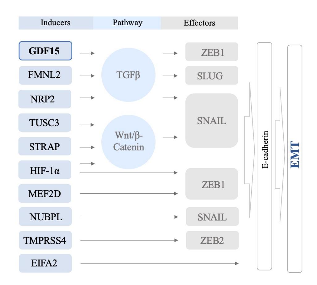
Fig. 1. Schematic of EMT induction
Adapted from “Regulation of EMT in Colorectal Cancer: A Culprit in Metastasis” by Vu T and Datta PK. Cancers (Basel). 2017 Dec 16;9(12):171 (1)
3. Oncogenic role of GDF-15 in Colorectal Cancer
Growth/differentiation factor-15, also known as macrophage inhibitory cytokine 1, nonsteroidal anti-inflammatory drug-activated gene-1, placental bone morphogenetic protein, prostate-derived factor or placental transforming growth factor (PTGFB), was identified more than two decades ago as a distant member of the transforming growth factor (TGF) superfamily (23). In situ hybridization revealed that the human GDF-15 gene is positioned on chromosome 19p13.1–13.2. The GDF-15 genes in humans, rats, and mice are comprised of two exons and one intron that interrupts the coding sequences in the pre-prodomain of their respective proteins (4).
GDF-15 has been shown to suppress early tumor promotion under normal physiological settings. However, aberrant expression of this gene in advanced malignancies results in proliferation, invasion, metastasis, cancer stem cell production, immune evasion, and a diminished response to treatment (24). GDF-15 has been shown to promote tumor cell proliferation, survival, invasion, metastasis, and chemoresistance (12), and has been associated with the development of several malignancies, including cervical cancer, colorectal cancer, and multiple myeloma (8).
A recent study examined the impact of an inflammatory microenvironment on colon cancer invasion and metastasis in vitro and in vivo. Anti-GDF-15 antibody therapy inhibited colon cancer cell invasion and migration in vitro, demonstrating that GDF-15 participates in inflammation-induced invasiveness. Lipopolysaccharide-induced inflammation elevated GDF-15 production in vivo, while c-Fos deletion inhibited colon cancer cell lung metastasis in a mouse model. Similarly, colon cancer metastases and TNM stages were shown to be linked with c-Fos expression in patient samples. As a result, the study established that GDF-15 stimulated colon cancer invasion and metastasis through the extracellular signal-regulated kinase 1/2 (Erk1/2)/cFos signaling pathway (25).
The results of a multivariate Cox analysis revealed that serum GDF-15 is an independent prognostic factor for CRC. Cancer-specific survival was higher for individuals with lower GDF-15 serum levels, according to the results of another study (26).
Conversely, other findings suggest that GDF-15 may function as a tumor suppressor gene in the context of colon cancer (27).
4. Circulating GDF-15 as a biomarker
The AJCC TNM classification is the best predictor of CRC prognosis. Other prognostic indicators include poorly differentiated or high histological grade, vascular or lymphatic invasion, intestinal obstruction or perforation at diagnosis, and increased preoperative serum carcinoembryonic antigen presence (CEA) (28). However, due to the great degree of individual heterogeneity, an individual’s chance of recurrence following therapy cannot be predicted properly (29). Allele loss of chromosome 18, microsatellite instability-deficient mismatch repair (MSI/dMMR) , and mutations in KRAS have been shown to negatively affect prognosis in cancer patients (30, 31). According to the research on EMT biomarkers, low expression of E-cadherin (32) and high expression of N-cadherin (21), Slug, and Vimentin (33) have been associated with a worse prognosis in CRC (34).
Several studies have attributed GDF-15 plasma levels to CRC diagnosis and death (11, 35, 36). High plasma levels of GDF-15 before diagnosis of CRC are related with an increased risk of CRC incidence and CRC-specific mortality, according to two significant cohorts evaluated in a prospective study, even after adjusting for other inflammatory indicators, risk factors, and clinical and pathological features (age, sex, tumor location, polyp history, tumor grade, or disease stage) (37).
Another study revealed that surgical excision of CRC decreases serum GDF-15, which was elevated in the context of tumor recurrence (26). Moreover, monitoring serum levels after surgery might help predict cancer recurrence. Further examination of the clinical significance of GDF-15 showed that serum levels are complementary to CEA and that GDF-15 is associated with the development or recurrence of CRC with liver metastases. However, GDF-15 blood concentrations were shown to be extremely variable in individuals with benign illness, indicating that serum levels should be read with caution (38), especially given the possibility of acute inflammation in CRC patients during chemotherapy (39).
Numerous articles have established that GDF-15 plays a critical function in tumor growth suppression, although they did not specifically study CRC.
One study tested the serum level of GDF-15 in 260 healthy blood donors as well as in 193 patients with adenomatous polyps or colon cancer. The study found that the TumorNode-Metastasis (TNM) stage was associated with the level of GDF-15 serum (4, 40).
Both the pro-domain and mature domains of GDF-15 may have various biological functions, depending on the context in which they are expressed (41, 42). It’s possible that during different phases of tumor evolution, these diverse forms may have contradictory purposes. In addition, GDF-15 may have varied roles in cancer depending on its intracellular location.
5. Resistance to chemotherapy
The conventional treatment for colon cancer is chemotherapy based on 5-fluorouracil (5-FU). Resistance to 5-FU is a major problem in clinical treatment, with much still unknown about the processes that allow colon cancer cells to become resistant to 5-FU treatment.
An investigation into whether 5-FU-resistant colon cancer cells undergo EMT and apoptosis, as well as the involvement of GDF-15 in controlling these processes revealed that 5-FU resistance in colon cancer was induced by suppression of GDF-15 and the downstream Smad signaling pathway. 5-FU-resistant human colon cancer cells/FU cells were created by exposing them to steadily increasing doses of 5-FU over time. Subsequently, HCT-15/FU cells resistant to 5-FU could be made susceptible to 5-FU by overexpressing GDF-15. Moreover, HCT-15/FU with decreased GDF-15 expression showed a more robust proliferation ability and metastatic propensity in vivo.
The findings suggest that the suppression of the Smad signaling pathway caused EMT in HCT-15/FU cells (via the modification of Ncadherin, E-cadherin, and MMP14 expression). The lack of E-cadherin in the HCT-15/FU group was further verified by in vivo tests (27).
Abbreviations
5-FU – 5-fluorouracil
AJCC-American Joint Committee on Cancer
CEA – carcinoembryonic antigen
CRC – colorectal cancer
EIF5A2 – eukaryotic initiation factor 5A2
EMT – epithelial-to-mesenchymal transition
Erk1/2 – extracellular signal-regulated kinase 1/2
FMNL2 – formin-like2).
GDF-15 – growth/differentiation factor-15
HIF-1 – hypoxia-inducible factor 1 alpha
MEF2D – myocyte enhancer factor 2D
MIC-1 – macrophage inhibitory cytokine 1
MSI/dMMR – microsatellite instability-deficient mismatch repair
MTA-1 – metastasis-associated protein 1
NAG-1 – nonsteroidal anti-inflammatory drug-activated gene-1
NRP2 – neuropilin-2
NUBPL – nucleotide binding protein-like
PDF – prostate-derived factor
PLAB – placental bone morphogenetic protein
PTGFB – placental transforming growth factor
STRAP – serine–threonine kinase receptor-associated protein
TGF – transforming growth factor
TMPRSS4 – transmembrane protease/serine 4
TNM – Tumor, Node, Metastasis
TUCS3 – tumor suppressor candidate 3
6. Conclusion and outlook
The EMT is one of the most widely acknowledged of the pathways that lead to metastasis, which accounts for the majority of cancer-related fatalities. To complete the process of EMT, a complex network of molecular signaling channels and regulators must operate together.
Although GDF-15 has antitumor action in the early stages of carcinogenesis, it has been shown to significantly increase the rate of cancer invasion and metastasis in advanced stages of cancer. Functional investigations of GDF-15’s role in malignancy are scarce and disputed: on one hand, the literature demonstrates overexpression of GDF-15 in cancers; on the other, GDF-15 has been found to act as a tumor suppressor.
To create optimal treatment methods for CRC, it is necessary to understand the fundamental underlying mechanisms behind these transformations, especially regarding how CRC cells gain invasive and metastatic features. Cancer treatment might benefit from a better comprehension of the EMT regulating mechanism. As yet, the involvement of GDF-15 in CRC metastasis and EMT is unclear.
Statements
Author Contributions: CL and CVL conceived of the presented idea. EAM, SMD, SR collected the data. CL and CVL drafted the manuscript. DS performed critical revision of the article.
Consent for publication: As the corresponding author, I confirm that the manuscript has been read and approved for submission by all co-authors
Funding:This study did not receive any specific grant from funding agencies in the public, commercial, or not-for-profit sector.
Conflicts of Interest: The authors declare no conflict of interest.
References
- Vu T, Datta P. Regulation of EMT in Colorectal Cancer: A Culprit in Metastasis. Cancers. 2017;9(12):171.
- Chen T, You Y, Jiang H, Wang ZZ. Epithelial-mesenchymal transition (EMT): A biological process in the development, stem cell differentiation, and tumorigenesis. J Cell Physiol. 2017 Dec;232(12):3261-3272.
- Lu W, Kang Y. Epithelial-Mesenchymal Plasticity in Cancer Progression and Metastasis. Dev Cell. 2019 May 6;49(3):361-374.
- Emmerson PJ, Duffin KL, Chintharlapalli S, Wu X. GDF-15 and Growth Control. Front Physiol. 2018 Nov 27;9:1712.
- Staff AC, Trovik J, Eriksson AG, Wik E, Wollert KC, Kempf T, Salvesen HB. Elevated plasma growth differentiation factor-15 correlates with lymph node metastases and poor survival in endometrial cancer. Clin Cancer Res. 2011 Jul 15;17(14):4825-33.
- Brown DA, Lindmark F, Stattin P, Bälter K, Adami HO, Zheng SL, Xu J, Isaacs WB, Grönberg H, Breit SN, Wiklund FE. Macrophage inhibitory cytokine 1: a new prognostic marker in prostate cancer. Clin Cancer Res. 2009 Nov 1;15(21):6658-64.
- Wang X, Li Y, Tian H, Qi J, Li M, Fu C, Wu F, Wang Y, Cheng D, Zhao W, Zhang C, Wang T, Rao J, Zhang W. Macrophage inhibitory cytokine 1 (MIC-1/GDF-15) as a novel diagnostic serum biomarker in pancreatic ductal adenocarcinoma. BMC Cancer. 2014 Aug 8;14:578.
- Mehta RS, Song M, Bezawada N, Wu K, Garcia-Albeniz X, Morikawa T, Fuchs CS, Ogino S, Giovannucci EL, Chan AT. A prospective study of macrophage inhibitory cytokine-1 (MIC-1/GDF-15) and risk of colorectal cancer. J Natl Cancer Inst. 2014;106(4).
- Yang MH, Kim J, Khan IA, Walker LA, Khan SI. Nonsteroidal anti-inflammatory drug activated gene-1 (NAG-1) modulators from natural products as anti-cancer agents. Life Sci. 2014 Apr 1;100(2):75-84.
- Zhong Y, Krisanapun C, Lee SH, Nualsanit T, Sams C, Peungvicha P, Baek SJ. Molecular targets of apigenin in colorectal cancer cells: involvement of p21, NAG-1 and p53. Eur J Cancer. 2010 Dec;46(18):3365-74.
- Wang X, Baek SJ, Eling TE. The diverse roles of nonsteroidal anti-inflammatory drug activated gene (NAG-1/GDF-15) in cancer. Biochem Pharmacol. 2013 Mar 1;85(5):597-606.
- Mimeault M, Batra SK. Divergent molecular mechanisms underlying the pleiotropic functions of macrophage inhibitory cytokine-1 in cancer. J Cell Physiol. 2010 Sep;224(3):626-35.
- Shook D, Keller R. Mechanisms, mechanics and function of epithelial-mesenchymal transitions in early development. Mech Dev. 2003 Nov;120(11):1351-83.
- Kalluri R. EMT: when epithelial cells decide to become mesenchymal-like cells. J Clin Invest. 2009 Jun;119(6):1417-9.
- Kang Y, Massagué J. Epithelial-mesenchymal transitions: twist in development and metastasis. Cell. 2004 Aug 6;118(3):277-9.
- Singh A, Settleman J. EMT, cancer stem cells and drug resistance: an emerging axis of evil in the war on cancer. Oncogene. 2010 Aug 26;29(34):4741-51.
- Thiery JP, Acloque H, Huang RY, Nieto MA. Epithelial-mesenchymal transitions in development and disease. Cell. 2009 Nov 25;139(5):871-90.
- Tsai JH, Yang J. Epithelial-mesenchymal plasticity in carcinoma metastasis. Genes Dev. 2013 Oct 15;27(20):2192-206.
- Yun JA, Kim SH, Hong HK, Yun SH, Kim HC, Chun HK, Cho YB, Lee WY. Loss of E-Cadherin expression is associated with a poor prognosis in stage III colorectal cancer. Oncology. 2014;86(5-6):318-28.
- Saito N, Nishimura H, Kameoka S. Clinical significance of fibronectin expression in colorectal cancer. Mol Med Rep. 2008;1(1):77-81.
- Yan X, Yan L, Liu S, Shan Z, Tian Y, Jin Z. N-cadherin, a novel prognostic biomarker, drives malignant progression of colorectal cancer. Mol Med Rep. 2015 Aug;12(2):2999-3006.
- Dongre A, Weinberg RA. New insights into the mechanisms of epithelial-mesenchymal transition and implications for cancer. Nat Rev Mol Cell Biol. 2019 Feb;20(2):69-84.
- Bootcov MR, Bauskin AR, Valenzuela SM, Moore AG, Bansal M, He XY, Zhang HP, Donnellan M, Mahler S, Pryor K, Walsh BJ, Nicholson RC, Fairlie WD, Por SB, Robbins JM, Breit SN. MIC-1, a novel macrophage inhibitory cytokine, is a divergent member of the TGF-beta superfamily. Proc Natl Acad Sci U S A. 1997 Oct 14;94(21):11514-9.
- Fang L, Li F, Gu C. GDF-15: A Multifunctional Modulator and Potential Therapeutic Target in Cancer. Curr Pharm Des. 2019;25(6):654-662.
- Ding Y, Hao K, Li Z, Ma R, Zhou Y, Zhou Z, Wei M, Liao Y, Dai Y, Yang Y, Zhang X, Zhao L. c-Fos separation from Lamin A/C by GDF-15 promotes colon cancer invasion and metastasis in inflammatory microenvironment. J Cell Physiol. 2020 May;235(5):4407-4421.
- Wang X, Yang Z, Tian H, Li Y, Li M, Zhao W, Zhang C, Wang T, Liu J, Zhang A, Shen D, Zheng C, Qi J, Zhao D, Shi J, Jin L, Rao J, Zhang W. Circulating MIC-1/GDF-15 is a complementary screening biomarker with CEA and correlates with liver metastasis and poor survival in colorectal cancer. Oncotarget. 2017 Apr 11;8(15):24892-24901.
- Wang B, Ma N, Zheng X, Li X, Ma X, Hu J, Cao B. GDF-15 Repression Contributes to 5-Fluorouracil Resistance in Human Colon Cancer by Regulating Epithelial-Mesenchymal Transition and Apoptosis. Biomed Res Int. 2020 Sep 28;2020:2826010.
- Compton CC, Fielding LP, Burgart LJ, Conley B, Cooper HS, Hamilton SR, Hammond ME, Henson DE, Hutter RV, Nagle RB, Nielsen ML, Sargent DJ, Taylor CR, Welton M, Willett C. Prognostic factors in colorectal cancer. College of American Pathologists Consensus Statement 1999. Arch Pathol Lab Med. 2000 Jul;124(7):979-94.
- Zafar SN, Hu CY, Snyder RA, Cuddy A, You YN, Lowenstein LM, Volk RJ, Chang GJ. Predicting Risk of Recurrence After Colorectal Cancer Surgery in the United States: An Analysis of a Special Commission on Cancer National Study. Ann Surg Oncol. 2020 Aug;27(8):2740-2749.
- Kawakami H, Zaanan A, Sinicrope FA. Microsatellite instability testing and its role in the management of colorectal cancer. Curr Treat Options Oncol. 2015 Jul;16(7):30.
- Deng Y, Wang L, Tan S, Kim GP, Dou R, Chen D, Cai Y, Fu X, Wang L, Zhu J, Wang J. KRAS as a predictor of poor prognosis and benefit from postoperative FOLFOX chemotherapy in patients with stage II and III colorectal cancer. Mol Oncol. 2015 Aug;9(7):1341-7.
- Christou N, Perraud A, Blondy S, Jauberteau MO, Battu S, Mathonnet M. E-cadherin: A potential biomarker of colorectal cancer prognosis. Oncol Lett. 2017 Jun;13(6):4571-4576.
- Toiyama Y, Yasuda H, Saigusa S, Tanaka K, Inoue Y, Goel A, Kusunoki M. Increased expression of Slug and Vimentin as novel predictive biomarkers for lymph node metastasis and poor prognosis in colorectal cancer. Carcinogenesis. 2013 Nov;34(11):2548-57.
- Parisi E, Sorolla A, Montal R, González-Resina R, Novell A, Salud A, Sorolla MA. Prognostic Factors Involved in the Epithelial-Mesenchymal Transition Process in Colorectal Cancer Have a Preponderant Role in Oxidative Stress: A Systematic Review and Meta-Analysis. Cancers (Basel). 2020 Nov 11;12(11):3330.
- Li C, Wang X, Casal I, Wang J, Li P, Zhang W, Xu E, Lai M, Zhang H. Growth differentiation factor 15 is a promising diagnostic and prognostic biomarker in colorectal cancer. J Cell Mol Med. 2016 Aug;20(8):1420-6.
- Wallin U, Glimelius B, Jirström K, Darmanis S, Nong RY, Pontén F, Johansson C, Påhlman L, Birgisson H. Growth differentiation factor 15: a prognostic marker for recurrence in colorectal cancer. Br J Cancer. 2011 May 10;104(10):1619-27.
- Mehta RS, Chong DQ, Song M, Meyerhardt JA, Ng K, Nishihara R, Qian Z, Morikawa T, Wu K, Giovannucci EL, Fuchs CS, Ogino S, Chan AT. Association Between Plasma Levels of Macrophage Inhibitory Cytokine-1 Before Diagnosis of Colorectal Cancer and Mortality. Gastroenterology. 2015 Sep;149(3):614-22.
- Kim JM, Kosak JP, Kim JK, Kissling G, Germolec DR, Zeldin DC, Bradbury JA, Baek SJ, Eling TE. NAG-1/GDF-15 transgenic mouse has less white adipose tissue and a reduced inflammatory response. Mediators Inflamm. 2013;2013:641851.
- Lee HN, Na HK, Surh YJ. Resolution of inflammation as a novel chemopreventive strategy. Semin Immunopathol. 2013 Mar;35(2):151-61.
- Brown DA, Ward RL, Buckhaults P, Liu T, Romans KE, Hawkins NJ, Bauskin AR, Kinzler KW, Vogelstein B, Breit SN. MIC-1 serum level and genotype: associations with progress and prognosis of colorectal carcinoma. Clin Cancer Res. 2003 Jul;9(7):2642-50.
- Bauskin AR, Zhang HP, Fairlie WD, He XY, Russell PK, Moore AG, Brown DA, Stanley KK, Breit SN. The propeptide of macrophage inhibitory cytokine (MIC-1), a TGF-beta superfamily member, acts as a quality control determinant for correctly folded MIC-1. EMBO J. 2000 May 15;19(10):2212-20.
- Bauskin AR, Brown DA, Junankar S, Rasiah KK, Eggleton S, Hunter M, Liu T, Smith D, Kuffner T, Pankhurst GJ, Johnen H, Russell PJ, Barret W, Stricker PD, Grygiel JJ, Kench JG, Henshall SM, Sutherland RL, Breit SN. The propeptide mediates formation of stromal stores of PROMIC-1: role in determining prostate cancer outcome. Cancer Res. 2005 Mar 15;65(6):2330-6.